|
Milky Spots
Milky spots are very small white-coloured areas of lymphoid tissue, found in the peritoneal, pleural and pericardial cavities. These are the three coelomic cavities that form as separate compartments from the intraembryonic coelom. More milky spots are found on the greater omentum in the peritoneal cavity than anywhere else. The milky spots are made up of mesenchymal cells and are covered in a layer of mesothelium. These structures surround the small blood vessels. The enclosing mesothelium contains macrophages, lymphocytes and mast cell A mast cell (also known as a mastocyte or a labrocyte) is a resident cell of connective tissue that contains many granules rich in histamine and heparin. Specifically, it is a type of granulocyte derived from the myeloid stem cell that is a par ...s. They are also known as secondary lymphoid organs. Most milky spots contain extremely thin-walled lymphatic capillaries. The macrophages are usually located on the edge of the milky spot and the l ... [...More Info...] [...Related Items...] OR: [Wikipedia] [Google] [Baidu] |
Lymphoid Tissue
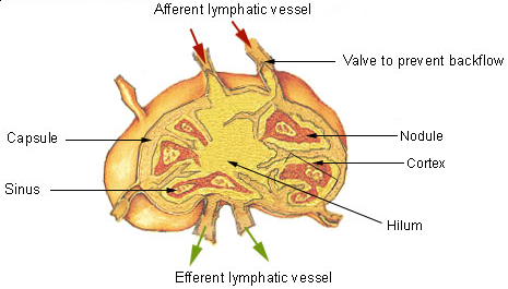
The lymphatic system, or lymphoid system, is an organ system in vertebrates that is part of the immune system, and complementary to the circulatory system. It consists of a large network of lymphatic vessels, lymph nodes, lymphatic or lymphoid organs, and lymphoid tissues. The vessels carry a clear fluid called lymph (the Latin word ''lympha'' refers to the deity of fresh water, "Lympha") back towards the heart, for re-circulation. Unlike the circulatory system that is a closed system, the lymphatic system is open. The human circulatory system processes an average of 20 litres of blood per day through capillary filtration, which removes plasma from the blood. Roughly 17 litres of the filtered blood is reabsorbed directly into the blood vessels, while the remaining three litres are left in the interstitial fluid. One of the main functions of the lymphatic system is to provide an accessory return route to the blood for the surplus three litres. The other main function is that of i ... [...More Info...] [...Related Items...] OR: [Wikipedia] [Google] [Baidu] |
Peritoneal
The peritoneum is the serous membrane forming the lining of the abdominal cavity or coelom in amniotes and some invertebrates, such as annelids. It covers most of the intra-abdominal (or coelomic) organs, and is composed of a layer of mesothelium supported by a thin layer of connective tissue. This peritoneal lining of the cavity supports many of the abdominal organs and serves as a conduit for their blood vessels, lymphatic vessels, and nerves. The abdominal cavity (the space bounded by the vertebrae, abdominal muscles, diaphragm, and pelvic floor) is different from the intraperitoneal space (located within the abdominal cavity but wrapped in peritoneum). The structures within the intraperitoneal space are called "intraperitoneal" (e.g., the stomach and intestines), the structures in the abdominal cavity that are located behind the intraperitoneal space are called "retroperitoneal" (e.g., the kidneys), and those structures below the intraperitoneal space are called "subperi ... [...More Info...] [...Related Items...] OR: [Wikipedia] [Google] [Baidu] |
Pleural
The pleural cavity, pleural space, or interpleural space is the potential space between the pleurae of the pleural sac that surrounds each lung. A small amount of serous pleural fluid is maintained in the pleural cavity to enable lubrication between the membranes, and also to create a pressure gradient. The serous membrane that covers the surface of the lung is the visceral pleura and is separated from the outer membrane the parietal pleura by just the film of pleural fluid in the pleural cavity. The visceral pleura follows the fissures of the lung and the root of the lung structures. The parietal pleura is attached to the mediastinum, the upper surface of the diaphragm, and to the inside of the ribcage. Structure In humans, the left and right lungs are completely separated by the mediastinum, and there is no communication between their pleural cavities. Therefore, in cases of a unilateral pneumothorax, the contralateral lung will remain functioning normally unless there is a ... [...More Info...] [...Related Items...] OR: [Wikipedia] [Google] [Baidu] |
Pericardial Cavity
The pericardium, also called pericardial sac, is a double-walled sac containing the heart and the roots of the great vessels. It has two layers, an outer layer made of strong connective tissue (fibrous pericardium), and an inner layer made of serous membrane (serous pericardium). It encloses the pericardial cavity, which contains pericardial fluid, and defines the middle mediastinum. It separates the heart from interference of other structures, protects it against infection and blunt trauma, and lubricates the heart's movements. The English name originates from the Ancient Greek prefix "''peri-''" (περί; "around") and the suffix "''-cardion''" (κάρδιον; "heart"). Anatomy The pericardium is a tough fibroelastic sac which covers the heart from all sides except at the cardiac root (where the great vessels join the heart) and the bottom (where only the serous pericardium exists to cover the upper surface of the central tendon of diaphragm, central tendon of thoracic d ... [...More Info...] [...Related Items...] OR: [Wikipedia] [Google] [Baidu] |
Coelom
The coelom (or celom) is the main body cavity in most animals and is positioned inside the body to surround and contain the digestive tract and other organs. In some animals, it is lined with mesothelium. In other animals, such as molluscs, it remains undifferentiated. In the past, and for practical purposes, coelom characteristics have been used to classify bilaterian animal phyla into informal groups. Etymology The term ''coelom'' derives from the Ancient Greek word (), meaning 'cavity'. Structure Development The coelom is the mesodermally lined cavity between the gut and the outer body wall. During the development of the embryo, coelom formation begins in the gastrulation stage. The developing digestive tube of an embryo forms as a blind pouch called the archenteron. In Protostomes, the coelom forms by a process known as schizocoely. The archenteron initially forms, and the mesoderm splits into two layers: the first attaches to the body wall or ectoderm, forming ... [...More Info...] [...Related Items...] OR: [Wikipedia] [Google] [Baidu] |
Body Cavity
A body cavity is any space or compartment, or potential space, in an animal body. Cavities accommodate organs and other structures; cavities as potential spaces contain fluid. The two largest human body cavities are the ventral body cavity, and the dorsal body cavity. In the dorsal body cavity the brain and spinal cord are located. The membranes that surround the central nervous system organs (the brain and the spinal cord, in the cranial and spinal cavities) are the three meninges. The differently lined spaces contain different types of fluid. In the meninges for example the fluid is cerebrospinal fluid; in the abdominal cavity the fluid contained in the peritoneum is a serous fluid. In amniotes and some invertebrates the peritoneum lines their largest body cavity called the coelom. Mammals Mammalian embryos develop two body cavities: the intraembryonic coelom and the extraembryonic coelom (or chorionic cavity). The intraembryonic coelom is lined by somatic and splanchnic ... [...More Info...] [...Related Items...] OR: [Wikipedia] [Google] [Baidu] |
Intraembryonic Coelom
In the development of the human embryo the intraembryonic coelom (or somatic coelom) is a portion of the conceptus forming in the mesoderm during the third week of development. During the third week of development, the lateral plate mesoderm splits into a dorsal somatic mesoderm (somatopleure) and a ventral splanchnic mesoderm (splanchnopleure). The resulting cavity between the somatopleure and splanchnopleure is called the intraembryonic coelom. This space will give rise to the thoracic and abdominal cavities. The coelomic spaces in the lateral mesoderm and cardiogenic area are isolated. The isolated coelom begins to organize into a horseshoe shape. The spaces soon join together and form a single horseshoe-shaped cavity: the intraembryonic coelom. It then separates the mesoderm into two layers. It briefly has a connection with the extraembryonic coelom. See also * Cavitation (embryology) Cavitation is a process in early embryonic development that follows cleavage. Cavitation ... [...More Info...] [...Related Items...] OR: [Wikipedia] [Google] [Baidu] |
Greater Omentum
The greater omentum (also the great omentum, omentum majus, gastrocolic omentum, epiploon, or, especially in animals, caul) is a large apron-like fold of visceral peritoneum that hangs down from the stomach. It extends from the greater curvature of the stomach, passing in front of the small intestines and doubles back to ascend to the transverse colon before reaching to the posterior abdominal wall. The greater omentum is larger than the lesser omentum, which hangs down from the liver to the lesser curvature. The common anatomical term "epiploic" derives from "epiploon", from the Greek ''epipleein'', meaning to float or sail on, since the greater omentum appears to float on the surface of the intestines. It is the first structure observed when the abdominal cavity is opened anteriorly (from the front). Structure The greater omentum is the larger of the two peritoneal folds. It consists of a double sheet of peritoneum, folded on itself so that it has four layers. The two layers o ... [...More Info...] [...Related Items...] OR: [Wikipedia] [Google] [Baidu] |
Mesenchymal
Mesenchyme () is a type of loosely organized animal embryonic connective tissue of undifferentiated cells that give rise to most tissues, such as skin, blood or bone. The interactions between mesenchyme and epithelium help to form nearly every organ in the developing embryo. Vertebrates Structure Mesenchyme is characterized morphologically by a prominent ground substance matrix containing a loose aggregate of reticular fibers and unspecialized mesenchymal stem cells. Mesenchymal cells can migrate easily (in contrast to epithelial cells, which lack mobility), are organized into closely adherent sheets, and are polarized in an apical-basal orientation. Development The mesenchyme originates from the mesoderm. From the mesoderm, the mesenchyme appears as an embryologically primitive "soup". This "soup" exists as a combination of the mesenchymal cells plus serous fluid plus the many different tissue proteins. Serous fluid is typically stocked with the many serous elements, such a ... [...More Info...] [...Related Items...] OR: [Wikipedia] [Google] [Baidu] |
Mesothelium
The mesothelium is a membrane composed of simple squamous epithelial cells of mesodermal origin, which forms the lining of several body cavities: the pleura (pleural cavity around the lungs), peritoneum (abdominopelvic cavity including the mesentery, omenta, falciform ligament and the perimetrium) and pericardium (around the heart). Mesothelial tissue also surrounds the male testis (as the tunica vaginalis) and occasionally the spermatic cord (in a patent processus vaginalis). Mesothelium that covers the internal organs is called visceral mesothelium, while one that covers the surrounding body walls is called the parietal mesothelium. The mesothelium that secretes serous fluid as a main function is also known as a serosa. Origin Mesothelium derives from the embryonic mesoderm cell layer, that lines the coelom (body cavity) in the embryo. It develops into the layer of cells that covers and protects most of the internal organs of the body. Structure The mesothelium forms a ... [...More Info...] [...Related Items...] OR: [Wikipedia] [Google] [Baidu] |
Blood Vessels
The blood vessels are the components of the circulatory system that transport blood throughout the human body. These vessels transport blood cells, nutrients, and oxygen to the tissues of the body. They also take waste and carbon dioxide away from the tissues. Blood vessels are needed to sustain life, because all of the body's tissues rely on their functionality. There are five types of blood vessels: the arteries, which carry the blood away from the heart; the arterioles; the capillaries, where the exchange of water and chemicals between the blood and the tissues occurs; the venules; and the veins, which carry blood from the capillaries back towards the heart. The word ''vascular'', meaning relating to the blood vessels, is derived from the Latin ''vas'', meaning vessel. Some structures – such as cartilage, the epithelium, and the lens and cornea of the eye – do not contain blood vessels and are labeled ''avascular''. Etymology * artery: late Middle English; from Latin ' ... [...More Info...] [...Related Items...] OR: [Wikipedia] [Google] [Baidu] |
Macrophage
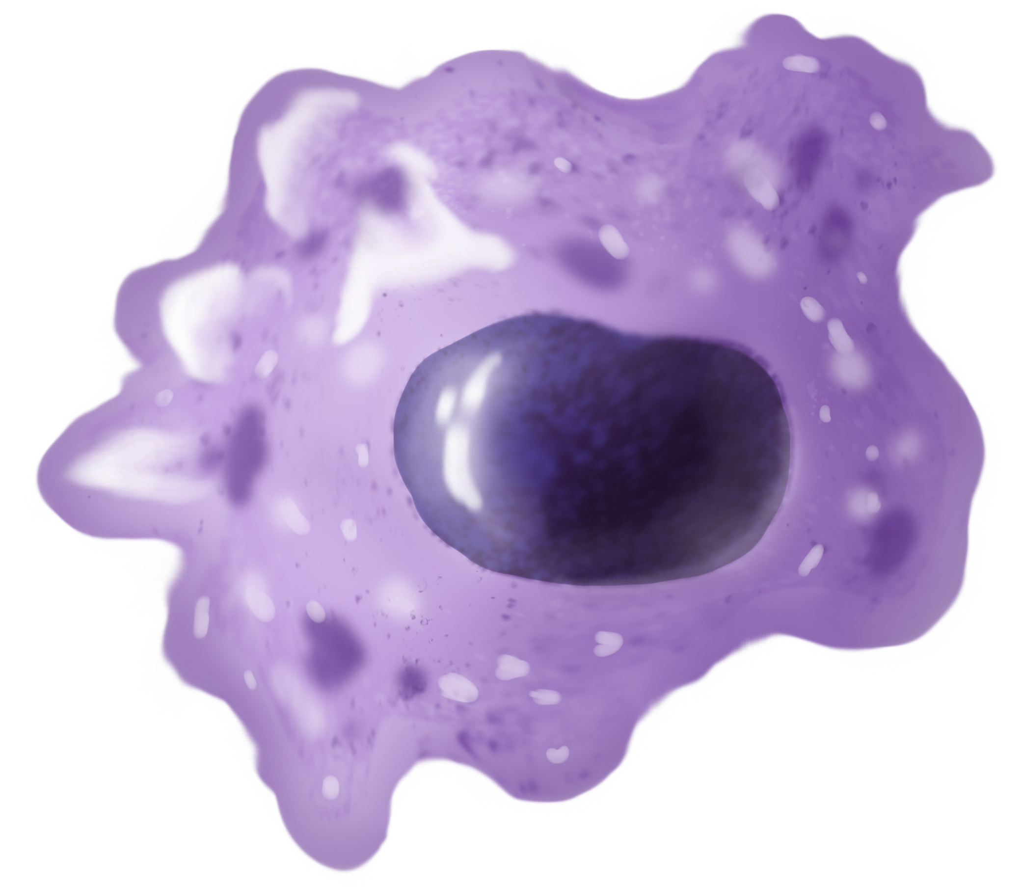
Macrophages (abbreviated as M φ, MΦ or MP) ( el, large eaters, from Greek ''μακρός'' (') = large, ''φαγεῖν'' (') = to eat) are a type of white blood cell of the immune system that engulfs and digests pathogens, such as cancer cells, microbes, cellular debris, and foreign substances, which do not have proteins that are specific to healthy body cells on their surface. The process is called phagocytosis, which acts to defend the host against infection and injury. These large phagocytes are found in essentially all tissues, where they patrol for potential pathogens by amoeboid movement. They take various forms (with various names) throughout the body (e.g., histiocytes, Kupffer cells, alveolar macrophages, microglia, and others), but all are part of the mononuclear phagocyte system. Besides phagocytosis, they play a critical role in nonspecific defense (innate immunity) and also help initiate specific defense mechanisms (adaptive immunity) by recruiting other immune ... [...More Info...] [...Related Items...] OR: [Wikipedia] [Google] [Baidu] |


