|
Lymphoid Tissue
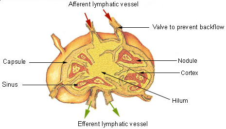
The lymphatic system, or lymphoid system, is an organ system in vertebrates that is part of the immune system, and complementary to the circulatory system. It consists of a large network of lymphatic vessels, lymph nodes, lymphatic or lymphoid organs, and lymphoid tissues. The vessels carry a clear fluid called lymph (the Latin word ''lympha'' refers to the deity of fresh water, "Lympha") back towards the heart, for re-circulation. Unlike the circulatory system that is a closed system, the lymphatic system is open. The human circulatory system processes an average of 20 litres of blood per day through capillary filtration, which removes plasma from the blood. Roughly 17 litres of the filtered blood is reabsorbed directly into the blood vessels, while the remaining three litres are left in the interstitial fluid. One of the main functions of the lymphatic system is to provide an accessory return route to the blood for the surplus three litres. The other main function is that of i ... [...More Info...] [...Related Items...] OR: [Wikipedia] [Google] [Baidu] |
Organ System
An organ system is a biological system consisting of a group of organs that work together to perform one or more functions. Each organ has a specialized role in a plant or animal body, and is made up of distinct tissues. Plants Plants have two major organ systems. Vascular plants have two distinct organ systems: a shoot system, and a root system. The shoot system consists stems, leaves, and the reproductive parts of the plant (flowers and fruits). The shoot system generally grows above ground, where it absorbs the light needed for photosynthesis. The root system, which supports the plants and absorbs water and minerals, is usually underground. Animals Other animals have similar organ systems to humans although simpler animals may have fewer organs in an organ system or even fewer organ systems. Humans There are 11 distinct organ systems in human beings, which form the basis of human anatomy and physiology. The 11 organ systems include the respiratory system, dige ... [...More Info...] [...Related Items...] OR: [Wikipedia] [Google] [Baidu] |
Lymphocyte
A lymphocyte is a type of white blood cell (leukocyte) in the immune system of most vertebrates. Lymphocytes include natural killer cells (which function in cell-mediated, cytotoxic innate immunity), T cells (for cell-mediated, cytotoxic adaptive immunity), and B cells (for humoral, antibody-driven adaptive immunity). They are the main type of cell found in lymph, which prompted the name "lymphocyte". Lymphocytes make up between 18% and 42% of circulating white blood cells. Types The three major types of lymphocyte are T cells, B cells and natural killer (NK) cells. Lymphocytes can be identified by their large nucleus. T cells and B cells T cells (thymus cells) and B cells ( bone marrow- or bursa-derived cells) are the major cellular components of the adaptive immune response. T cells are involved in cell-mediated immunity, whereas B cells are primarily responsible for humoral immunity (relating to antibodies). The function of T cells and B cells is to recognize sp ... [...More Info...] [...Related Items...] OR: [Wikipedia] [Google] [Baidu] |
Olaus Rudbeck
Olaus Rudbeck (also known as Olof Rudbeck the Elder, to distinguish him from his son, and occasionally with the surname Latinized as ''Olaus Rudbeckius'') (13 September 1630 – 12 December 1702) was a Swedish scientist and writer, professor of medicine at Uppsala University, and for several periods ''rector magnificus'' of the same university. He was born in Västerås, the son of Bishop Johannes Rudbeckius, who was personal chaplain to King Gustavus Adolphus, and the father of botanist Olof Rudbeck the Younger. Rudbeck is primarily known for his contributions in two fields: human anatomy and linguistics, but he was also accomplished in many other fields including music and botany. He established the first botanical garden in Sweden at Uppsala, called Rudbeck's Garden, but which was renamed a hundred years later for his son's student, the botanist Carl Linnaeus. Human anatomy Rudbeck was one of the pioneers in the study of lymphatic vessels. According to his supporters in Sw ... [...More Info...] [...Related Items...] OR: [Wikipedia] [Google] [Baidu] |
Lymph Heart
A lymph heart is an organ which pumps lymph in lungfishes, amphibians, reptiles, and flightless birds back into the circulatory system. In some amphibian species, lymph hearts are in pairs, and may number as many as 200 in one animal the size of a worm, while salamanders have as many as 23 pairs of lymph hearts. Lymph hearts are thought to have evolved in ''Rhipidistia''. Mammals have lost the lymph heart as a centralized organ, instead having the lymph vessel themselves contract to pump lymph. and other amphibians The lymphatic system of a frog consists of lymph, lymph vessels, lymph heart, lymph spaces and spleen. Some mast cells can also be found in the lymphatics of the tongue of some of the frog species. Lymphatics and lymph As lymph is a filtrate of blood, it closely resembles the plasma in its water content. Lymph also contains a small amount of metabolic waste and a much smaller amount of protein than that of blood. Lymph vessels carry the lymph and, in the frog, open ... [...More Info...] [...Related Items...] OR: [Wikipedia] [Google] [Baidu] |
Subclavian Vein
The subclavian vein is a paired large vein, one on either side of the body, that is responsible for draining blood from the upper extremities, allowing this blood to return to the heart. The left subclavian vein plays a key role in the absorption of lipids, by allowing products that have been carried by lymph in the thoracic duct to enter the bloodstream. The diameter of the subclavian veins is approximately 1–2 cm, depending on the individual. Structure Each subclavian vein is a continuation of the axillary vein and runs from the outer border of the first rib to the medial border of anterior scalene muscle. From here it joins with the internal jugular vein to form the brachiocephalic vein (also known as "innominate vein"). The angle of union is termed the venous angle. The subclavian vein follows the subclavian artery and is separated from the subclavian artery by the insertion of anterior scalene. Thus, the subclavian vein lies anterior to the anterior scalene while the su ... [...More Info...] [...Related Items...] OR: [Wikipedia] [Google] [Baidu] |
Thoracic Duct
In human anatomy, the thoracic duct is the larger of the two lymph ducts of the lymphatic system. It is also known as the ''left lymphatic duct'', ''alimentary duct'', ''chyliferous duct'', and ''Van Hoorne's canal''. The other duct is the right lymphatic duct. The thoracic duct carries chyle, a liquid containing both lymph and emulsified fats, rather than pure lymph. It also collects most of the lymph in the body other than from the right thorax, arm, head, and neck (which are drained by the right lymphatic duct). The thoracic duct usually starts from the level of the twelfth thoracic vertebra (T12) and extends to the root of the neck. It drains into the systemic (blood) circulation at the junction of the left subclavian and internal jugular veins, at the commencement of the brachiocephalic vein. When the duct ruptures, the resulting flood of liquid into the pleural cavity is known as chylothorax. Structure In adults, the thoracic duct is typically 38–45 cm in length an ... [...More Info...] [...Related Items...] OR: [Wikipedia] [Google] [Baidu] |
Right Lymphatic Duct
The right lymphatic duct is an important lymphatic vessel that drains the right upper quadrant of the body. It forms various combinations with the right subclavian vein and right internal jugular vein. Structure The right lymphatic duct courses along the medial border of the anterior scalene at the root of the neck. The right lymphatic duct forms various combinations with the right subclavian vein and right internal jugular vein. It is approximately 1.25 cm long. Variations A right lymphatic duct that enters directly into the junction of the internal jugular and subclavian veins is uncommon. Function The right duct drains lymph fluid from: * the upper right section of the trunk, (right thoracic cavity, via the right bronchomediastinal trunk ), * the right arm (via the right subclavian trunk ), * and right side of the head and neck (via the right jugular trunk), * also, in some individuals, the lower lobe of the left lung. All other sections of the human body are d ... [...More Info...] [...Related Items...] OR: [Wikipedia] [Google] [Baidu] |
Lymph Duct
A lymph duct is a great lymphatic vessel that empties lymph into one of the subclavian veins. There are two lymph ducts in the body—the right lymphatic duct and the thoracic duct. The right lymphatic duct drains lymph from the right upper limb, right side of thorax and right halves of head and neck. The thoracic duct drains lymph into the circulatory system at the left brachiocephalic vein between the left subclavian and left internal jugular veins. See also * Lymphatic system * Right lymphatic duct * Thoracic duct In human anatomy, the thoracic duct is the larger of the two lymph ducts of the lymphatic system. It is also known as the ''left lymphatic duct'', ''alimentary duct'', ''chyliferous duct'', and ''Van Hoorne's canal''. The other duct is the right ... References Lymphatic system {{lymphatic-stub ... [...More Info...] [...Related Items...] OR: [Wikipedia] [Google] [Baidu] |
Mucosa-associated Lymphoid Tissue
The mucosa-associated lymphoid tissue (MALT), also called mucosa-associated lymphatic tissue, is a diffuse system of small concentrations of lymphoid tissue found in various submucosal membrane sites of the body, such as the gastrointestinal tract, nasopharynx, thyroid, breast, lung, salivary glands, eye, and skin. MALT is populated by lymphocytes such as T cells and B cells, as well as plasma cells and macrophages, each of which is well situated to encounter antigens passing through the mucosal epithelium. In the case of intestinal MALT, M cells are also present, which sample antigen from the lumen and deliver it to the lymphoid tissue. MALT constitute about 50% of the lymphoid tissue in human body. Immune responses that occur at mucous membranes are studied by mucosal immunology. Categorization The components of MALT are sometimes subdivided into the following: * GALT (gut-associated lymphoid tissue. Peyer's patches are a component of GALT found in the lining of the small in ... [...More Info...] [...Related Items...] OR: [Wikipedia] [Google] [Baidu] |
Mucosa
A mucous membrane or mucosa is a membrane that lines various cavities in the body of an organism and covers the surface of internal organs. It consists of one or more layers of epithelial cells overlying a layer of loose connective tissue. It is mostly of endodermal origin and is continuous with the skin at body openings such as the eyes, eyelids, ears, inside the nose, inside the mouth, lips, the genital areas, the urethral opening and the anus. Some mucous membranes secrete mucus, a thick protective fluid. The function of the membrane is to stop pathogens and dirt from entering the body and to prevent bodily tissues from becoming dehydrated. Structure The mucosa is composed of one or more layers of epithelial cells that secrete mucus, and an underlying lamina propria of loose connective tissue. The type of cells and type of mucus secreted vary from organ to organ and each can differ along a given tract. Mucous membranes line the digestive, respiratory and reproductive trac ... [...More Info...] [...Related Items...] OR: [Wikipedia] [Google] [Baidu] |
Stromal Cell
Stromal cells, or mesenchymal stromal cells, are differentiating cells found in abundance within bone marrow but can also be seen all around the body. Stromal cells can become connective tissue cells of any organ, for example in the uterine mucosa (endometrium), prostate, bone marrow, lymph node and the ovary. They are cells that support the function of the parenchymal cells of that organ. The most common stromal cells include fibroblasts and pericytes. The term ''stromal'' comes from Latin , "bed covering", and Ancient Greek , , "bed". Stromal cells are an important part of the body's immune response and modulate inflammation through multiple pathways. They also aid in differentiation of hematopoietic cells and forming necessary blood elements. The interaction between stromal cells and tumor cells is known to play a major role in cancer growth and progression. In addition, by regulating local cytokine networks (e.g. M-CSF, LIF), bone marrow stromal cells have been described to be ... [...More Info...] [...Related Items...] OR: [Wikipedia] [Google] [Baidu] |
Bone Marrow
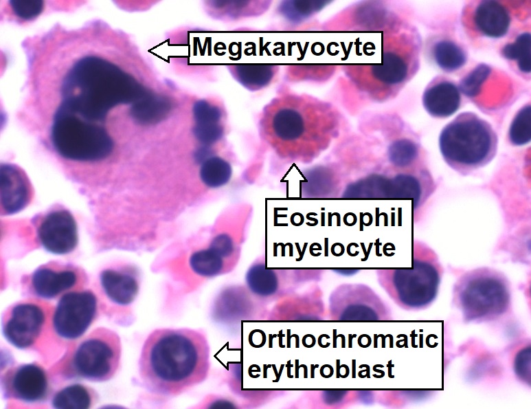
Bone marrow is a semi-solid tissue found within the spongy (also known as cancellous) portions of bones. In birds and mammals, bone marrow is the primary site of new blood cell production (or haematopoiesis). It is composed of hematopoietic cells, marrow adipose tissue, and supportive stromal cells. In adult humans, bone marrow is primarily located in the ribs, vertebrae, sternum, and bones of the pelvis. Bone marrow comprises approximately 5% of total body mass in healthy adult humans, such that a man weighing 73 kg (161 lbs) will have around 3.7 kg (8 lbs) of bone marrow. Human marrow produces approximately 500 billion blood cells per day, which join the systemic circulation via permeable vasculature sinusoids within the medullary cavity. All types of hematopoietic cells, including both myeloid and lymphoid lineages, are created in bone marrow; however, lymphoid cells must migrate to other lymphoid organs (e.g. thymus) in order to complete maturation. ... [...More Info...] [...Related Items...] OR: [Wikipedia] [Google] [Baidu] |


