|
Microglia
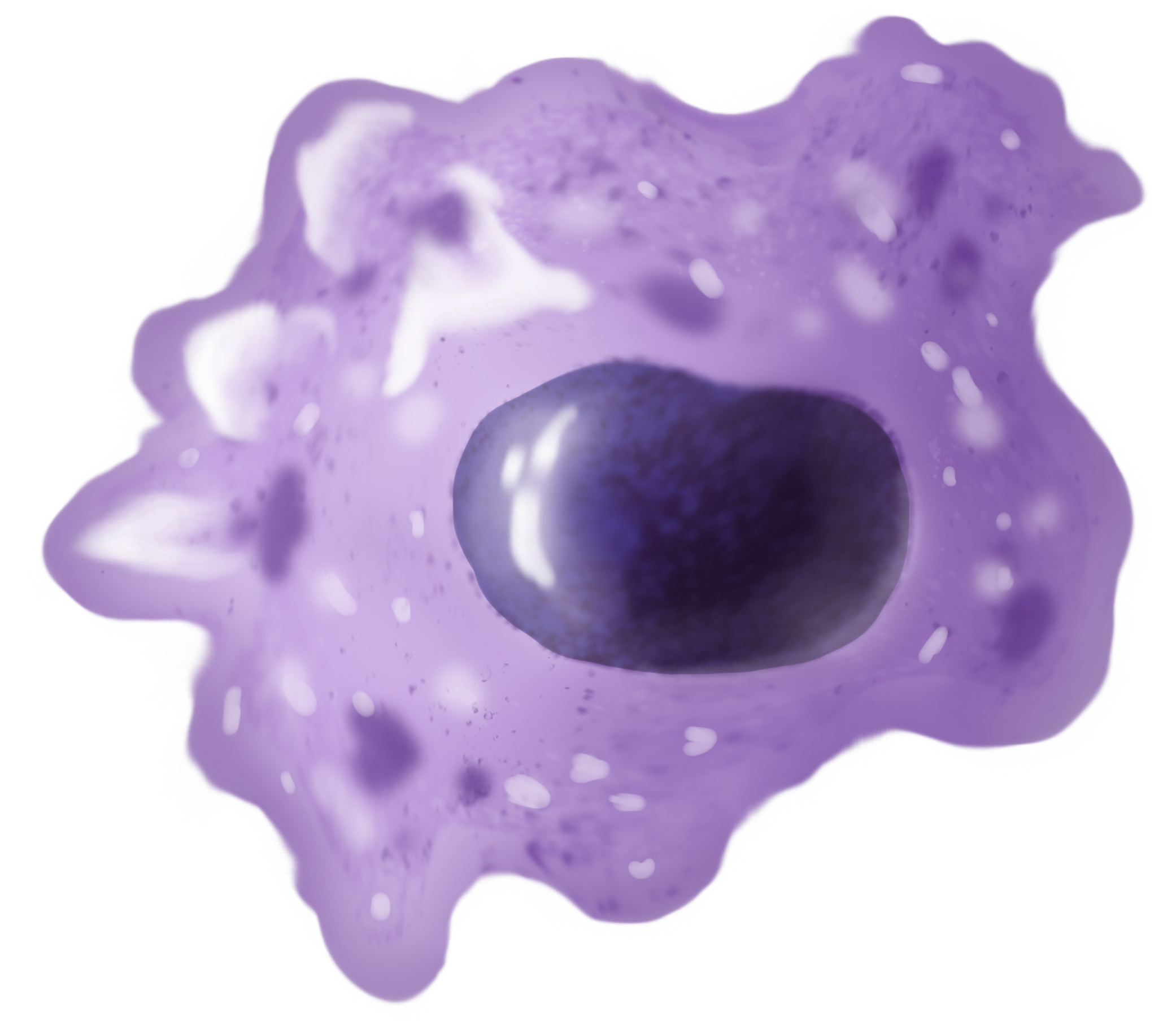
Microglia are a type of glia, glial cell located throughout the brain and spinal cord of the central nervous system (CNS). Microglia account for about around 5–10% of cells found within the brain. As the resident macrophage cells, they act as the first and main form of active immune defense in the CNS. Microglia originate in the yolk sac under tightly regulated molecular conditions. These cells (and other neuroglia including astrocytes) are distributed in large non-overlapping regions throughout the CNS. Microglia are key cells in overall brain maintenancethey are constantly scavenging the CNS for senile plaques, plaques, damaged or unnecessary neurons and synapses, and infectious agents. Since these processes must be efficient to prevent potentially fatal damage, microglia are extremely sensitive to even small pathological changes in the CNS. This sensitivity is achieved in part by the presence of unique potassium channels that respond to even small changes in extracellular pota ... [...More Info...] [...Related Items...] OR: [Wikipedia] [Google] [Baidu] |
Glia
Glia, also called glial cells (gliocytes) or neuroglia, are non-neuronal cells in the central nervous system (the brain and the spinal cord) and in the peripheral nervous system that do not produce electrical impulses. The neuroglia make up more than one half the volume of neural tissue in the human body. They maintain homeostasis, form myelin, and provide support and protection for neurons. In the central nervous system, glial cells include oligodendrocytes (that produce myelin), astrocytes, ependymal cells and microglia, and in the peripheral nervous system they include Schwann cells (that produce myelin), and satellite cells. Function They have four main functions: * to surround neurons and hold them in place * to supply nutrients and oxygen to neurons * to insulate one neuron from another * to destroy pathogens and remove dead neurons. They also play a role in neurotransmission and synaptic connections, and in physiological processes such as breathing. While glia we ... [...More Info...] [...Related Items...] OR: [Wikipedia] [Google] [Baidu] |
Neurons
A neuron (American English), neurone (British English), or nerve cell, is an membrane potential#Cell excitability, excitable cell (biology), cell that fires electric signals called action potentials across a neural network (biology), neural network in the nervous system. They are located in the nervous system and help to receive and conduct impulses. Neurons communicate with other cells via synapses, which are specialized connections that commonly use minute amounts of chemical neurotransmitters to pass the electric signal from the presynaptic neuron to the target cell through the synaptic gap. Neurons are the main components of nervous tissue in all Animalia, animals except sponges and placozoans. Plants and fungi do not have nerve cells. Molecular evidence suggests that the ability to generate electric signals first appeared in evolution some 700 to 800 million years ago, during the Tonian period. Predecessors of neurons were the peptidergic secretory cells. They eventually ga ... [...More Info...] [...Related Items...] OR: [Wikipedia] [Google] [Baidu] |
Pío Del Río Hortega
Pío del Río Hortega (1882 – 1945) was a Spanish neuroscientist who discovered microglia. Biography Río Hortega was born in Portillo, Valladolid on 5 May 1882. He studied locally and qualified to practice medicine in 1905. He obtained his doctorate at the Complutense University of Madrid, University of Madrid by researching the pathology of brain tumours. In 1913, he was funded to study research histology in France and Germany but the outbreak of war between them forced him to return to Spain. He worked with the histologist Santiago Ramón y Cajal and briefly with Wilder Penfield. Ramón y Cajal discovered neurons, Penfield helped explain oligodendroglia, whilst Rio Hortega discovered microglia, which are the cells that protect the brain from infection. He managed to identify microglia between 1919 and 1921 by staining the cells with silver carbonate. His method of staining also led to the discovery of oligodendroglia in 1921, which both he and Penfield are now credited with ... [...More Info...] [...Related Items...] OR: [Wikipedia] [Google] [Baidu] |
Macrophage
Macrophages (; abbreviated MPhi, φ, MΦ or MP) are a type of white blood cell of the innate immune system that engulf and digest pathogens, such as cancer cells, microbes, cellular debris and foreign substances, which do not have proteins that are specific to healthy body cells on their surface. This self-protection method can be contrasted with that employed by Natural killer cell, Natural Killer cells. This process of engulfment and digestion is called phagocytosis; it acts to defend the host against infection and injury. Macrophages are found in essentially all tissues, where they patrol for potential pathogens by amoeboid movement. They take various forms (with various names) throughout the body (e.g., histiocytes, Kupffer cells, alveolar macrophages, microglia, and others), but all are part of the mononuclear phagocyte system. Besides phagocytosis, they play a critical role in nonspecific defense (innate immunity) and also help initiate specific defense mechanisms (adapti ... [...More Info...] [...Related Items...] OR: [Wikipedia] [Google] [Baidu] |
Antigen Presentation
Antigen presentation is a vital immune process that is essential for T cell immune response triggering. Because T cells recognize only fragmented antigens displayed on cell surfaces, antigen processing must occur before the antigen fragment can be recognized by a T-cell receptor. Specifically, the fragment, bound to the major histocompatibility complex (MHC), is transported to the surface of the antigen-presenting cell, a process known as presentation. If there has been an infection with viruses or bacteria, the antigen-presenting cell will present an endogenous or exogenous peptide fragment derived from the antigen by MHC molecules. There are two types of MHC molecules which differ in the behaviour of the antigens: MHC class I molecules (MHC-I) bind peptides from the cell cytosol, while peptides generated in the endocytic vesicles after internalisation are bound to MHC class II (MHC-II). Cellular membranes separate these two cellular environments - intracellular and extracell ... [...More Info...] [...Related Items...] OR: [Wikipedia] [Google] [Baidu] |
Phagocytosis
Phagocytosis () is the process by which a cell (biology), cell uses its plasma membrane to engulf a large particle (≥ 0.5 μm), giving rise to an internal compartment called the phagosome. It is one type of endocytosis. A cell that performs phagocytosis is called a phagocyte. In a Multicellular organism, multicellular organism's immune system, phagocytosis is a major mechanism used to remove pathogens and cell debris. The ingested material is then digested in the phagosome. Bacteria, dead tissue cells, and small mineral particles are all examples of objects that may be phagocytized. Some protozoa use phagocytosis as means to obtain nutrients. The two main cells that do this are the Macrophages and the Neutrophils of the immune system. Where phagocytosis is used as a means of feeding and provides the organism part or all of its nourishment, it is called phagotrophy and is distinguished from osmotrophy, which is nutrition taking place by absorption. History The history of phag ... [...More Info...] [...Related Items...] OR: [Wikipedia] [Google] [Baidu] |
MHC Class II
MHC Class II molecules are a class of major histocompatibility complex (MHC) molecules normally found only on professional antigen-presenting cells such as dendritic cells, macrophages, some endothelial cells, thymic epithelial cells, and B cells. These cells are important in initiating immune responses. Antigens presented by MHC class II molecules are exogenous, originating from extracellular proteins rather than cytosolic and endogenous sources like those presented by MHC class I. The loading of a MHC class II molecule occurs by phagocytosis. Extracellular proteins are endocytosed into a phagosome, which subsequently fuses with a lysosome to create a phagolysosome. Within the phagolysosome, lysosomal enzymes degrade the proteins into peptide fragments. These fragments are then loaded into the peptide-binding groove of the MHC class II molecule. Once loaded, the MHC class II-peptide complexes are transported to the plasma membrane via vesicular transport, where they prese ... [...More Info...] [...Related Items...] OR: [Wikipedia] [Google] [Baidu] |
White Matter
White matter refers to areas of the central nervous system that are mainly made up of myelinated axons, also called Nerve tract, tracts. Long thought to be passive tissue, white matter affects learning and brain functions, modulating the distribution of action potentials, acting as a relay and coordinating communication between different brain regions. White matter is named for its relatively light appearance resulting from the lipid content of myelin. Its white color in prepared specimens is due to its usual preservation in formaldehyde. It appears pinkish-white to the naked eye otherwise, because myelin is composed largely of lipid tissue veined with capillaries. Structure White matter White matter is composed of bundles, which connect various grey matter areas (the locations of nerve cell bodies) of the brain to each other, and carry nerve impulses between neurons. Myelin acts as an insulator, which allows saltatory conduction, electrical signals to jump, rather than cour ... [...More Info...] [...Related Items...] OR: [Wikipedia] [Google] [Baidu] |
Franz Nissl
Franz Alexander Nissl (9 September 1860, in Frankenthal – 11 August 1919, in Munich) was a German psychiatrist and medical researcher. He was a noted neuropathologist. Early life Nissl was born in Frankenthal to Theodor Nissl and Maria Haas. Theodor taught Latin in a Catholic school and wanted Franz to become a priest. However Franz entered the Ludwig Maximilian University of Munich to study medicine. Later, he specialized in Psychiatry. One of Nissl's university professors was Bernhard von Gudden. His assistant, Sigbert Josef Maria Ganser suggested that Nissl write an essay on the pathology of the cells of the cortex of the brain. When the medical faculty offered a competition for a prize in neurology in 1884, Nissl undertook the brain-cortex study. He used alcohol as a fixative and developed a staining technique that allowed the demonstration of several new nerve-cell constituents. Nissl won the prize, and wrote his doctoral dissertation on the same topic in 1885. Ca ... [...More Info...] [...Related Items...] OR: [Wikipedia] [Google] [Baidu] |


