|
Antigen Presentation
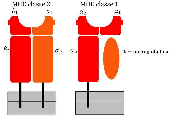
Antigen presentation is a vital immune process that is essential for T cell immune response triggering. Because T cells recognize only fragmented antigens displayed on cell surfaces, antigen processing must occur before the antigen fragment can be recognized by a T-cell receptor. Specifically, the fragment, bound to the major histocompatibility complex (MHC), is transported to the surface of the antigen-presenting cell, a process known as presentation. If there has been an infection with viruses or bacteria, the antigen-presenting cell will present an endogenous or exogenous peptide fragment derived from the antigen by MHC molecules. There are two types of MHC molecules which differ in the behaviour of the antigens: MHC class I molecules (MHC-I) bind peptides from the cell cytosol, while peptides generated in the endocytic vesicles after internalisation are bound to MHC class II (MHC-II). Cellular membranes separate these two cellular environments - intracellular and extracell ... [...More Info...] [...Related Items...] OR: [Wikipedia] [Google] [Baidu] |
Programmed Cell Death
Programmed cell death (PCD) sometimes referred to as cell, or cellular suicide is the death of a cell (biology), cell as a result of events inside of a cell, such as apoptosis or autophagy. PCD is carried out in a biological process, which usually confers advantage during an organism's biological life cycle, lifecycle. For example, the Limb development, differentiation of fingers and toes in a developing human embryo occurs because cells between the fingers apoptose; the result is that the digits are separate. PCD serves fundamental functions during both plant and animal tissue development. Apoptosis and autophagy are both forms of programmed cell death. Necrosis is the death of a cell caused by external factors such as trauma or infection and occurs in several different forms. Necrosis was long seen as a non-physiological process that occurs as a result of infection or injury, but in the 2000s, a form of programmed necrosis, called necroptosis, was recognized as an alternative f ... [...More Info...] [...Related Items...] OR: [Wikipedia] [Google] [Baidu] |
Dendritic Cell
A dendritic cell (DC) is an antigen-presenting cell (also known as an ''accessory cell'') of the mammalian immune system. A DC's main function is to process antigen material and present it on the cell surface to the T cells of the immune system. They act as messengers between the innate and adaptive immune systems. Dendritic cells are present in tissues that are in contact with the body's external environment, such as the skin, and the inner lining of the nose, lungs, stomach and intestines. They can also be found in an immature and mature state in the blood. Once activated, they migrate to the lymph nodes, where they interact with T cells and B cells to initiate and shape the adaptive immune response. At certain development stages they grow branched projections, the '' dendrites,'' that give the cell its name (δένδρον or déndron being Greek for 'tree'). While similar in appearance to the dendrites of neurons, these are structures distinct from them. Immature dendr ... [...More Info...] [...Related Items...] OR: [Wikipedia] [Google] [Baidu] |
Cd4+ T Cells
The T helper cells (Th cells), also known as CD4+ cells or CD4-positive cells, are a type of T cell that play an important role in the adaptive immune system. They aid the activity of other immune cells by releasing cytokines. They are considered essential in B cell antibody class switching, breaking cross-tolerance in dendritic cells, in the activation and growth of cytotoxic T cells, and in maximizing bactericidal activity of phagocytes such as macrophages and neutrophils. CD4+ cells are mature Th cells that express the surface protein CD4. Genetic variation in regulatory elements expressed by CD4+ cells determines susceptibility to a broad class of autoimmune diseases. Structure and function Th cells contain and release cytokines to aid other immune cells. Cytokines are small protein mediators that alter the behavior of target cells that express receptors for those cytokines. These cells help polarize the immune response depending on the nature of the immunological in ... [...More Info...] [...Related Items...] OR: [Wikipedia] [Google] [Baidu] |
MHC2
The major histocompatibility complex (MHC) is a large locus on vertebrate DNA containing a set of closely linked polymorphic genes that code for cell surface proteins essential for the adaptive immune system. These cell surface proteins are called MHC molecules. Its name comes from its discovery during the study of transplanted tissue compatibility. Later studies revealed that tissue rejection due to incompatibility is only a facet of the full function of MHC molecules, which is to bind an antigen derived from self-proteins, or from pathogens, and bring the antigen presentation to the cell surface for recognition by the appropriate T-cells. MHC molecules mediate the interactions of leukocytes, also called white blood cells (WBCs), with other leukocytes or with body cells. The MHC determines donor compatibility for organ transplant, as well as one's susceptibility to autoimmune diseases. In a cell, protein molecules of the host's own phenotype or of other biologic entities are c ... [...More Info...] [...Related Items...] OR: [Wikipedia] [Google] [Baidu] |
Plasmacytoid Dendritic Cell
Plasmacytoid dendritic cells (pDCs) are a rare type of immune cell that are known to secrete large quantities of type 1 interferon (IFNs) in response to a viral infection. They circulate in the blood and are found in peripheral lymphoid organs. They develop from bone marrow hematopoietic stem cells and constitute 2% of nucleated cells) and bone marrow and evidence (i.e. cytopenias) of bone marrow failure. Blastic plasmacytoid dendritic cell neoplasm has a high rate of recurrence following initial treatments with various chemotherapy regimens. In consequence, the disease has a poor overall prognosis and newer chemotherapeutic and novel non-chemotherapeutic drug regimens to improve the situation are under study. Role in immunity Upon stimulation and subsequent activation of TLR7 and TLR9, these cells produce large amounts (up to 1,000 times more than other cell type) of type I interferon (mainly IFN-α and IFN-β), which are critical anti-viral compounds mediating a wide range ... [...More Info...] [...Related Items...] OR: [Wikipedia] [Google] [Baidu] |
Cytotoxicity
Cytotoxicity is the quality of being toxic to cells. Examples of toxic agents are toxic metals, toxic chemicals, microbe neurotoxins, radiation particles and even specific neurotransmitters when the system is out of balance. Also some types of drugs, e.g alcohol, and some venom, e.g. from the puff adder (''Bitis arietans'') or brown recluse spider (''Loxosceles reclusa'') are toxic to cells. Cell physiology Treating cells with the cytotoxic compound can result in a variety of prognoses. The cells may undergo necrosis, in which they lose membrane integrity and die rapidly as a result of cell lysis. The cells can stop actively growing and dividing (a decrease in cell viability), or the cells can activate a genetic program of controlled cell death (apoptosis). Cells undergoing necrosis typically exhibit rapid swelling, lose membrane integrity, shut down metabolism, and release their contents into the environment. Cells that undergo rapid necrosis in vitro do not have sufficient ... [...More Info...] [...Related Items...] OR: [Wikipedia] [Google] [Baidu] |
Cross-presentation
Cross-presentation is the ability of certain professional antigen-presenting cells (mostly dendritic cells) to take up, process and present ''extracellular'' antigens with MHC class I molecules to CD8 T cells (cytotoxic T cells). Cross-priming, the result of this process, describes the stimulation of naive cytotoxic CD8+ T cells into activated cytotoxic CD8+ T cells. This process is necessary for immunity against most tumors and against viruses that infect dendritic cells and sabotage their presentation of virus antigens. Cross presentation is also required for the induction of cytotoxic immunity by vaccination with protein antigens, for example, tumour vaccination. Cross-presentation is of particular importance, because it permits the presentation of exogenous antigens, which are normally presented by MHC II on the surface of dendritic cells, to also be presented through the MHC I pathway. The MHC I pathway is normally used to present endogenous antigens that have infected a ... [...More Info...] [...Related Items...] OR: [Wikipedia] [Google] [Baidu] |
CD8+ T Cells
A cytotoxic T cell (also known as TC, cytotoxic T lymphocyte, CTL, T-killer cell, cytolytic T cell, CD8+ T-cell or killer T cell) is a T lymphocyte (a type of white blood cell) that kills cancer cells, cells that are infected by intracellular pathogens such as viruses or bacteria, or cells that are damaged in other ways. Most cytotoxic T cells express T-cell receptors (TCRs) that can recognize a specific antigen. An antigen is a molecule capable of stimulating an immune response and is often produced by cancer cells, viruses, bacteria or intracellular signals. Antigens inside a cell are bound to class I MHC molecules, and brought to the surface of the cell by the class I MHC molecule, where they can be recognized by the T cell. If the TCR is specific for that antigen, it binds to the complex of the class I MHC molecule and the antigen, and the T cell destroys the cell. In order for the TCR to bind to the class I MHC molecule, the former must be accompanied by a glycoprotein ca ... [...More Info...] [...Related Items...] OR: [Wikipedia] [Google] [Baidu] |
Tapasin
TAP-associated glycoprotein, also known as tapasin or TAPBP, is a protein that in humans is encoded by the ''TAPBP'' gene. Function The ''TAPBP'' gene encodes a transmembrane glycoprotein that mediates interaction between newly assembled major histocompatibility complex ( MHC) class I molecules and the transporter associated with antigen processing (TAP), which is required for the transport of antigenic peptides across the endoplasmic reticulum membrane. This interaction facilitates optimal peptide loading on the MHC class I molecule. Up to four complexes of MHC class I and tapasin may be bound to a single TAP molecule. Tapasin contains a C-terminal double-lysine motif (KKKAE) known to maintain membrane proteins in the endoplasmic reticulum. In humans, the tapasin gene lies within the major histocompatibility complex on chromosome 6. Alternative splicing results in three transcript variants encoding different isoforms. Tapasin is a MHC class I antigen-processing molecule pre ... [...More Info...] [...Related Items...] OR: [Wikipedia] [Google] [Baidu] |
Calreticulin
Calreticulin also known as calregulin, CRP55, CaBP3, calsequestrin-like protein, and endoplasmic reticulum resident protein 60 (ERp60) is a protein that in humans is encoded by the ''CALR'' gene. Calreticulin is a multifunctional soluble protein that binds Ca2+ ions (a second messenger in signal transduction), rendering them inactive. The Ca2+ is bound with low affinity, but high capacity, and can be released on a signal (see inositol trisphosphate). Calreticulin is located in storage compartments associated with the endoplasmic reticulum and is considered an ER resident protein. The term "Mobilferrin" is considered to be the same as calreticulin by some sources. Function Calreticulin binds to misfolded proteins and prevents them from being exported from the endoplasmic reticulum to the Golgi apparatus. A similar quality-control molecular chaperone, calnexin, performs the same service for soluble proteins as does calreticulin, however it is a membrane-bound protein. ... [...More Info...] [...Related Items...] OR: [Wikipedia] [Google] [Baidu] |
Calnexin
Calnexin (CNX) is a 67kDa integral protein (that appears variously as a 90kDa, 80kDa, or 75kDa band on western blotting depending on the source of the antibody) of the endoplasmic reticulum (ER). It consists of a large (50 kDa) N-terminal calcium- binding lumenal domain, a single transmembrane helix and a short (90 residues), acidic cytoplasmic tail. In humans, calnexin is encoded by the gene ''CANX''. Function Calnexin is a chaperone, characterized by assisting protein folding and quality control, ensuring that only properly folded and assembled proteins proceed further along the secretory pathway. It specifically acts to retain unfolded or unassembled N-linked glycoproteins in the ER. Calnexin binds only those ''N''-glycoproteins that have GlcNAc2Man9Glc1 oligosaccharides. These monoglucosylated oligosaccharides result from the trimming of two glucose residues by the sequential action of two glucosidases, I and II. Glucosidase II can also remove the third and last glu ... [...More Info...] [...Related Items...] OR: [Wikipedia] [Google] [Baidu] |


