|
Mediastinitis
Mediastinitis is inflammation of the tissues in the mid-chest, or mediastinum. It can be either Acute (medical), acute or Chronic (medical), chronic. It is thought to be due to four different etiologies: * direct contamination * hematogenous or Lymphatic system, lymphatic spread * extension of infection from the neck or Retroperitoneal space, retroperitoneum * extension from the lung or pleura Acute mediastinitis is usually caused by bacteria and is most often due to perforation of the esophagus. As the infection can progress rapidly, this is considered a serious condition. Chronic Sclerosis (medicine), sclerosing (or fibrosis, fibrosing) mediastinitis, while potentially serious, is caused by a long-standing inflammation of the mediastinum, leading to growth of acellular collagen and fibrous tissue within the chest and around the central vessels and airways. It has a different cause, treatment, and prognosis than acute infectious mediastinitis. Space infections: Pretracheal space ... [...More Info...] [...Related Items...] OR: [Wikipedia] [Google] [Baidu] |
Mediastinum
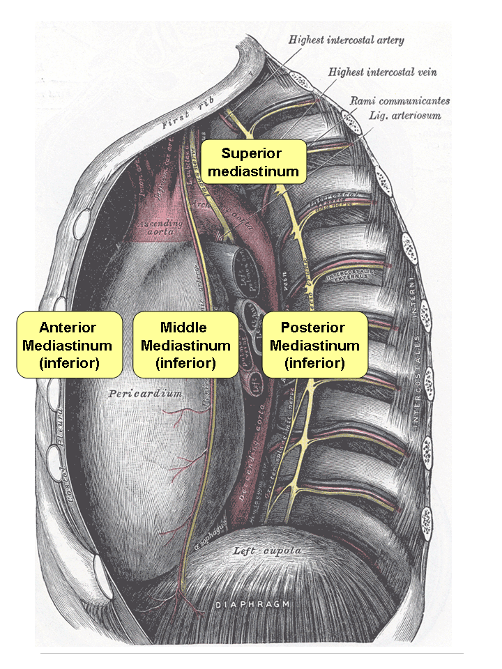
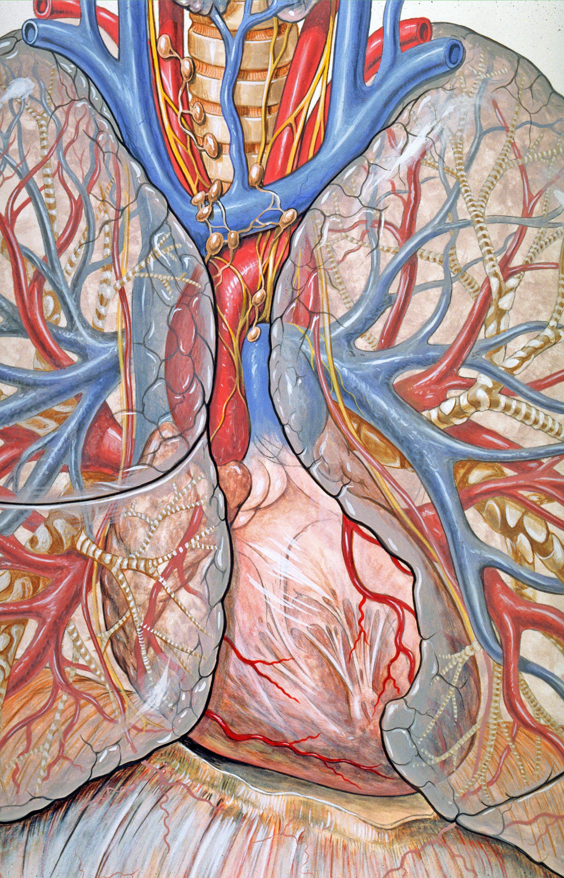
The mediastinum (from ) is the central compartment of the thoracic cavity. Surrounded by loose connective tissue, it is an undelineated region that contains a group of structures within the thorax, namely the heart and its vessels, the esophagus, the trachea, the phrenic nerve, phrenic and cardiac nerves, the thoracic duct, the thymus and the lymph nodes of the central chest. Anatomy The mediastinum lies within the thorax and is enclosed on the right and left by pulmonary pleurae, pleurae. It is surrounded by the chest wall in front, the lungs to the sides and the Spine (anatomy), spine at the back. It extends from the sternum in front to the vertebral column behind. It contains all the organs of the thorax except the lungs. It is continuous with the loose connective tissue of the neck. The mediastinum can be divided into an upper (or superior) and lower (or inferior) part: * The superior mediastinum starts at the superior thoracic aperture and ends at the #Thoracic plane, t ... [...More Info...] [...Related Items...] OR: [Wikipedia] [Google] [Baidu] |
Inflammation
Inflammation (from la, wikt:en:inflammatio#Latin, inflammatio) is part of the complex biological response of body tissues to harmful stimuli, such as pathogens, damaged cells, or Irritation, irritants, and is a protective response involving immune cells, blood vessels, and molecular mediators. The function of inflammation is to eliminate the initial cause of cell injury, clear out necrotic cells and tissues damaged from the original insult and the inflammatory process, and initiate tissue repair. The five cardinal signs are heat, pain, redness, swelling, and Functio laesa, loss of function (Latin ''calor'', ''dolor'', ''rubor'', ''tumor'', and ''functio laesa''). Inflammation is a generic response, and therefore it is considered as a mechanism of innate immune system, innate immunity, as compared to adaptive immune system, adaptive immunity, which is specific for each pathogen. Too little inflammation could lead to progressive tissue destruction by the harmful stimulus (e.g. b ... [...More Info...] [...Related Items...] OR: [Wikipedia] [Google] [Baidu] |
X-ray
An X-ray, or, much less commonly, X-radiation, is a penetrating form of high-energy electromagnetic radiation. Most X-rays have a wavelength ranging from 10 picometers to 10 nanometers, corresponding to frequencies in the range 30 petahertz to 30 exahertz ( to ) and energies in the range 145 eV to 124 keV. X-ray wavelengths are shorter than those of UV rays and typically longer than those of gamma rays. In many languages, X-radiation is referred to as Röntgen radiation, after the German scientist Wilhelm Conrad Röntgen, who discovered it on November 8, 1895. He named it ''X-radiation'' to signify an unknown type of radiation.Novelline, Robert (1997). ''Squire's Fundamentals of Radiology''. Harvard University Press. 5th edition. . Spellings of ''X-ray(s)'' in English include the variants ''x-ray(s)'', ''xray(s)'', and ''X ray(s)''. The most familiar use of X-rays is checking for fractures (broken bones), but X-rays are also used in other ways. ... [...More Info...] [...Related Items...] OR: [Wikipedia] [Google] [Baidu] |
Gram-negative Bacteria
Gram-negative bacteria are bacteria that do not retain the crystal violet stain used in the Gram staining method of bacterial differentiation. They are characterized by their cell envelopes, which are composed of a thin peptidoglycan cell wall sandwiched between an inner cytoplasmic cell membrane and a bacterial outer membrane. Gram-negative bacteria are found in virtually all environments on Earth that support life. The gram-negative bacteria include the model organism ''Escherichia coli'', as well as many pathogenic bacteria, such as ''Pseudomonas aeruginosa'', '' Chlamydia trachomatis'', and ''Yersinia pestis''. They are a significant medical challenge as their outer membrane protects them from many antibiotics (including penicillin), detergents that would normally damage the inner cell membrane, and lysozyme, an antimicrobial enzyme produced by animals that forms part of the innate immune system. Additionally, the outer leaflet of this membrane comprises a complex lipopol ... [...More Info...] [...Related Items...] OR: [Wikipedia] [Google] [Baidu] |
Anaerobic Organism
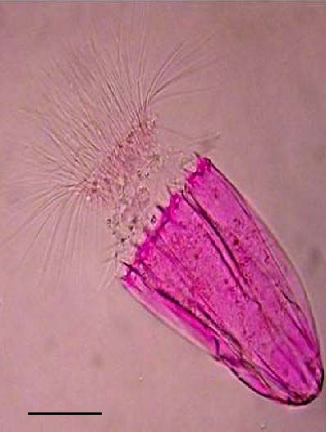
An anaerobic organism or anaerobe is any organism that does not require molecular oxygen for growth. It may react negatively or even die if free oxygen is present. In contrast, an aerobic organism (aerobe) is an organism that requires an oxygenated environment. Anaerobes may be unicellular (e.g. protozoans, bacteria) or multicellular. Most fungi are obligate aerobes, requiring oxygen to survive. However, some species, such as the Chytridiomycota that reside in the rumen of cattle, are obligate anaerobes; for these species, anaerobic respiration is used because oxygen will disrupt their metabolism or kill them. Deep waters of the ocean are a common anoxic environment. First observation In his letter of 14 June 1680 to The Royal Society, Antonie van Leeuwenhoek described an experiment he carried out by filling two identical glass tubes about halfway with crushed pepper powder, to which some clean rain water was added. Van Leeuwenhoek sealed one of the glass tubes using a flame an ... [...More Info...] [...Related Items...] OR: [Wikipedia] [Google] [Baidu] |
Gram-positive Bacteria
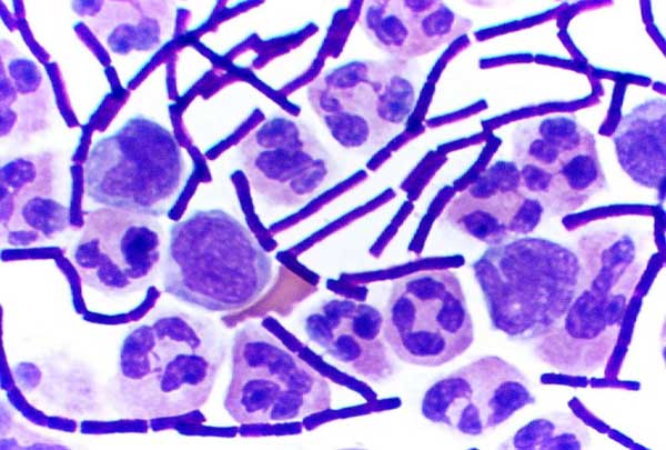
In bacteriology, gram-positive bacteria are bacteria that give a positive result in the Gram stain test, which is traditionally used to quickly classify bacteria into two broad categories according to their type of cell wall. Gram-positive bacteria take up the crystal violet stain used in the test, and then appear to be purple-coloured when seen through an optical microscope. This is because the thick peptidoglycan layer in the bacterial cell wall retains the stain after it is washed away from the rest of the sample, in the decolorization stage of the test. Conversely, gram-negative bacteria cannot retain the violet stain after the decolorization step; alcohol used in this stage degrades the outer membrane of gram-negative cells, making the cell wall more porous and incapable of retaining the crystal violet stain. Their peptidoglycan layer is much thinner and sandwiched between an inner cell membrane and a bacterial outer membrane, causing them to take up the counterstain (sa ... [...More Info...] [...Related Items...] OR: [Wikipedia] [Google] [Baidu] |
Epiglottitis
Epiglottitis is the inflammation of the epiglottis—the flap at the base of the tongue that prevents food entering the trachea (windpipe). Symptoms are usually rapid in onset and include trouble swallowing which can result in drooling, changes to the voice, fever, and an increased breathing rate. As the epiglottis is in the upper airway, swelling can interfere with breathing. People may lean forward in an effort to open the airway. As the condition worsens, stridor and bluish skin may occur. Epiglottitis was historically mostly caused by infection by '' H. influenzae type b'' (commonly referred to as "Hib"). With vaccination, it is now more often caused by other bacteria, most commonly ''Streptococcus pneumoniae'', ''Streptococcus pyogenes'', or ''Staphylococcus aureus''. Predisposing factors include burns and trauma to the area. The most accurate way to make the diagnosis is to look directly at the epiglottis. X-rays of the neck from the side may show a "thumbprint sign" bu ... [...More Info...] [...Related Items...] OR: [Wikipedia] [Google] [Baidu] |
Pharyngitis
Pharyngitis is inflammation of the back of the throat, known as the pharynx. It typically results in a sore throat and fever. Other symptoms may include a runny nose, cough, headache, difficulty swallowing, swollen lymph nodes, and a hoarse voice. Symptoms usually last 3–5 days, but can be longer depending on cause. Complications can include sinusitis and acute otitis media. Pharyngitis is a type of upper respiratory tract infection. Most cases are caused by a viral infection. Strep throat, a bacterial infection, is the cause in about 25% of children and 10% of adults. Uncommon causes include other bacteria such as ''gonococcus'', fungi, irritants such as smoke, allergies, and gastroesophageal reflux disease. Specific testing is not recommended in people who have clear symptoms of a viral infection, such as a cold. Otherwise, a rapid antigen detection test or throat swab is recommended. PCR testing is becoming commonly used as it is as good as taking a throat swab but gives a ... [...More Info...] [...Related Items...] OR: [Wikipedia] [Google] [Baidu] |
Peritonsillar Abscess
Peritonsillar abscess (PTA), also known as quinsy, is an accumulation of pus due to an infection behind the tonsil. Symptoms include fever, throat pain, trouble opening the mouth, and a change to the voice. Pain is usually worse on one side. Complications may include blockage of the airway or aspiration pneumonitis. PTA is typically due to infection by a number of types of bacteria. Often it follows streptococcal pharyngitis. They do not typically occur in those who have had a tonsillectomy. Diagnosis is usually based on the symptoms. Medical imaging may be done to rule out complications. Treatment is by removing the pus, antibiotics, sufficient fluids, and pain medication. Steroids may also be useful. Admission to hospital is generally not needed. In the United States about 3 per 10,000 people per year are affected. Young adults are most commonly affected. Signs and symptoms Physical signs of a peritonsillar abscess include redness and swelling in the tonsillar area of ... [...More Info...] [...Related Items...] OR: [Wikipedia] [Google] [Baidu] |
Behçet's Disease
Behçet's disease (BD) is a type of inflammatory disorder which affects multiple parts of the body. The most common symptoms include painful sores on the mucous membranes of the mouth and other parts of the body, inflammation of parts of the eye, and arthritis. The sores can last from a few days, up to a week or more. Less commonly there may be inflammation of the brain or spinal cord, blood clots, aneurysms, or blindness. Often, the symptoms come and go. The cause is unknown. It is believed to be partly genetic. Behçet's is not contagious. Diagnosis is based on at least three episodes of mouth sores in a year together with at least two of the following: genital sores, eye inflammation, skin sores, a positive skin prick test. There is no cure. Treatments may include immunosuppressive medication such as corticosteroids and lifestyle changes. Lidocaine mouthwash may help with the pain. Colchicine may decrease the frequency of attacks. While rare in the United States an ... [...More Info...] [...Related Items...] OR: [Wikipedia] [Google] [Baidu] |


.jpg)

