|
Mammograph
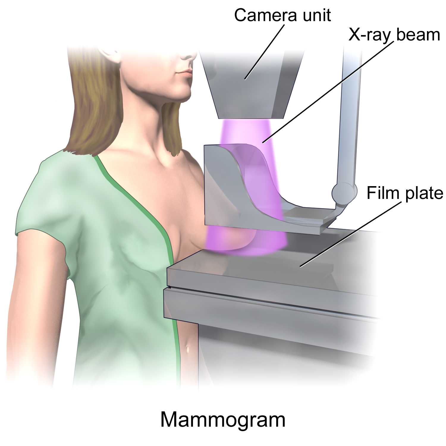
Mammography (also called mastography) is the process of using low-energy X-rays (usually around 30 kVp) to examine the human breast for diagnosis and screening. The goal of mammography is the early detection of breast cancer, typically through detection of characteristic masses or microcalcifications. As with all X-rays, mammograms use doses of ionizing radiation to create images. These images are then analyzed for abnormal findings. It is usual to employ lower-energy X-rays, typically Mo (K-shell X-ray energies of 17.5 and 19.6 keV) and Rh (20.2 and 22.7 keV) than those used for radiography of bones. Mammography may be 2D or 3D (tomosynthesis), depending on the available equipment and/or purpose of the examination. Ultrasound, ductography, positron emission mammography (PEM), and magnetic resonance imaging (MRI) are adjuncts to mammography. Ultrasound is typically used for further evaluation of masses found on mammography or palpable masses that may or may not be seen on mammogr ... [...More Info...] [...Related Items...] OR: [Wikipedia] [Google] [Baidu] |
Breast Cancer
Breast cancer is cancer that develops from breast tissue. Signs of breast cancer may include a lump in the breast, a change in breast shape, dimpling of the skin, milk rejection, fluid coming from the nipple, a newly inverted nipple, or a red or scaly patch of skin. In those with distant spread of the disease, there may be bone pain, swollen lymph nodes, shortness of breath, or yellow skin. Risk factors for developing breast cancer include obesity, a lack of physical exercise, alcoholism, hormone replacement therapy during menopause, ionizing radiation, an early age at first menstruation, having children late in life or not at all, older age, having a prior history of breast cancer, and a family history of breast cancer. About 5–10% of cases are the result of a genetic predisposition inherited from a person's parents, including BRCA1 and BRCA2 among others. Breast cancer most commonly develops in cells from the lining of milk ducts and the lobules that supply these ... [...More Info...] [...Related Items...] OR: [Wikipedia] [Google] [Baidu] |
Positron Emission Mammography
Positron emission mammography (PEM) is a nuclear medicine imaging modality used to detect or characterise breast cancer. Mammography typically refers to x-ray imaging of the breast, while PEM uses an injected positron emitting isotope and a dedicated scanner to locate breast tumors. Scintimammography is another nuclear medicine breast imaging technique, however it is performed using a gamma camera. Breasts can be imaged on standard whole-body PET scanners, however dedicated PEM scanners offer advantages including improved Image resolution, resolution. PEM is not recommended for routine use or for breast cancer screening, in part due to higher radiation dose compared to other modalities. Compared to breast MRI, PEM offers higher Sensitivity and specificity, specificity. Specific indications can include "high-risk patients with masses > 2 cm or aggressive malignancy and serum tumor marker elevation". Fludeoxyglucose (18F), 18F-FDG is the most common radiopharmaceutical used for PEM. ... [...More Info...] [...Related Items...] OR: [Wikipedia] [Google] [Baidu] |
United States Preventive Services Task Force
The United States Preventive Services Task Force (USPSTF) is "an independent panel of experts in primary care and prevention that systematically reviews the evidence of effectiveness and develops recommendations for clinical preventive services". The task force, a volunteer panel of primary care clinicians (including those from internal medicine, pediatrics, family medicine, obstetrics and gynecology, nursing, and psychology) with methodology experience including epidemiology, biostatistics, health services research, decision sciences, and health economics, is funded, staffed, and appointed by the U.S. Department of Health and Human Services' Agency for Healthcare Research and Quality. Intent The USPSTF evaluates scientific evidence to determine whether medical screenings, counseling, and preventive medications work for adults and children who have no symptoms. Methods The methods of evidence synthesis used by the Task Force have been described in detail. In 2007, their methods ... [...More Info...] [...Related Items...] OR: [Wikipedia] [Google] [Baidu] |
Dense Breast Tissue
Dense breast tissue, also known as dense breasts, is a condition of the breasts where a higher proportion of the breasts are made up of glandular tissue and fibrous tissue than fatty tissue. Around 40–50% of women have dense breast tissue and one of the main medical components of the condition is that mammograms are unable to differentiate tumorous tissue from the surrounding dense tissue. This increases the risk of late diagnosis of breast cancer in women with dense breast tissue. Additionally, women with such tissue have a higher likelihood of developing breast cancer in general, though the reasons for this are poorly understood. Definition Dense breast tissue is defined based on the amount of glandular and fibrous tissue as compared to the percentage of fatty tissue. The current mammography classifications split up the density of breasts into four categories. Approximately 10% of women have almost entirely fatty breasts, 40% with small pockets of dense tissue, 40% with even d ... [...More Info...] [...Related Items...] OR: [Wikipedia] [Google] [Baidu] |
Albert Salomon (surgeon)
Albert Salomon (1883–1976)Creativity and Its Imprint: Three Jewish Artists and Some Books About Them: Philip Guston, Charlotte Salomon, R. B. Kitaj , Leonard Gold, Rosaline and Myer Feinstein Lecture Series, 2001. Hosted by www.jewishlibraries.org. Retrieved 11 Jul 2011. was a Jewish-German surgeon at the Royal Surgical University Clinic in Berlin. He is best known for his study of early mastectomies that is considered the beginning of mammography. He was the father of the artist Charlotte Salomon, who was murdered in Auschwitz concentration camp during the Holocaust. Breast pathology In 1913, Salomon pe ...[...More Info...] [...Related Items...] OR: [Wikipedia] [Google] [Baidu] |
Tomosynthesis
Tomosynthesis, also digital tomosynthesis (DTS), is a method for performing high-resolution limited-angle tomography at radiation dose levels comparable with projectional radiography. It has been studied for a variety of clinical applications, including vascular imaging, dental imaging, orthopedic imaging, mammographic imaging, musculoskeletal imaging, and chest imaging. History The concept of tomosynthesis was derived from the work of Ziedses des Plantes, who developed methods of reconstructing an arbitrary number of planes from a set of projections. Though this idea was displaced by the advent of computed tomography, tomosynthesis later gained interest as a low-dose tomographic alternative to CT. Reconstruction Tomosynthesis reconstruction algorithms are similar to CT reconstructions, in that they are based on performing an inverse Radon transform. Due to partial data sampling with very few projections, approximation algorithms have to be used. Filtered back projection and itera ... [...More Info...] [...Related Items...] OR: [Wikipedia] [Google] [Baidu] |
Radiography
Radiography is an imaging technique using X-rays, gamma rays, or similar ionizing radiation and non-ionizing radiation to view the internal form of an object. Applications of radiography include medical radiography ("diagnostic" and "therapeutic") and industrial radiography. Similar techniques are used in airport security (where "body scanners" generally use backscatter X-ray). To create an image in conventional radiography, a beam of X-rays is produced by an X-ray generator and is projected toward the object. A certain amount of the X-rays or other radiation is absorbed by the object, dependent on the object's density and structural composition. The X-rays that pass through the object are captured behind the object by a detector (either photographic film or a digital detector). The generation of flat two dimensional images by this technique is called projectional radiography. In computed tomography (CT scanning) an X-ray source and its associated detectors rotate around the su ... [...More Info...] [...Related Items...] OR: [Wikipedia] [Google] [Baidu] |
Jacob Gershon-Cohen
Jacob Gershon-Cohen (January 9, 1899 in Philadelphia - February 6, 1971 in Philadelphia) was an American researcher and physician, known for the use of mammography for the early detection of breast cancer. Biography Gershon-Cohen was born in Philadelphia to Jewish immigrant parents. He was director of radiology at the Albert Einstein Medical Center, professor of radiology at the University of Pennsylvania, and Professor of Research Radiology at Temple University. in 1964, he developed mammography to detect breast cancer, which has been a significant step for more effective treatment of the disease. He is also known for the development of thermography Infrared thermography (IRT), thermal video and/or thermal imaging, is a process where a Thermographic camera, thermal camera captures and creates an image of an object by using infrared radiation emitted from the object in a process, which are .... Gershon-Cohen published more than 400 scientific works during his career. ... [...More Info...] [...Related Items...] OR: [Wikipedia] [Google] [Baidu] |
X-ray
An X-ray, or, much less commonly, X-radiation, is a penetrating form of high-energy electromagnetic radiation. Most X-rays have a wavelength ranging from 10 picometers to 10 nanometers, corresponding to frequencies in the range 30 petahertz to 30 exahertz ( to ) and energies in the range 145 eV to 124 keV. X-ray wavelengths are shorter than those of UV rays and typically longer than those of gamma rays. In many languages, X-radiation is referred to as Röntgen radiation, after the German scientist Wilhelm Conrad Röntgen, who discovered it on November 8, 1895. He named it ''X-radiation'' to signify an unknown type of radiation.Novelline, Robert (1997). ''Squire's Fundamentals of Radiology''. Harvard University Press. 5th edition. . Spellings of ''X-ray(s)'' in English include the variants ''x-ray(s)'', ''xray(s)'', and ''X ray(s)''. The most familiar use of X-rays is checking for fractures (broken bones), but X-rays are also used in other ways. ... [...More Info...] [...Related Items...] OR: [Wikipedia] [Google] [Baidu] |
Breast
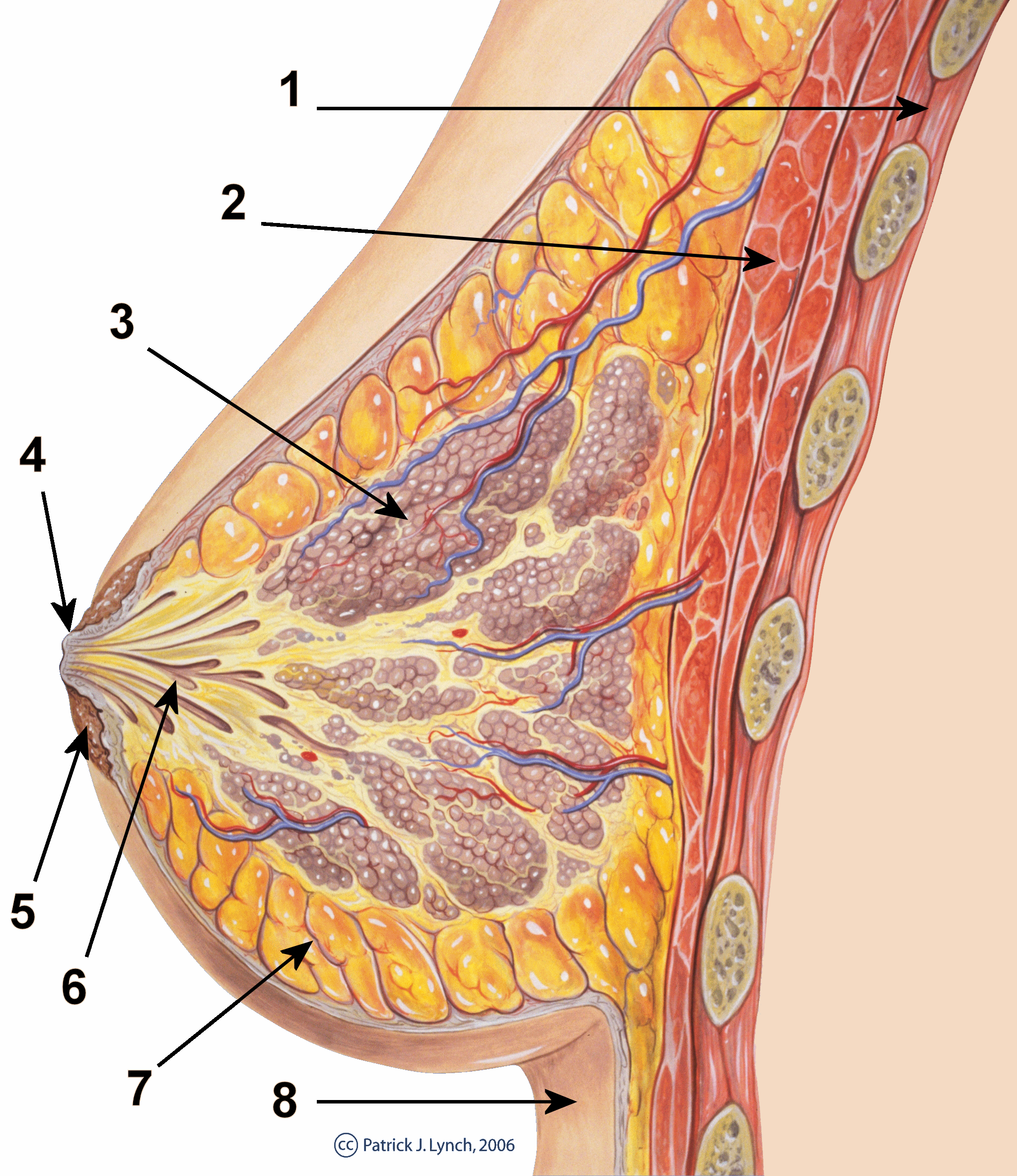
The breast is one of two prominences located on the upper ventral region of a primate's torso. Both females and males develop breasts from the same embryological tissues. In females, it serves as the mammary gland, which produces and secretes milk to feed infants. Subcutaneous fat covers and envelops a network of ducts that converge on the nipple, and these tissues give the breast its size and shape. At the ends of the ducts are lobules, or clusters of alveoli, where milk is produced and stored in response to hormonal signals. During pregnancy, the breast responds to a complex interaction of hormones, including estrogens, progesterone, and prolactin, that mediate the completion of its development, namely lobuloalveolar maturation, in preparation of lactation and breastfeeding. Humans are the only animals with permanent breasts. At puberty, estrogens, in conjunction with growth hormone, cause permanent breast growth in female humans. This happens only to a much lesser ... [...More Info...] [...Related Items...] OR: [Wikipedia] [Google] [Baidu] |
Microcalcification
Microcalcifications are tiny deposits of calcium salts that are too small to be felt but can be detected by imaging. They can be scattered throughout the mammary gland, or occur in clusters. Microcalcifications can be an early sign of breast cancer. Based on morphology, it is possible to classify by radiography how likely microcalcifications are to indicate cancer. Microcalcifications are made up of calcium oxalate and calcium phosphate The term calcium phosphate refers to a family of materials and minerals containing calcium ions (Ca2+) together with inorganic phosphate anions. Some so-called calcium phosphates contain oxide and hydroxide as well. Calcium phosphates are whi .... The mechanism of their formation is not known. Microcalcification was first described in 1913 by surgeon Albert Salomon. References Medical signs {{Med-sign-stub ... [...More Info...] [...Related Items...] OR: [Wikipedia] [Google] [Baidu] |
Mastitis
Mastitis is inflammation of the breast or udder, usually associated with breastfeeding. Symptoms typically include local pain and redness. There is often an associated fever and general soreness. Onset is typically fairly rapid and usually occurs within the first few months of delivery. Complications can include abscess formation. Risk factors include poor latch, cracked nipples, use of a breast pump, and weaning. The bacteria most commonly involved are '' Staphylococcus'' and '' Streptococci''. Diagnosis is typically based on symptoms. Ultrasound may be useful for detecting a potential abscess. Prevention is by proper breastfeeding techniques. When infection is present, antibiotics such as cephalexin may be recommended. Breastfeeding should typically be continued, as emptying the breast is important for healing. Tentative evidence supports benefits from probiotics. About 10% of breastfeeding women are affected. Types When it occurs in breastfeeding mothers, it is known as puer ... [...More Info...] [...Related Items...] OR: [Wikipedia] [Google] [Baidu] |


