|
Keratoderma Blennorrhagica
Keratoderma blennorrhagicum etymologically meaning keratinized (kerato-) skin (derma-) mucousy (blenno-) discharge (-rrhagia) (also called keratoderma blennorrhagica) are skin lesions commonly found on the palms and soles but which may spread to the scrotum, scalp and trunk. The lesions may resemble psoriasis.James, William; Berger, Timothy; Elston, Dirk (2005). ''Andrews' Diseases of the Skin: Clinical Dermatology''. (10th ed.). Saunders. . Keratoderma blennorrhagicum is commonly seen as an additional feature of reactive arthritis in almost 15% of male patients. The appearance is usually of a vesico-pustular waxy lesion with a yellow brown colour. These lesions may join to form larger crusty plaques with desquamating edges. See also * Keratoderma * Keratosis * Blennorrhea * List of cutaneous conditions Many skin conditions affect the human integumentary system—the organ system covering the entire surface of the body and composed of skin, hair, nails, and related muscle ... [...More Info...] [...Related Items...] OR: [Wikipedia] [Google] [Baidu] |
Reactive Arthritis
Reactive arthritis, also known as Reiter's syndrome, is a form of inflammatory arthritis that develops in response to an infection in another part of the body (cross-reactivity). Coming into contact with bacteria and developing an infection can trigger the disease. By the time the patient presents with symptoms, often the "trigger" infection has been cured or is in remission in chronic cases, thus making determination of the initial cause difficult. The arthritis often is coupled with other characteristic symptoms; this has been called Reiter's syndrome, Reiter's disease or Reiter's arthritis. The term "reactive arthritis" is increasingly used as a substitute for this designation because of Hans Reiter's war crimes with the Nazi Party. The manifestations of reactive arthritis include the following triad of symptoms: an inflammatory arthritis of large joints, inflammation of the eyes in the form of conjunctivitis or uveitis, and urethritis in men or cervicitis in women. Arthrit ... [...More Info...] [...Related Items...] OR: [Wikipedia] [Google] [Baidu] |
Blennorrhea
Blennorrhea is mucous discharge, especially from the urethra or vagina (that is, mucus vaginal discharge). Blennorrhagia is an excess of such discharge, citing: * McGraw-Hill Dictionary of Scientific & Technical Terms, 6E, Copyright 2003 often specifically referring to that seen in . In fact, blennorrhagia is also a German
German(s) may refer to:
* Germany (of or related to)
**Germania (historical use)
* Germans, citizens of Germany, people of German ancestry, or native speakers of the German language
** For citizens ...
[...More Info...] [...Related Items...] OR: [Wikipedia] [Google] [Baidu] |
Skin Lesions
A skin condition, also known as cutaneous condition, is any medical condition that affects the integumentary system—the organ system that encloses the body and includes skin, nails, and related muscle and glands. The major function of this system is as a barrier against the external environment. Conditions of the human integumentary system constitute a broad spectrum of diseases, also known as dermatoses, as well as many nonpathologic states (like, in certain circumstances, melanonychia and racquet nails). While only a small number of skin diseases account for most visits to the physician, thousands of skin conditions have been described. Classification of these conditions often presents many nosological challenges, since underlying causes and pathogenetics are often not known. Therefore, most current textbooks present a classification based on location (for example, conditions of the mucous membrane), morphology ( chronic blistering conditions), cause (skin conditions result ... [...More Info...] [...Related Items...] OR: [Wikipedia] [Google] [Baidu] |
Hand
A hand is a prehensile, multi-fingered appendage located at the end of the forearm or forelimb of primates such as humans, chimpanzees, monkeys, and lemurs. A few other vertebrates such as the koala (which has two opposable thumbs on each "hand" and fingerprints extremely similar to human fingerprints) are often described as having "hands" instead of paws on their front limbs. The raccoon is usually described as having "hands" though opposable thumbs are lacking. Some evolutionary anatomists use the term ''hand'' to refer to the appendage of digits on the forelimb more generally—for example, in the context of whether the three digits of the bird hand involved the same homologous loss of two digits as in the dinosaur hand. The human hand usually has five digits: four fingers plus one thumb; these are often referred to collectively as five fingers, however, whereby the thumb is included as one of the fingers. It has 27 bones, not including the sesamoid bone, the number o ... [...More Info...] [...Related Items...] OR: [Wikipedia] [Google] [Baidu] |
Sole (foot)
The sole is the bottom of the foot. In humans the sole of the foot is anatomically referred to as the plantar aspect. Structure The glabrous skin on the sole of the foot lacks the hair and pigmentation found elsewhere on the body, and it has a high concentration of sweat pores. The sole contains the thickest layers of skin on the body due to the weight that is continually placed on it. It is crossed by a set of creases that form during the early stages of embryonic development. Like those of the palm, the sweat pores of the sole lack sebaceous glands. The sole is a sensory organ by which we can perceive the ground while standing and walking. The subcutaneous tissue in the sole has adapted to deal with the high local compressive forces on the heel and the ball (between the toes and the arch) by developing a system of "pressure chambers." Each chamber is composed of internal fibrofatty tissue covered by external collagen connective tissue. The septa (internal walls) ... [...More Info...] [...Related Items...] OR: [Wikipedia] [Google] [Baidu] |
Scrotum
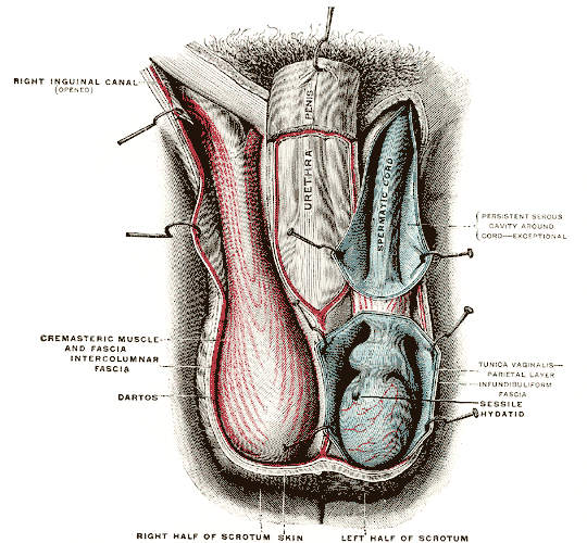
The scrotum or scrotal sac is an anatomical male reproductive structure located at the base of the penis that consists of a suspended dual-chambered sac of skin and smooth muscle. It is present in most terrestrial male mammals. The scrotum contains the external spermatic fascia, testes, epididymis, and ductus deferens. It is a distention of the perineum and carries some abdominal tissues into its cavity including the testicular artery, testicular vein, and pampiniform plexus. The perineal raphe is a small, vertical, slightly raised ridge of scrotal skin under which is found the scrotal septum. It appears as a thin longitudinal line that runs front to back over the entire scrotum. In humans and some other mammals the scrotum becomes covered with pubic hair at puberty. The scrotum will usually tighten during penile erection and when exposed to cold temperatures. One testis is typically lower than the other to avoid compression in the event of an impact. The scrotum is biologicall ... [...More Info...] [...Related Items...] OR: [Wikipedia] [Google] [Baidu] |
Scalp
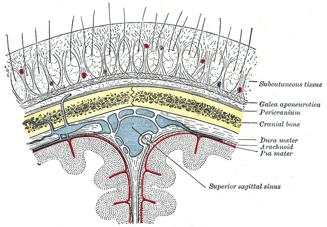
The scalp is the anatomical area bordered by the human face at the front, and by the neck at the sides and back. Structure The scalp is usually described as having five layers, which can conveniently be remembered as a mnemonic: * S: The skin on the head from which head hair grows. It contains numerous sebaceous glands and hair follicles. * C: Connective tissue. A dense subcutaneous layer of fat and fibrous tissue that lies beneath the skin, containing the nerves and vessels of the scalp. * A: The aponeurosis called epicranial aponeurosis (or galea aponeurotica) is the next layer. It is a tough layer of dense fibrous tissue which runs from the frontalis muscle anteriorly to the occipitalis posteriorly. * L: The loose areolar connective tissue layer provides an easy plane of separation between the upper three layers and the pericranium. In scalping the scalp is torn off through this layer. It also provides a plane of access in craniofacial surgery and neurosurgery. This layer i ... [...More Info...] [...Related Items...] OR: [Wikipedia] [Google] [Baidu] |
Torso
The torso or trunk is an anatomical term for the central part, or the core, of the body of many animals (including humans), from which the head, neck, limbs, tail and other appendages extend. The tetrapod torso — including that of a human — is usually divided into the ''thoracic'' segment (also known as the upper torso, where the forelimbs extend), the ''abdominal'' segment (also known as the "mid-section" or "midriff"), and the ''pelvic'' and '' perineal'' segments (sometimes known together with the abdomen as the lower torso, where the hindlimbs extend). Anatomy Major organs In humans, most critical organs, with the notable exception of the brain, are housed within the torso. In the upper chest, the heart and lungs are protected by the rib cage, and the abdomen contains most of the organs responsible for digestion: the stomach, which breaks down partially digested food via gastric acid; the liver, which respectively produces bile necessary for digestion; the large and ... [...More Info...] [...Related Items...] OR: [Wikipedia] [Google] [Baidu] |
Psoriasis
Psoriasis is a long-lasting, noncontagious autoimmune disease characterized by raised areas of abnormal skin. These areas are red, pink, or purple, dry, itchy, and scaly. Psoriasis varies in severity from small, localized patches to complete body coverage. Injury to the skin can trigger psoriatic skin changes at that spot, which is known as the Koebner phenomenon. The five main types of psoriasis are plaque, guttate, inverse, pustular, and erythrodermic. Plaque psoriasis, also known as psoriasis vulgaris, makes up about 90% of cases. It typically presents as red patches with white scales on top. Areas of the body most commonly affected are the back of the forearms, shins, navel area, and scalp. Guttate psoriasis has drop-shaped lesions. Pustular psoriasis presents as small, noninfectious, pus-filled blisters. Inverse psoriasis forms red patches in skin folds. Erythrodermic psoriasis occurs when the rash becomes very widespread, and can develop from any of the other types. ... [...More Info...] [...Related Items...] OR: [Wikipedia] [Google] [Baidu] |
Desquamation
Desquamation occurs when the outermost layer of a tissue, such as the skin, is shed. The term is . Physiologic desquamation Keratinocytes are the predominant cells of the epidermis, the outermost layer of the skin. Living keratinocytes reside in the basal, spinous, or granular layers of the epidermis. The outermost layer of the epidermis is called the Stratum corneum and it is composed of terminally differentiated keratinocytes, the Corneocytes. In the absence of disease, desquamation occurs when corneocytes are individually shed unnoticeably from the surface of the skin. Typically the time it takes for a corneocyte to be formed and then shed is about 14 weeks but this time can vary depending on the anatomical location that the skin is covering. For example, desquamation occurs more slowly at acral (palm and sole) surfaces and more rapidly where the skin is thin, such as the eyelids. Normal desquamation can be visualized by immersing skin in warm or hot water. This induces the out ... [...More Info...] [...Related Items...] OR: [Wikipedia] [Google] [Baidu] |
Keratoderma
Keratoderma is a hornlike skin condition. Classification The keratodermas are classified into the following subgroups:Freedberg, et al. (2003). ''Fitzpatrick's Dermatology in General Medicine''. (6th ed.). McGraw-Hill. . Congenital * Simple keratodermas ** Diffuse palmoplantar keratodermas *** Diffuse epidermolytic palmoplantar keratoderma *** Diffuse nonepidermolytic palmoplantar keratoderma *** mal de Meleda ** Focal palmoplantar keratoderma *** Striate palmoplantar keratoderma ** Punctate palmoplantar keratoderma *** Keratosis punctata palmaris et plantaris *** Spiny keratoderma *** Focal acral hyperkeratosis * Complex keratodermas ** Diffuse palmoplantar keratoderma *** Erythrokeratodermia variabilis *** Palmoplantar keratoderma of Sybert *** Olmsted syndrome *** Naegeli–Franceschetti–Jadassohn syndrome ** Focal palmoplantar keratoderma *** Papillon–Lefèvre syndrome *** Pachyonychia congenita type I *** Pachyonychia congenita type II *** Focal palmoplantar ... [...More Info...] [...Related Items...] OR: [Wikipedia] [Google] [Baidu] |
Keratosis
Keratosis (from '' kerat-'' + '' -osis'') is a growth of keratin on the skin or on mucous membranes stemming from keratinocytes, the prominent cell type in the epidermis. More specifically, it can refer to: * actinic keratosis (also known as solar keratosis), a premalignant condition * chronic scar keratosis * hydrocarbon keratosis * keratosis pilaris (KP, also known as follicular keratosis) * seborrheic keratosis, ''not'' premalignant See also * Folliculitis * Keratoderma Keratoderma is a hornlike skin condition. Classification The keratodermas are classified into the following subgroups:Freedberg, et al. (2003). ''Fitzpatrick's Dermatology in General Medicine''. (6th ed.). McGraw-Hill. . Congenital * Simple ker ... References External links Dermatologic terminology {{cutaneous-condition-stub ... [...More Info...] [...Related Items...] OR: [Wikipedia] [Google] [Baidu] |


