|
Interpeduncular Fossa
The interpeduncular fossa is a deep depression of the ventral surface of the midbrain between the two crura cerebri. It has been found in humans and macaques, but not in rats or mice, showing that this is a relatively new evolutionary region. Anatomy The interpeduncular fossa is a somewhat rhomboid-shaped area of the base of the brain. Features The lateral wall of the interpeduncular fossa bears a groove - the oculomotor sulcus - from which rootlets of the oculomotor nerve emerge from the substance of the brainstem and aggregate into a single fascicle. Anatomical relations The ventral tegmental area lies at the depth of the interpeduncular fossa. Boundaries The interpeduncular fossa is in front by the optic chiasma, behind by the antero-superior surface of the pons, antero-laterally by the converging optic tracts, and postero-laterally by the diverging cerebral peduncles. The floor of interpeduncular fossa, from behind forward, are the posterior perforated substance, ... [...More Info...] [...Related Items...] OR: [Wikipedia] [Google] [Baidu] |
Brain
A brain is an organ that serves as the center of the nervous system in all vertebrate and most invertebrate animals. It is located in the head, usually close to the sensory organs for senses such as vision. It is the most complex organ in a vertebrate's body. In a human, the cerebral cortex contains approximately 14–16 billion neurons, and the estimated number of neurons in the cerebellum is 55–70 billion. Each neuron is connected by synapses to several thousand other neurons. These neurons typically communicate with one another by means of long fibers called axons, which carry trains of signal pulses called action potentials to distant parts of the brain or body targeting specific recipient cells. Physiologically, brains exert centralized control over a body's other organs. They act on the rest of the body both by generating patterns of muscle activity and by driving the secretion of chemicals called hormones. This centralized control allows rapid and coordinated respon ... [...More Info...] [...Related Items...] OR: [Wikipedia] [Google] [Baidu] |
Interpeduncular Cistern
The interpeduncular cistern of Sweeney is the subarachnoid cistern that encloses the cerebral peduncles and the structures contained in the interpeduncular fossa and contains the arterial circle of Willis as well as the oculomotor nerve The oculomotor nerve, also known as the third cranial nerve, cranial nerve III, or simply CN III, is a cranial nerve that enters the orbit through the superior orbital fissure and innervates extraocular muscles that enable most movements of ... (CN3). References Meninges {{neuroanatomy-stub ... [...More Info...] [...Related Items...] OR: [Wikipedia] [Google] [Baidu] |
Neurocutaneous Melanosis
Neurocutaneous melanosis is a congenital disorder characterized by the presence of congenital melanocytic nevi on the skin and melanocytic tumors in the leptomeninges of the central nervous system. These lesions may occur in the amygdala, cerebellum, cerebrum, pons and spinal cord of patients. Although typically asymptomatic, malignancy occurs in the form of leptomeningeal melanoma in over half of patients. Regardless of the presence of malignancy, patients with symptomatic neurocutaneous melanosis generally have a poor prognosis with few treatment options. The pathogenesis of neurocutaneous melanosis is believed to be related to the abnormal postzygotic development of melanoblasts and mutations of the '' NRAS'' gene. Signs and symptoms Neurocutaneous melanosis is associated with the presence of either giant congenital melanocytic nevi or non-giant nevi of the skin. It is estimated that neurocutaneous melanosis is present in 2% to 45% of patients with giant congenital melanoc ... [...More Info...] [...Related Items...] OR: [Wikipedia] [Google] [Baidu] |
Great Cerebral Vein
The great cerebral vein is one of the large blood vessels in the skull draining the cerebrum of the brain. It is also known as the "vein of Galen", named for its discoverer, the Greek physician Galen. However, it is not the only vein with this eponym. Structure The great cerebral vein is considered one of the deep cerebral veins. Other deep cerebral veins are the internal cerebral veins, formed by the union of the superior thalamostriate vein and the superior choroid vein at the interventricular foramina. The internal cerebral veins can be seen on the superior surfaces of the caudate nuclei and thalami just under the corpus callosum. The veins at the anterior poles of the thalami merge posterior to the pineal gland to form the great cerebral vein. Most of the blood in the deep cerebral veins collects into the great cerebral vein. This comes from the inferior side of the posterior end of the corpus callosum and empties ie similarities, there are also differences between these ... [...More Info...] [...Related Items...] OR: [Wikipedia] [Google] [Baidu] |
Basal Vein
The basal vein is a vein in the brain. It is formed at the anterior perforated substance by the union of * (a) a ''small anterior cerebral vein'' which accompanies the anterior cerebral artery and supplies the medial surface of the frontal lobe by the fronto-basal vein. * (b) the ''deep middle cerebral vein'' (''deep Sylvian vein''), which receives tributaries from the insula and neighboring gyri, and runs in the lower part of the lateral cerebral fissure, and * (c) the ''inferior striate veins'', which leave the corpus striatum through the anterior perforated substance. The basal vein passes backward around the cerebral peduncle, and ends in the great cerebral vein; it receives tributaries from the interpeduncular fossa, the inferior horn of the lateral ventricle, the hippocampal gyrus, and the mid-brain The midbrain or mesencephalon is the forward-most portion of the brainstem and is associated with vision, hearing, motor control, sleep and wakefulness, arousal (alertness) ... [...More Info...] [...Related Items...] OR: [Wikipedia] [Google] [Baidu] |
Circle Of Willis
The circle of Willis (also called Willis' circle, loop of Willis, cerebral arterial circle, and Willis polygon) is a circulatory anastomosis that supplies blood to the brain and surrounding structures in reptiles, birds and mammals, including humans. It is named after Thomas Willis (1621–1675), an English physician. Structure The circle of Willis is a part of the cerebral circulation and is composed of the following arteries: * Anterior cerebral artery (left and right) * Anterior communicating artery * Internal carotid artery (left and right) * Posterior cerebral artery (left and right) * Posterior communicating artery (left and right) The middle cerebral arteries, supplying the brain, are not considered part of the circle of Willis. Origin of arteries The left and right internal carotid arteries arise from the left and right common carotid arteries. The posterior communicating artery is given off as a branch of the internal carotid artery just before it divides into its termi ... [...More Info...] [...Related Items...] OR: [Wikipedia] [Google] [Baidu] |
Pituitary Gland
In vertebrate anatomy, the pituitary gland, or hypophysis, is an endocrine gland, about the size of a chickpea and weighing, on average, in humans. It is a protrusion off the bottom of the hypothalamus at the base of the brain. The hypophysis rests upon the hypophyseal fossa of the sphenoid bone in the center of the middle cranial fossa and is surrounded by a small bony cavity (sella turcica) covered by a dural fold (diaphragma sellae). The anterior pituitary (or adenohypophysis) is a lobe of the gland that regulates several physiological processes including stress, growth, reproduction, and lactation. The intermediate lobe synthesizes and secretes melanocyte-stimulating hormone. The posterior pituitary (or neurohypophysis) is a lobe of the gland that is functionally connected to the hypothalamus by the median eminence via a small tube called the pituitary stalk (also called the infundibular stalk or the infundibulum). Hormones secreted from the pituitary gland ... [...More Info...] [...Related Items...] OR: [Wikipedia] [Google] [Baidu] |
Tuber Cinereum
The tuber cinereum is a hollow eminence of the middle–ventral hypothalamus, specifically the arcuate nucleus, situated between the mammillary bodies and the optic chiasm. In addition to the ventral hypothalamus, the tuber cinereum includes the median eminence and pituitary gland. Together with the hollow itself, it is sometimes referred to as the pituitary stalk. Structure The tuber cinereum is an inferior distention of the floor of the third ventricle; the conical hollow formed by the distention (a continuation of the ventricle itself), is known as the infundibulum (''funnel''). Thus, the tuber cinereum is anteriorly continuous with the lamina terminalis, while laterally it is continuous with the anterior perforated substances of the hypothalamus. The inferior end adjoins the posterior lobe of the pituitary gland. Capillaries of the tuber cinereum are specialized and confluent to enable rapid communication via brain- or blood-borne factors between compartments of the t ... [...More Info...] [...Related Items...] OR: [Wikipedia] [Google] [Baidu] |
Midbrain
The midbrain or mesencephalon is the forward-most portion of the brainstem and is associated with vision, hearing, motor control, sleep and wakefulness, arousal (alertness), and temperature regulation. The name comes from the Greek ''mesos'', "middle", and ''enkephalos'', "brain". Structure The principal regions of the midbrain are the tectum, the cerebral aqueduct, tegmentum, and the cerebral peduncles. Rostrally the midbrain adjoins the diencephalon (thalamus, hypothalamus, etc.), while caudally it adjoins the hindbrain (pons, medulla and cerebellum). In the rostral direction, the midbrain noticeably splays laterally. Sectioning of the midbrain is usually performed axially, at one of two levels – that of the superior colliculi, or that of the inferior colliculi. One common technique for remembering the structures of the midbrain involves visualizing these cross-sections (especially at the level of the superior colliculi) as the upside-down face of a be ... [...More Info...] [...Related Items...] OR: [Wikipedia] [Google] [Baidu] |
Corpora Mamillaria
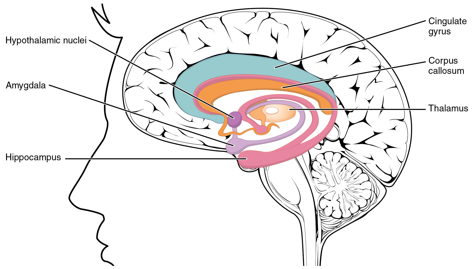
The mammillary bodies are a pair of small round bodies, located on the undersurface of the brain that, as part of the diencephalon, form part of the limbic system. They are located at the ends of the anterior arches of the fornix. They consist of two groups of nuclei, the medial mammillary nuclei and the lateral mammillary nuclei. Neuroanatomists have often categorized the mammillary bodies as part of the posterior part of hypothalamus. Structure Connections They are connected to other parts of the brain (as shown in the schematic, below left), and act as a relay for impulses coming from the amygdalae and hippocampi, via the mamillo-thalamic tract to the thalamus. Function File:Slide5dd.JPG, Mammillary body Mammillary bodies, and their projections to the anterior thalamus via the mammillothalamic tract, are important for recollective memory. The damage of medial mammillary nucleus leads to spatial memory deficit, according to observations in rats with mammillary ... [...More Info...] [...Related Items...] OR: [Wikipedia] [Google] [Baidu] |
Posterior Perforated Substance
The depressed area between the crura is termed the interpeduncular fossa, and consists of a layer of gray matter, the posterior perforated substance, which is pierced by small apertures for the transmission of blood vessels; its lower part lies on the ventral aspect of the medial portions of the tegmenta, and contains a nucleus named the interpeduncular ganglion; its upper part assists in forming the floor of the third ventricle The third ventricle is one of the four connected ventricles of the ventricular system within the mammalian brain. It is a slit-like cavity formed in the diencephalon between the two thalami, in the midline between the right and left lateral .... See also * Anterior perforated substance Additional images File:Human brainstem anterior view 2 description.JPG, Human brainstem anterior view References External links * * Central nervous system {{Portal bar, Anatomy ... [...More Info...] [...Related Items...] OR: [Wikipedia] [Google] [Baidu] |


