|
Iridocyclitis
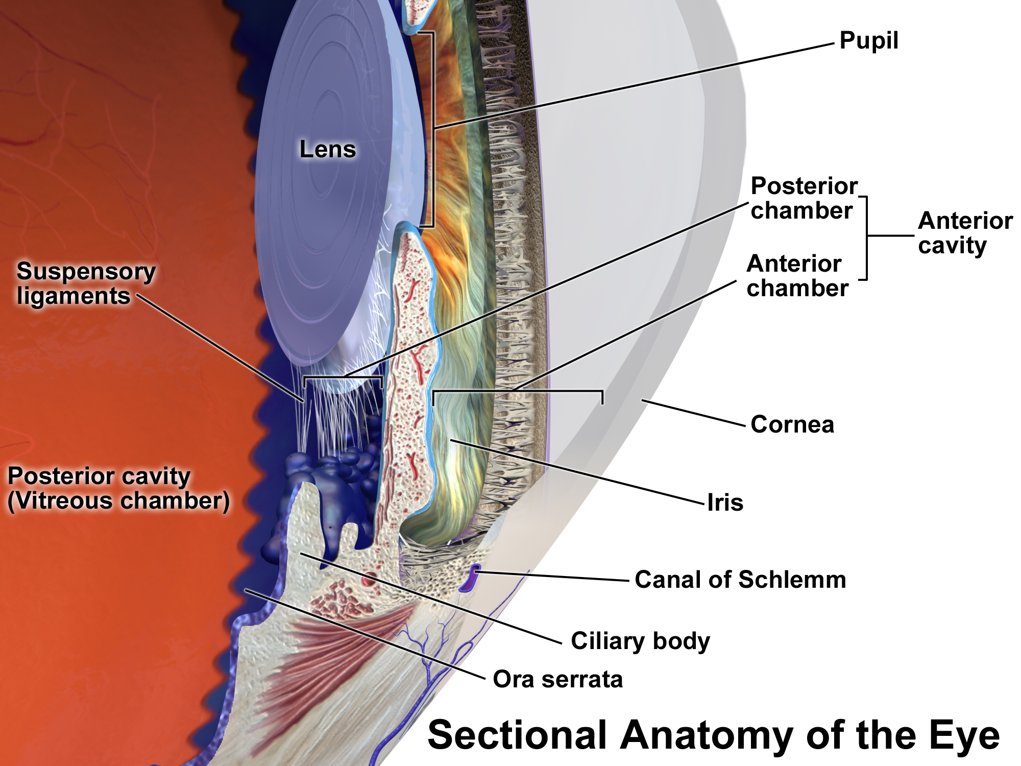
Uveitis () is inflammation of the uvea, the pigmented layer of the eye between the inner retina and the outer fibrous layer composed of the sclera and cornea. The uvea consists of the middle layer of pigmented vascular structures of the eye and includes the iris, ciliary body, and choroid. Uveitis is described anatomically, by the part of the eye affected, as anterior, intermediate or posterior, or panuveitic if all parts are involved. Anterior uveitis ( iridocyclytis) is the most common, with the incidence of uveitis overall affecting approximately 1:4500, most commonly those between the ages of 20-60. Symptoms include eye pain, eye redness, floaters and blurred vision, and ophthalmic examination may show dilated ciliary blood vessels and the presence of cells in the anterior chamber. Uveitis may arise spontaneously, have a genetic component, or be associated with an autoimmune disease or infection. While the eye is a relatively protected environment, its immune mechanisms may ... [...More Info...] [...Related Items...] OR: [Wikipedia] [Google] [Baidu] |
Ophthalmology
Ophthalmology ( ) is a surgical subspecialty within medicine that deals with the diagnosis and treatment of eye disorders. An ophthalmologist is a physician who undergoes subspecialty training in medical and surgical eye care. Following a medical degree, a doctor specialising in ophthalmology must pursue additional postgraduate residency training specific to that field. This may include a one-year integrated internship that involves more general medical training in other fields such as internal medicine or general surgery. Following residency, additional specialty training (or fellowship) may be sought in a particular aspect of eye pathology. Ophthalmologists prescribe medications to treat eye diseases, implement laser therapy, and perform surgery when needed. Ophthalmologists provide both primary and specialty eye care - medical and surgical. Most ophthalmologists participate in academic research on eye diseases at some point in their training and many include research as part ... [...More Info...] [...Related Items...] OR: [Wikipedia] [Google] [Baidu] |
Fuchs Heterochromic Iridocyclitis
Fuchs heterochromic iridocyclitis (FHI) is a chronic unilateral uveitis appearing with the triad of heterochromia, predisposition to cataract and glaucoma, and keratitic precipitates on the posterior corneal surface. Patients are often asymptomatic and the disease is often discovered through investigation of the cause of the heterochromia or cataract. Neovascularisation (growth of new abnormal vessels) is possible and any eye surgery, such as cataract surgery, can cause bleeding from the fragile vessels in the atrophic iris causing accumulation of blood in the anterior chamber of the eye, also known as hyphema. Presentation This condition is usually unilateral, and its symptoms vary from none to mild blurring and discomfort. Signs include diffuse iris atrophy and small white keratic precipitates (deposits on the inner surface of the cornea), cells presenting in the anterior chamber as well as the anterior vitreous. Glaucoma and cataract occur frequently. Complications * Glaucoma * ... [...More Info...] [...Related Items...] OR: [Wikipedia] [Google] [Baidu] |
Juvenile Idiopathic Arthritis
Juvenile idiopathic arthritis (JIA) is the most common, chronic rheumatic disease of childhood, affecting approximately one per 1,000 children. ''Juvenile'', in this context, refers to disease onset before 16 years of age, while ''idiopathic'' refers to a condition with no defined cause, and '' arthritis'' is inflammation within the joint. JIA is an autoimmune, noninfective, inflammatory joint disease, the cause of which remains poorly understood. It is characterised by chronic joint inflammation. JIA is a subset of childhood arthritis, but unlike other, more transient forms of childhood arthritis, JIA persists for at least six weeks, and in some children is a lifelong condition. It differs significantly from forms of arthritis commonly seen in adults (osteoarthritis, rheumatoid arthritis), in terms of cause, disease associations, and prognosis. The prognosis for children with JIA has improved dramatically over recent decades, particularly with the introduction of biological the ... [...More Info...] [...Related Items...] OR: [Wikipedia] [Google] [Baidu] |
Uvea
The uvea (; Lat. ''uva'', "grape"), also called the ''uveal layer'', ''uveal coat'', ''uveal tract'', ''vascular tunic'' or ''vascular layer'' is the pigmented middle of the three concentric layers that make up an eye. History and etymology The originally medieval Latin term comes from the Latin word ''uva'' ("grape") and is a reference to its grape-like appearance (reddish-blue or almost black colour, wrinkled appearance and grape-like size and shape when stripped intact from a cadaveric eye). In fact, it is a partial loan translation of the Ancient Greek term for the choroid, which literally means “covering resembling a grape”. Its use as a technical term for part of the eye is ancient, but it only referred to the choroid in Middle English and before. Structure Regions The uvea is the vascular middle layer of the eye. It is traditionally divided into three areas, from front to back, the: * Iris * Ciliary body * Choroid Function The prime functions of the uveal tra ... [...More Info...] [...Related Items...] OR: [Wikipedia] [Google] [Baidu] |
Toxoplasmic Chorioretinitis
Toxoplasma chorioretinitis, more simply known as ocular toxoplasmosis, is possibly the most common cause of infections in the back of the eye (posterior segment) worldwide. The causitive agent is ''Toxoplasma gondii'', and in the United States, most cases are acquired congenitally. The most common symptom is decreased visual acuity in one eye. The diagnosis is made by examination of the eye, using ophthalmoscopy. Sometimes serologic testing is used to rule out the disease, but due to high rates of false positives, serologies are not diagnostic of toxoplasmic retinitis. If vision is not compromised, treatment may not be necessary. When vision is affected or threatened, treatment consists of pyrimethamine, sulfadiazine, and folinic acid for 4–6 weeks. Prednisone is sometimes used to decrease inflammation. Signs and symptoms A unilateral decrease in visual acuity is the most common symptom of toxoplasmic retinitis. Under ophthalmic examination, toxoplasmic chorioretinitis ... [...More Info...] [...Related Items...] OR: [Wikipedia] [Google] [Baidu] |
Intermediate Uveitis
Intermediate uveitis is a form of uveitis localized to the vitreous and peripheral retina. Primary sites of inflammation include the vitreous of which other such entities as pars planitis, posterior cyclitis, and hyalitis are encompassed. Intermediate uveitis may either be an isolated eye disease or associated with the development of a systemic disease such as multiple sclerosis or sarcoidosis. As such, intermediate uveitis may be the first expression of a systemic condition. Infectious causes of intermediate uveitis include Epstein–Barr virus infection, Lyme disease, HTLV-1 virus infection, cat scratch disease, and hepatitis C. Permanent loss of vision is most commonly seen in patients with chronic cystoid macular edema (CME). Every effort must be made to eradicate CME when present. Other less common causes of visual loss include retinal detachment, glaucoma, band keratopathy, cataract, vitreous hemorrhage, epiretinal membrane and choroidal neovascularization. Signs and symptoms ... [...More Info...] [...Related Items...] OR: [Wikipedia] [Google] [Baidu] |
Choroid
The choroid, also known as the choroidea or choroid coat, is a part of the uvea, the vascular layer of the eye, and contains connective tissues, and lies between the retina and the sclera. The human choroid is thickest at the far extreme rear of the eye (at 0.2 mm), while in the outlying areas it narrows to 0.1 mm. The choroid provides oxygen and nourishment to the outer layers of the retina. Along with the ciliary body and iris, the choroid forms the uveal tract. The structure of the choroid is generally divided into four layers (classified in order of furthest away from the retina to closest): *Haller's layer - outermost layer of the choroid consisting of larger diameter blood vessels; *Sattler's layer - layer of medium diameter blood vessels; * Choriocapillaris - layer of capillaries; and *Bruch's membrane (synonyms: Lamina basalis, Complexus basalis, Lamina vitra) - innermost layer of the choroid. Blood supply There are two circulations of the eye: the retin ... [...More Info...] [...Related Items...] OR: [Wikipedia] [Google] [Baidu] |
Ciliary Body
The ciliary body is a part of the eye that includes the ciliary muscle, which controls the shape of the lens, and the ciliary epithelium, which produces the aqueous humor. The aqueous humor is produced in the non-pigmented portion of the ciliary body. The ciliary body is part of the uvea, the layer of tissue that delivers oxygen and nutrients to the eye tissues. The ciliary body joins the ora serrata of the choroid to the root of the iris.Cassin, B. and Solomon, S. ''Dictionary of Eye Terminology''. Gainesville, Florida: Triad Publishing Company, 1990. Structure The ciliary body is a ring-shaped thickening of tissue inside the eye that divides the posterior chamber from the vitreous body. It contains the ciliary muscle, vessels, and fibrous connective tissue. Folds on the inner ciliary epithelium are called ciliary processes, and these secrete aqueous humor into the posterior chamber. The aqueous humor then flows through the pupil into the anterior chamber. The ciliary bo ... [...More Info...] [...Related Items...] OR: [Wikipedia] [Google] [Baidu] |
Iris (anatomy)
In humans and most mammals and birds, the iris (plural: ''irides'' or ''irises'') is a thin, annular structure in the eye, responsible for controlling the diameter and size of the pupil, and thus the amount of light reaching the retina. Eye color is defined by the iris. In optical terms, the pupil is the eye's aperture, while the iris is the diaphragm. Structure The iris consists of two layers: the front pigmented fibrovascular layer known as a stroma and, beneath the stroma, pigmented epithelial cells. The stroma is connected to a sphincter muscle (sphincter pupillae), which contracts the pupil in a circular motion, and a set of dilator muscles ( dilator pupillae), which pull the iris radially to enlarge the pupil, pulling it in folds. The sphincter pupillae is the opposing muscle of the dilator pupillae. The pupil's diameter, and thus the inner border of the iris, changes size when constricting or dilating. The outer border of the iris does not change size. The constricti ... [...More Info...] [...Related Items...] OR: [Wikipedia] [Google] [Baidu] |
Vascular
The blood vessels are the components of the circulatory system that transport blood throughout the human body. These vessels transport blood cells, nutrients, and oxygen to the tissues of the body. They also take waste and carbon dioxide away from the tissues. Blood vessels are needed to sustain life, because all of the body's tissues rely on their functionality. There are five types of blood vessels: the arteries, which carry the blood away from the heart; the arterioles; the capillaries, where the exchange of water and chemicals between the blood and the tissues occurs; the venules; and the veins, which carry blood from the capillaries back towards the heart. The word ''vascular'', meaning relating to the blood vessels, is derived from the Latin ''vas'', meaning vessel. Some structures – such as cartilage, the epithelium, and the lens and cornea of the eye – do not contain blood vessels and are labeled ''avascular''. Etymology * artery: late Middle English; from Latin ' ... [...More Info...] [...Related Items...] OR: [Wikipedia] [Google] [Baidu] |
Cornea
The cornea is the transparent front part of the eye that covers the iris, pupil, and anterior chamber. Along with the anterior chamber and lens, the cornea refracts light, accounting for approximately two-thirds of the eye's total optical power. In humans, the refractive power of the cornea is approximately 43 dioptres. The cornea can be reshaped by surgical procedures such as LASIK. While the cornea contributes most of the eye's focusing power, its focus is fixed. Accommodation (the refocusing of light to better view near objects) is accomplished by changing the geometry of the lens. Medical terms related to the cornea often start with the prefix "'' kerat-''" from the Greek word κέρας, ''horn''. Structure The cornea has unmyelinated nerve endings sensitive to touch, temperature and chemicals; a touch of the cornea causes an involuntary reflex to close the eyelid. Because transparency is of prime importance, the healthy cornea does not have or need blood vessels with ... [...More Info...] [...Related Items...] OR: [Wikipedia] [Google] [Baidu] |


