|
Intersegmental Artery
The intersegmental arteries are a set of 30 arteries arising from the embryonic dorsal aorta, with each artery providing blood supply to one somite and its derivatives. Cervical intersegmental arteries The cervical intersegmental arteries merge into the vertebral artery with the exception of the 7th (or possibly the 6th) cervical intersegmental artery, which becomes the subclavian artery. The confusion arises because the vertebral artery drains into the subclavian artery following the disappearance of the dorsal aortae in part of the cervical region. Thoracic intersegmental arteries The thoracic intersegmental arteries all develop into the intercostal arteries. Lumbar intersegmental arteries The lumbar intersegmental arteries develop into the lumbar arteries, with the exception of the 5th (last) lumbar intersegmental artery, which becomes the common iliac arteries. Sacral intersegmental arteries These arteries merge into the lateral sacral artery The lateral sacral arte ... [...More Info...] [...Related Items...] OR: [Wikipedia] [Google] [Baidu] |
Dorsal Aorta
The dorsal aortae are paired (left and right) embryological vessels which progress to form the descending aorta. The paired dorsal aortae arise from aortic arches that in turn arise from the aortic sac. The primary dorsal aorta is located deep to the lateral plate of mesoderm and move from lateral to medial position with development and eventually will fuse with the other dorsal aorta to form the descending aorta. Each primitive aorta anteriorly receives the vitelline vein from the yolk-sac, and is prolonged backward on the lateral aspect of the notochord under the name of the dorsal aorta. The dorsal aortae give branches to the yolk-sac, and are continued backward through the body-stalk as the umbilical arteries to the villi of the chorion The chorion is the outermost fetal membrane around the embryo in mammals, birds and reptiles (amniotes). It develops from an outer fold on the surface of the yolk sac, which lies outside the zona pellucida (in mammals), known as the v ... [...More Info...] [...Related Items...] OR: [Wikipedia] [Google] [Baidu] |
Somite
The somites (outdated term: primitive segments) are a set of bilaterally paired blocks of paraxial mesoderm that form in the embryonic stage of somitogenesis, along the head-to-tail axis in segmented animals. In vertebrates, somites subdivide into the dermatomes, myotomes, sclerotomes and syndetomes that give rise to the vertebrae of the vertebral column, rib cage, part of the occipital bone, skeletal muscle, cartilage, tendons, and skin (of the back). The word ''somite'' is sometimes also used in place of the word '' metamere''. In this definition, the somite is a homologously-paired structure in an animal body plan, such as is visible in annelids and arthropods. Development The mesoderm forms at the same time as the other two germ layers, the ectoderm and endoderm. The mesoderm at either side of the neural tube is called paraxial mesoderm. It is distinct from the mesoderm underneath the neural tube which is called the chordamesoderm that becomes the notochord. The pa ... [...More Info...] [...Related Items...] OR: [Wikipedia] [Google] [Baidu] |
Vertebral Artery
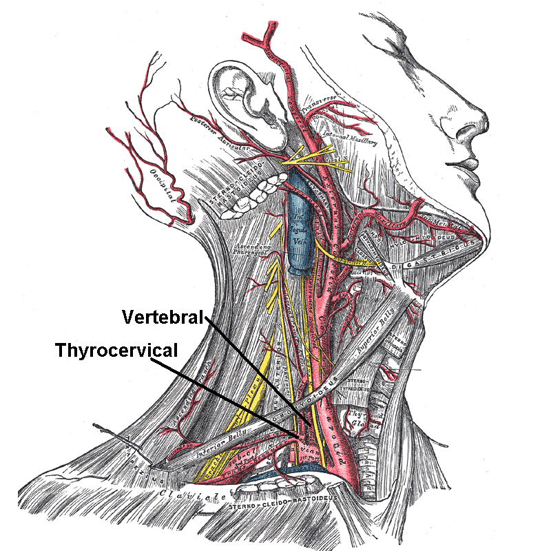
The vertebral arteries are major arteries An artery (plural arteries) () is a blood vessel in humans and most animals that takes blood away from the heart to one or more parts of the body (tissues, lungs, brain etc.). Most arteries carry oxygenated blood; the two exceptions are the pu ... of the neck. Typically, the vertebral arteries originate from the subclavian arteries. Each vessel courses superiorly along each side of the neck, merging within the skull to form the single, midline basilar artery. As the supplying component of the ''vertebrobasilar vascular system'', the vertebral arteries supply blood to the upper spinal cord, brainstem, cerebellum, and Cerebral circulation#Posterior cerebral circulation, posterior part of brain. Structure The vertebral arteries usually arise from the posterosuperior aspect of the central subclavian arteries on each side of the body, then enter deep to the transverse process at the level of the 6th cervical vertebrae (C6), or occasio ... [...More Info...] [...Related Items...] OR: [Wikipedia] [Google] [Baidu] |
Subclavian Artery
In human anatomy, the subclavian arteries are paired major arteries of the upper thorax, below the clavicle. They receive blood from the aortic arch. The left subclavian artery supplies blood to the left arm and the right subclavian artery supplies blood to the right arm, with some branches supplying the head and thorax. On the left side of the body, the subclavian comes directly off the aortic arch, while on the right side it arises from the relatively short brachiocephalic artery when it bifurcates into the subclavian and the right common carotid artery. The usual branches of the subclavian on both sides of the body are the vertebral artery, the internal thoracic artery, the thyrocervical trunk, the costocervical trunk and the dorsal scapular artery, which may branch off the transverse cervical artery, which is a branch of the thyrocervical trunk. The subclavian becomes the axillary artery at the lateral border of the first rib. Structure From its origin, the subclavian artery t ... [...More Info...] [...Related Items...] OR: [Wikipedia] [Google] [Baidu] |
Intercostal Arteries
The intercostal arteries are a group of arteries that supply the area between the ribs ("costae"), called the intercostal space. The highest intercostal artery (supreme intercostal artery or superior intercostal artery) is an artery in the human body that usually gives rise to the first and second posterior intercostal arteries, which supply blood to their corresponding intercostal space. It usually arises from the costocervical trunk, which is a branch of the subclavian artery. Some anatomists may contend that there is no supreme intercostal artery, only a supreme intercostal vein. The anterior intercostal branches of internal thoracic artery supply the upper five or six intercostal spaces. The internal thoracic artery (previously called as internal mammary artery) then divides into the superior epigastric artery and musculophrenic artery. The latter gives out the remaining anterior intercostal branches. Two in number in each space, these small vessels pass lateralward, one l ... [...More Info...] [...Related Items...] OR: [Wikipedia] [Google] [Baidu] |
Lumbar Arteries
The lumbar arteries are arteries located in the lower back or lumbar region. The lumbar arteries are in parallel with the intercostals. They are usually four in number on either side, and arise from the back of the aorta, opposite the bodies of the upper four lumbar vertebrae. A fifth pair, small in size, is occasionally present: they arise from the middle sacral artery. They run lateralward and backward on the bodies of the lumbar vertebrae, behind the sympathetic trunk, to the intervals between the adjacent transverse processes, and are then continued into the abdominal wall. The arteries of the right side pass behind the inferior vena cava, and the upper two on each side run behind the corresponding crus of the diaphragm. The arteries of both sides pass beneath the tendinous arches which give origin to the psoas major, and are then continued behind this muscle and the lumbar plexus. They now cross the quadratus lumborum, the upper three arteries running behind, the las ... [...More Info...] [...Related Items...] OR: [Wikipedia] [Google] [Baidu] |
Common Iliac Artery
The common iliac artery is a large artery of the abdomen paired on each side. It originates from the aortic bifurcation at the level of the 4th lumbar vertebra. It ends in front of the sacroiliac joint, one on either side, and each bifurcates into the external and internal iliac arteries. Structure The common iliac artery are about 4 cm long in adults and more than a centimeter in diameter. It begins as a branch of the aorta. This is at the level of the 4th lumbar vertebra. It runs inferolaterally, along the medial border of the psoas muscles. It bifurcates into the external iliac artery and the internal iliac artery at the pelvic brim, in front of the sacroiliac joints. The common iliac artery, and all of its branches, exist as paired structures (that is to say, there is one on the left side and one on the right). The distribution of the common iliac artery is basically the pelvis and lower limb (as the femoral artery) on the corresponding side. Relations Both common il ... [...More Info...] [...Related Items...] OR: [Wikipedia] [Google] [Baidu] |
Lateral Sacral Artery
The lateral sacral arteries is an artery in the pelvis that arises from the posterior division of the internal iliac artery. It later splits into two smaller branches, a superior and an inferior. Structure The lateral sacral artery is the second branch of the posterior division of the internal iliac artery. It is a parietal branch. Superior The superior, of large size, passes medialward, and, after anastomosing with branches from the middle sacral, enters the first or second anterior sacral foramen, supplies branches to the contents of the sacral canal, and, escaping by the corresponding posterior sacral foramen, is distributed to the skin and muscles on the dorsum of the sacrum, anastomosing with the superior gluteal. Inferior The inferior runs obliquely across the front of the piriformis and the sacral nerves to the medial side of the anterior sacral foramina, descends on the front of the sacrum, and anastomoses over the coccyx with the middle sacral and opposite latera ... [...More Info...] [...Related Items...] OR: [Wikipedia] [Google] [Baidu] |


