|
Internal Capsule
The internal capsule is a white matter structure situated in the inferomedial part of each cerebral hemisphere of the brain. It carries information past the basal ganglia, separating the caudate nucleus and the thalamus from the putamen and the globus pallidus. The internal capsule contains both ascending and descending axons, going to and coming from the cerebral cortex. It also separates the caudate nucleus and the putamen in the dorsal striatum, a brain region involved in motor and reward pathways. The corticospinal tract constitutes a large part of the internal capsule, carrying motor information from the primary motor cortex to the lower motor neurons in the spinal cord. Above the basal ganglia the corticospinal tract is a part of the corona radiata. Below the basal ganglia the tract is called cerebral crus (a part of the cerebral peduncle) and below the pons it is referred to as the corticospinal tract. Structure The internal capsule consists of three parts and is V-s ... [...More Info...] [...Related Items...] OR: [Wikipedia] [Google] [Baidu] |
White Matter
White matter refers to areas of the central nervous system (CNS) that are mainly made up of myelinated axons, also called tracts. Long thought to be passive tissue, white matter affects learning and brain functions, modulating the distribution of action potentials, acting as a relay and coordinating communication between different brain regions. White matter is named for its relatively light appearance resulting from the lipid content of myelin. However, the tissue of the freshly cut brain appears pinkish-white to the naked eye because myelin is composed largely of lipid tissue veined with capillaries. Its white color in prepared specimens is due to its usual preservation in formaldehyde. Structure White matter White matter is composed of bundles, which connect various grey matter areas (the locations of nerve cell bodies) of the brain to each other, and carry nerve impulses between neurons. Myelin acts as an insulator, which allows electrical signals to jump, rather ... [...More Info...] [...Related Items...] OR: [Wikipedia] [Google] [Baidu] |
Corona Radiata
In neuroanatomy, the corona radiata is a white matter sheet that continues inferiorly as the internal capsule and superiorly as the centrum semiovale. This sheet of both ascending and descending axons carries most of the neural traffic from and to the cerebral cortex. The corona radiata is associated with the corticopontine tract, the corticobulbar tract, and the corticospinal tract. Structure Projection fibers are afferents carrying information to the cerebral cortex, and efferents carrying information away from it. The most prominent projection fibers are the corona radiata, which radiate out from the cortex and then come together in the brain stem. The projection fibers that make up the corona radiata also radiate out of the brain stem via the internal capsule. Cerebral white matter is commonly regarded today as an intricately organized system of fasciculi that facilitate the highest expression of cerebral activity. Function Motor pathway Evidence from subcortical small ... [...More Info...] [...Related Items...] OR: [Wikipedia] [Google] [Baidu] |
Corticobulbar Tract
In neuroanatomy, the corticobulbar (or corticonuclear) tract is a two-neuron white matter motor pathway connecting the motor cortex in the cerebral cortex to the medullary pyramids, which are part of the brainstem's medulla oblongata (also called "bulbar") region, and are primarily involved in carrying the motor function of the non-oculomotor cranial nerves. The corticobulbar tract is one of the pyramidal tracts, the other being the corticospinal tract. Structure The corticobulbar tract originates in the primary motor cortex of the frontal lobe, just superior to the lateral fissure and rostral to the central sulcus in the precentral gyrus (Brodmann area 4). The tract descends through the corona radiata and genu of the internal capsule with a few fibers in the posterior limb of the internal capsule, as it passes from the cortex down to the midbrain. In the midbrain, the internal capsule becomes the cerebral peduncles. The white matter is located in the ventral portion of the ... [...More Info...] [...Related Items...] OR: [Wikipedia] [Google] [Baidu] |
Cranial Nerve
Cranial nerves are the nerves that emerge directly from the brain (including the brainstem), of which there are conventionally considered twelve pairs. Cranial nerves relay information between the brain and parts of the body, primarily to and from regions of the head and neck, including the special senses of vision, taste, smell, and hearing. The cranial nerves emerge from the central nervous system above the level of the first vertebra of the vertebral column. Each cranial nerve is paired and is present on both sides. There are conventionally twelve pairs of cranial nerves, which are described with Roman numerals I–XII. Some considered there to be thirteen pairs of cranial nerves, including cranial nerve zero. The numbering of the cranial nerves is based on the order in which they emerge from the brain and brainstem, from front to back. The terminal nerves (0), olfactory nerves (I) and optic nerves (II) emerge from the cerebrum, and the remaining ten pairs arise from ... [...More Info...] [...Related Items...] OR: [Wikipedia] [Google] [Baidu] |
Decussation
Decussation is used in biological contexts to describe a crossing (due to the shape of the Roman numeral for ten, an uppercase 'X' (), ). In Latin anatomical terms, the form is used, e.g. . Similarly, the anatomical term chiasma is named after the Greek uppercase 'Χ' (chi). Whereas a decussation refers to a crossing within the central nervous system, various kinds of crossings in the peripheral nervous system are called chiasma. Examples include: * In the brain, where nerve fibers obliquely cross from one lateral side of the brain to the other, that is to say they cross at a level other than their origin. See for examples Decussation of pyramids and sensory decussation. In neuroanatomy, the term ''chiasma'' is reserved for crossing of- or within nerves such as in the optic chiasm. * In botanical leaf taxology, the word ''decussate'' describes an opposite pattern of leaves which has successive pairs at right angles to each other (i.e. rotated 90 degrees along the st ... [...More Info...] [...Related Items...] OR: [Wikipedia] [Google] [Baidu] |
Cerebrospinal Fibers
The cerebrospinal fibers, derived from the cells of the motor area of the cerebral cortex, occupy the middle three-fifths of the base; they are continued partly to the nuclei of the motor cranial nerves, but mainly into the pyramids of the medulla oblongata The medulla oblongata or simply medulla is a long stem-like structure which makes up the lower part of the brainstem. It is anterior and partially inferior to the cerebellum. It is a cone-shaped neuronal mass responsible for autonomic (involun .... References Central nervous system {{Portal bar, Anatomy ... [...More Info...] [...Related Items...] OR: [Wikipedia] [Google] [Baidu] |
Primary Auditory Cortex
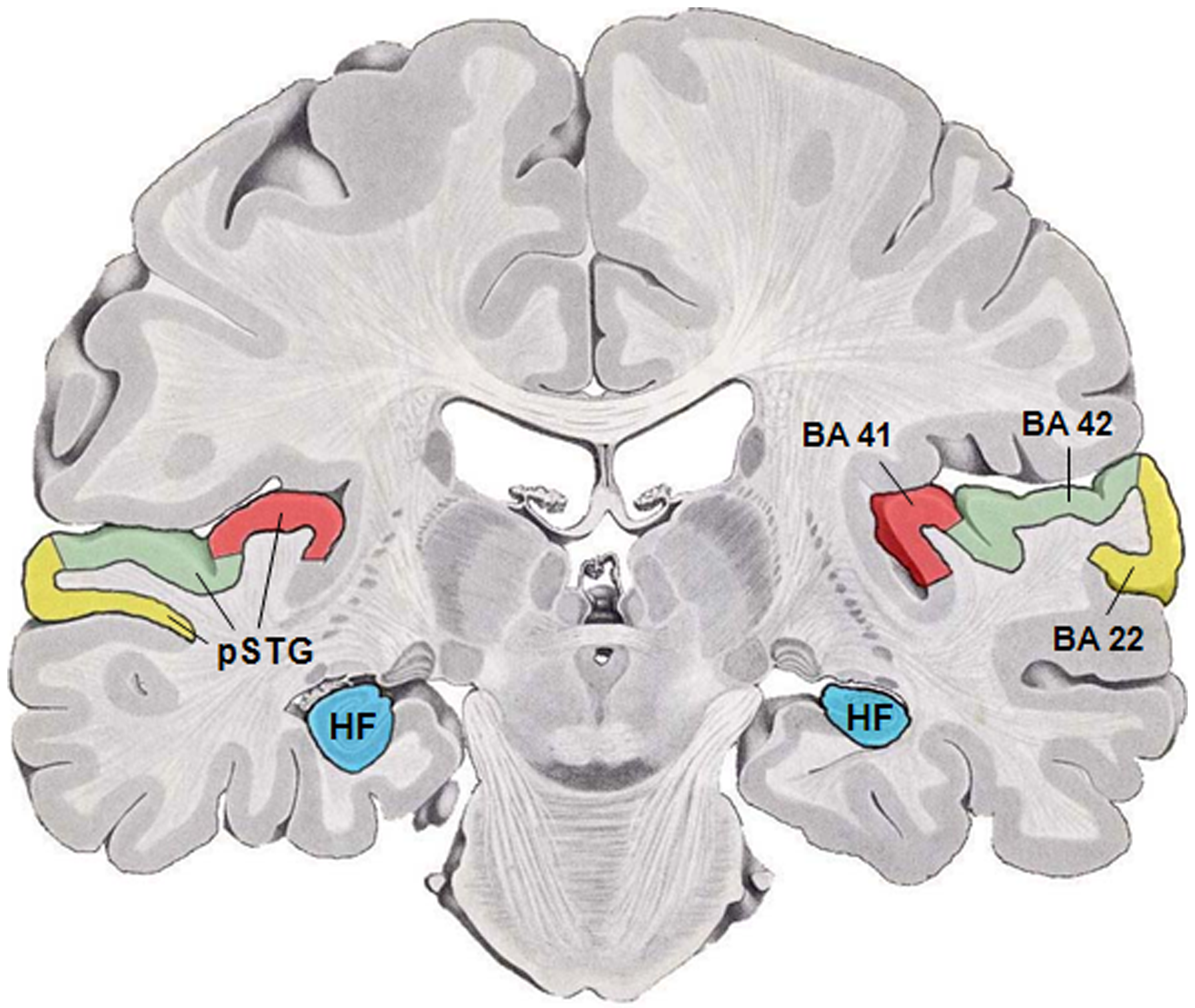
The auditory cortex is the part of the temporal lobe that processes auditory information in humans and many other vertebrates. It is a part of the auditory system, performing basic and higher functions in hearing, such as possible relations to language switching.Cf. Pickles, James O. (2012). ''An Introduction to the Physiology of Hearing'' (4th ed.). Bingley, UK: Emerald Group Publishing Limited, p. 238. It is located bilaterally, roughly at the upper sides of the temporal lobes – in humans, curving down and onto the medial surface, on the superior temporal plane, within the lateral sulcus and comprising parts of the transverse temporal gyri, and the superior temporal gyrus, including the planum polare and planum temporale (roughly Brodmann areas 41 and 42, and partially 22). The auditory cortex takes part in the spectrotemporal, meaning involving time and frequency, analysis of the inputs passed on from the ear. The cortex then filters and passes on the information to ... [...More Info...] [...Related Items...] OR: [Wikipedia] [Google] [Baidu] |
Medial Geniculate Nucleus
The medial geniculate nucleus (MGN) or medial geniculate body (MGB) is part of the auditory thalamus and represents the thalamic relay between the inferior colliculus (IC) and the auditory cortex (AC). It is made up of a number of sub-nuclei that are distinguished by their neuronal morphology and density, by their afferent and efferent connections, and by the coding properties of their neurons. It is thought that the MGN influences the direction and maintenance of attention. Divisions The MGN has three major divisions; ventral (VMGN), dorsal (DMGN) and medial (MMGN). Whilst the VMGN is specific to auditory information processing, the DMGN and MMGN also receive information from non-auditory pathways. Ventral subnucleus Cell types There are two main cell types in the ventral subnucleus of the medial geniculate body (VMGN): * Thalamocortical relay cells (or principal neurons): The dendritic Dendrite derives from the Greek word "dendron" meaning ( "tree-like"), and may refer to: ... [...More Info...] [...Related Items...] OR: [Wikipedia] [Google] [Baidu] |
Optic Radiation
In neuroanatomy, the optic radiation (also known as the geniculocalcarine tract, the geniculostriate pathway, and posterior thalamic radiation) are axons from the neurons in the lateral geniculate nucleus to the primary visual cortex. The optic radiation receives blood through deep branches of the middle cerebral artery and posterior cerebral artery. They carry visual information through two divisions (called upper and lower division) to the visual cortex (also called ''striate cortex'') along the calcarine fissure. There is one set of upper and lower divisions on each side of the brain. If a lesion only exists in one unilateral division of the optic radiation, the consequence is called quadrantanopia, which implies that only the respective superior or inferior quadrant of the visual field is affected. If both divisions on one side of the brain are affected, the result is a contralateral homonymous hemianopsia. Structure The upper division: :* Projects to the upper bank of ... [...More Info...] [...Related Items...] OR: [Wikipedia] [Google] [Baidu] |
Lenticular Nucleus
The lentiform nucleus, or lenticular nucleus, comprises the putamen and the globus pallidus within the basal ganglia. With the caudate nucleus, it forms the dorsal striatum. It is a large, lens-shaped mass of gray matter just lateral to the internal capsule. Structure When divided horizontally, it exhibits, to some extent, the appearance of a biconvex lens, while a coronal section of its central part presents a somewhat triangular outline. It is shorter than the caudate nucleus and does not extend as far forward. Boundaries It is lateral to the caudate nucleus and thalamus, and is seen only in sections of the hemisphere. It is bounded laterally by a lamina of white substance called the external capsule, and lateral to this is a thin layer of gray substance termed the claustrum. Its anterior end is continuous with the lower part of the head of the caudate nucleus and with the anterior perforated substance. Components In a coronal section through the middle of the lenti ... [...More Info...] [...Related Items...] OR: [Wikipedia] [Google] [Baidu] |
Genu Of The Internal Capsule
The internal capsule is a white matter structure situated in the inferomedial part of each cerebral hemisphere of the brain. It carries information past the basal ganglia, separating the caudate nucleus and the thalamus from the putamen and the globus pallidus. The internal capsule contains both ascending and descending axons, going to and coming from the cerebral cortex. It also separates the caudate nucleus and the putamen in the dorsal striatum, a brain region involved in motor and reward pathways. The corticospinal tract constitutes a large part of the internal capsule, carrying motor information from the primary motor cortex to the lower motor neurons in the spinal cord. Above the basal ganglia the corticospinal tract is a part of the corona radiata. Below the basal ganglia the tract is called cerebral crus (a part of the cerebral peduncle) and below the pons it is referred to as the corticospinal tract. Structure The internal capsule consists of three parts and is V-sha ... [...More Info...] [...Related Items...] OR: [Wikipedia] [Google] [Baidu] |
Transverse Plane
The transverse plane (also known as the horizontal plane, axial plane and transaxial plane) is an anatomical plane that divides the body into superior and inferior sections. It is perpendicular to the coronal and sagittal planes. List of clinically relevant anatomical planes * Transverse ''thoracic plane'' * '' Xiphosternal plane'' (or xiphosternal junction) * ''Transpyloric plane'' * ''Subcostal plane'' * '' Umbilical plane'' (or transumbilical plane) * ''Supracristal plane'' * ''Intertubercular plane'' (or transtubercular plane) * ''Interspinous plane'' Clinically relevant anatomical planes with associated structures * The transverse ''thoracic plane'' ** Plane through T4 & T5 vertebral junction and sternal angle of Louis. ** Marks the: *** Attachment of costal cartilage of rib 2 at the sternal angle; *** Aortic arch (beginning and end); *** Upper margin of SVC; *** Thoracic duct crossing; *** Tracheal bifurcation; *** Pulmonary trunk bifurcation; * The '' xiphosternal p ... [...More Info...] [...Related Items...] OR: [Wikipedia] [Google] [Baidu] |


