|
Hypophysectomy
Hypophysectomy is the surgical removal of the hypophysis (pituitary gland). It is most commonly performed to treat tumors, especially craniopharyngioma tumors. Sometimes it is used to treat Cushing's syndrome due to pituitary adenoma or Simmond's disease It is also applied in neurosciences (in experiments with lab animals) to understand the functioning of hypophysis. There are various ways a hypophysectomy can be carried out. These methods include transsphenoidal hypophysectomy, open craniotomy, and stereotactic radiosurgery. Medications that are given as hormone replacement therapy following a complete hypophysectomy (removal of the pituitary gland) are often glucocorticoids. Secondary Addison's and hyperlipidemia can occur. Thyroid hormone is useful in controlling cholesterol metabolism that has been affected by pituitary deletion. Methods of hypophysectomy Hypophysectomies can be performed in three ways. These include transsphenoidal hypophysectomy, open craniotomy, and ... [...More Info...] [...Related Items...] OR: [Wikipedia] [Google] [Baidu] |
Pituitary Gland
In vertebrate anatomy, the pituitary gland, or hypophysis, is an endocrine gland, about the size of a chickpea and weighing, on average, in humans. It is a protrusion off the bottom of the hypothalamus at the base of the brain. The hypophysis rests upon the hypophyseal fossa of the sphenoid bone in the center of the middle cranial fossa and is surrounded by a small bony cavity (sella turcica) covered by a dural fold (diaphragma sellae). The anterior pituitary (or adenohypophysis) is a lobe of the gland that regulates several physiological processes including stress, growth, reproduction, and lactation. The intermediate lobe synthesizes and secretes melanocyte-stimulating hormone. The posterior pituitary (or neurohypophysis) is a lobe of the gland that is functionally connected to the hypothalamus by the median eminence via a small tube called the pituitary stalk (also called the infundibular stalk or the infundibulum). Hormones secreted from the pituitary gland ... [...More Info...] [...Related Items...] OR: [Wikipedia] [Google] [Baidu] |
Magnetic Resonance Imaging
Magnetic resonance imaging (MRI) is a medical imaging technique used in radiology to form pictures of the anatomy and the physiological processes of the body. MRI scanners use strong magnetic fields, magnetic field gradients, and radio waves to generate images of the organs in the body. MRI does not involve X-rays or the use of ionizing radiation, which distinguishes it from CT and PET scans. MRI is a medical application of nuclear magnetic resonance (NMR) which can also be used for imaging in other NMR applications, such as NMR spectroscopy. MRI is widely used in hospitals and clinics for medical diagnosis, staging and follow-up of disease. Compared to CT, MRI provides better contrast in images of soft-tissues, e.g. in the brain or abdomen. However, it may be perceived as less comfortable by patients, due to the usually longer and louder measurements with the subject in a long, confining tube, though "Open" MRI designs mostly relieve this. Additionally, implants and oth ... [...More Info...] [...Related Items...] OR: [Wikipedia] [Google] [Baidu] |
List Of Surgeries By Type
Many surgical procedure names can be broken into parts to indicate the meaning. For example, in gastrectomy, "ectomy" is a suffix meaning the removal of a part of the body. "Gastro-" means stomach. Thus, ''gastrectomy'' refers to the surgical removal of the stomach (or sections thereof). "Otomy" means cutting into a part of the body; a ''gastrotomy'' would be cutting into, but not necessarily removing, the stomach. And also "pharyngo" means pharynx, "laryngo" means larynx, "esophag" means esophagus. Thus, "pharyngolaryngoesophagectomy" refers to the surgical removal of the three. The field of minimally invasive surgery has spawned another set of words, such as ''arthroscopic'' or ''laparoscopic'' surgery. These take the same form as above; an arthroscope is a device which allows the inside of the joint to be seen. List of common surgery terms Prefixes * ''mono-'' : one, from the Greek μόνος, ''monos'', "only, single" * ''angio-'' : related to a blood vessel, from the Gre ... [...More Info...] [...Related Items...] OR: [Wikipedia] [Google] [Baidu] |
Self-image
Self-image is the mental picture, generally of a kind that is quite resistant to change, that depicts not only details that are potentially available to an objective investigation by others (height, weight, hair color, etc.), but also items that have been learned by persons about themselves, either from personal experiences or by internalizing the judgments of others. Self-image may consist of six types: # Self-image resulting from how an individual sees oneself. # Self-image resulting from how others see the individual. # Self-image resulting from how the individual perceives the individual sees oneself. # Self-image resulting from how the individual perceives how others see the individual. # Self-image resulting from how others perceive how the individual sees oneself. # Self-image resulting from how others perceive how others see the individual. These six types may or may not be an accurate representation of the person. All, some, or none of them may be true. A more technical ... [...More Info...] [...Related Items...] OR: [Wikipedia] [Google] [Baidu] |
Cachexia
Cachexia () is a complex syndrome associated with an underlying illness, causing ongoing muscle loss that is not entirely reversed with nutritional supplementation. A range of diseases can cause cachexia, most commonly cancer, congestive heart failure, chronic obstructive pulmonary disease, chronic kidney disease, and AIDS. Systemic inflammation from these conditions can cause detrimental changes to metabolism and body composition. In contrast to weight loss from inadequate caloric intake, cachexia causes mostly muscle loss instead of fat loss. Diagnosis of cachexia can be difficult due to the lack of well-established diagnostic criteria. Cachexia can improve with treatment of the underlying illness but other treatment approaches have limited benefit. Cachexia is associated with increased mortality and poor quality of life. The term is from Greek κακός ''kakos'', "bad", and ἕξις ''hexis'', "condition". Causes Cachexia can be caused by diverse medical conditions, but i ... [...More Info...] [...Related Items...] OR: [Wikipedia] [Google] [Baidu] |
Asthenia
Weakness is a symptom of a number of different conditions. The causes are many and can be divided into conditions that have true or perceived muscle weakness. True muscle weakness is a primary symptom of a variety of skeletal muscle diseases, including muscular dystrophy and inflammatory myopathy. It occurs in neuromuscular junction disorders, such as myasthenia gravis. Pathophysiology Muscle cells work by detecting a flow of electrical impulses from the brain, which signals them to contract through the release of calcium by the sarcoplasmic reticulum. Fatigue (reduced ability to generate force) may occur due to the nerve, or within the muscle cells themselves. New research from scientists at Columbia University suggests that muscle fatigue is caused by calcium leaking out of the muscle cell. This makes less calcium available for the muscle cell. In addition, the Columbia researchers propose that an enzyme activated by this released calcium eats away at muscle fibers. Substr ... [...More Info...] [...Related Items...] OR: [Wikipedia] [Google] [Baidu] |
Adrenal Gland
The adrenal glands (also known as suprarenal glands) are endocrine glands that produce a variety of hormones including adrenaline and the steroids aldosterone and cortisol. They are found above the kidneys. Each gland has an outer cortex which produces steroid hormones and an inner medulla. The adrenal cortex itself is divided into three main zones: the zona glomerulosa, the zona fasciculata and the zona reticularis. The adrenal cortex produces three main types of steroid hormones: mineralocorticoids, glucocorticoids, and androgens. Mineralocorticoids (such as aldosterone) produced in the zona glomerulosa help in the regulation of blood pressure and electrolyte balance. The glucocorticoids cortisol and cortisone are synthesized in the zona fasciculata; their functions include the regulation of metabolism and immune system suppression. The innermost layer of the cortex, the zona reticularis, produces androgens that are converted to fully functional sex hormones in the gonads ... [...More Info...] [...Related Items...] OR: [Wikipedia] [Google] [Baidu] |
Thyroid
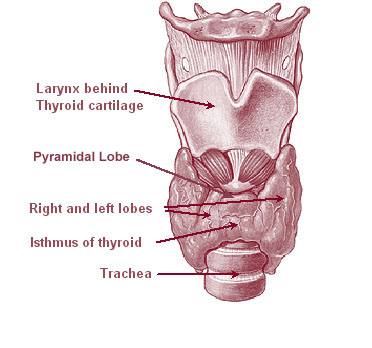
The thyroid, or thyroid gland, is an endocrine gland in vertebrates. In humans it is in the neck and consists of two connected lobes. The lower two thirds of the lobes are connected by a thin band of tissue called the thyroid isthmus. The thyroid is located at the front of the neck, below the Adam's apple. Microscopically, the functional unit of the thyroid gland is the spherical thyroid follicle, lined with follicular cells (thyrocytes), and occasional parafollicular cells that surround a lumen containing colloid. The thyroid gland secretes three hormones: the two thyroid hormones triiodothyronine (T3) and thyroxine (T4)and a peptide hormone, calcitonin. The thyroid hormones influence the metabolic rate and protein synthesis, and in children, growth and development. Calcitonin plays a role in calcium homeostasis. Secretion of the two thyroid hormones is regulated by thyroid-stimulating hormone (TSH), which is secreted from the anterior pituitary gland. TSH is regula ... [...More Info...] [...Related Items...] OR: [Wikipedia] [Google] [Baidu] |
Craniotomy
A craniotomy is a surgical operation in which a bone flap is temporarily removed from the skull to access the brain. Craniotomies are often critical operations, performed on patients who are suffering from brain lesions, such as tumors, blood clots, removal of foreign bodies such as bullets, or traumatic brain injury (TBI), and can also allow doctors to surgically implant devices, such as deep brain stimulators for the treatment of Parkinson's disease, epilepsy, and cerebellar tremor. The procedure is also used in epilepsy surgery to remove the parts of the brain that are causing epilepsy. Craniotomy is distinguished from craniectomy (in which the skull flap is not immediately replaced, allowing the brain to swell, thus reducing intracranial pressure) and from trepanation, the creation of a burr hole through the cranium in to the dura mater. Procedure Human craniotomy is usually performed under general anesthesia but can be also done with the patient awake using a local anaesthe ... [...More Info...] [...Related Items...] OR: [Wikipedia] [Google] [Baidu] |
Craniopharyngioma
A craniopharyngioma is a rare type of brain tumor derived from pituitary gland embryonic tissue that occurs most commonly in children, but also affects adults. It may present at any age, even in the prenatal and neonatal periods, but peak incidence rates are childhood-onset at 5–14 years and adult-onset at 50–74 years. People may present with bitemporal inferior quadrantanopia leading to bitemporal hemianopsia, as the tumor may compress the optic chiasm. It has a point prevalence around two per 1,000,000. Craniopharyngiomas are distinct from Rathke's cleft tumours and intrasellar arachnoid cysts. Symptoms and signs Craniopharyngiomas are almost always benign. However, as with many brain tumors, their treatment can be difficult, and significant morbidities are associated with both the tumor and treatment. * Headache (obstructive hydrocephalus) * Hypersomnia * Myxedema * Postsurgical weight gain * Polydipsia * Polyuria (diabetes insipidus) * Vision loss (bitemporal hemianopia ... [...More Info...] [...Related Items...] OR: [Wikipedia] [Google] [Baidu] |
Sphenoid Sinus
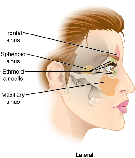
The sphenoid sinus is a paired paranasal sinus occurring within the within the body of the sphenoid bone. It represents one pair of the four paired paranasal sinuses.Illustrated Anatomy of the Head and Neck, Fehrenbach and Herring, Elsevier, 2012, page 64 The pair of sphenoid sinuses are separated in the middle by a septum of sphenoid sinuses. Each sphenoid sinus communicates with the nasal cavity via the opening of sphenoidal sinus. The two sphenoid sinuses vary in size and shape, and are usually asymmetrical. Anatomy On average, a sphenoid sinus measures 2.2 cm vertical height, 2 cm in transverse breadth; and 2.2 cm antero-posterior depth. Each spehoid sinus is contained within the body of sphenoid bone, being situated just inferior to the sella turcica. The two sphenoid sinuses are separated medially by the septum of sphenoidal sinuses (which is usually asymmetrical). An opening of sphenoidal sinus forms a passage between each sphenoidal sinus, and the nasal ca ... [...More Info...] [...Related Items...] OR: [Wikipedia] [Google] [Baidu] |


