Sphenoid sinus on:
[Wikipedia]
[Google]
[Amazon]
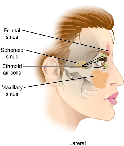
The sphenoid sinus is a paired paranasal sinus occurring within the within the body of the sphenoid bone. It represents one pair of the four paired paranasal sinuses.Illustrated Anatomy of the Head and Neck, Fehrenbach and Herring, Elsevier, 2012, page 64 The pair of sphenoid sinuses are separated in the middle by a septum of sphenoid sinuses. Each sphenoid sinus communicates with the nasal cavity via the opening of sphenoidal sinus. The two sphenoid sinuses vary in size and shape, and are usually asymmetrical.
 On average, a sphenoid sinus measures 2.2 cm vertical height, 2 cm in transverse breadth; and 2.2 cm antero-posterior depth.
Each spehoid sinus is contained within the body of sphenoid bone, being situated just inferior to the sella turcica. The two sphenoid sinuses are separated medially by the septum of sphenoidal sinuses (which is usually asymmetrical).
An opening of sphenoidal sinus forms a passage between each sphenoidal sinus, and the nasal cavity. Posteriorly, an opening of sphenoidal sinus opens into the sphenoidal sinus by an aperture high on the anterior wall the sinus; anteriorly, an opening of sphenoidal sinus opens into the roof of the nasal cavity via an aperture on the posterior wall of the sphenoethmoidal recess (occurring just superior the choana).Human Anatomy, Jacob, Elsevier, 2008, page 211
Anatomy
 On average, a sphenoid sinus measures 2.2 cm vertical height, 2 cm in transverse breadth; and 2.2 cm antero-posterior depth.
Each spehoid sinus is contained within the body of sphenoid bone, being situated just inferior to the sella turcica. The two sphenoid sinuses are separated medially by the septum of sphenoidal sinuses (which is usually asymmetrical).
An opening of sphenoidal sinus forms a passage between each sphenoidal sinus, and the nasal cavity. Posteriorly, an opening of sphenoidal sinus opens into the sphenoidal sinus by an aperture high on the anterior wall the sinus; anteriorly, an opening of sphenoidal sinus opens into the roof of the nasal cavity via an aperture on the posterior wall of the sphenoethmoidal recess (occurring just superior the choana).Human Anatomy, Jacob, Elsevier, 2008, page 211
Innervation
The mucous membrane receives sensory innervation by the posterior ethmoidal nerves (branch of the ophthalmic nerve), and postganglionic parasympathetic fibers of thefacial nerve
The facial nerve, also known as the seventh cranial nerve, cranial nerve VII, or simply CN VII, is a cranial nerve that emerges from the pons of the brainstem, controls the muscles of facial expression, and functions in the conveyance of ta ...
that synapse
In the nervous system, a synapse is a structure that permits a neuron (or nerve cell) to pass an electrical or chemical signal to another neuron or to the target effector cell.
Synapses are essential to the transmission of nervous impulses fr ...
d at the pterygopalatine ganglion which controls secretion 440px
Secretion is the movement of material from one point to another, such as a secreted chemical substance from a cell or gland. In contrast, excretion is the removal of certain substances or waste products from a cell or organism. The classica ...
of mucus
Mucus ( ) is a slippery aqueous secretion produced by, and covering, mucous membranes. It is typically produced from cells found in mucous glands, although it may also originate from mixed glands, which contain both serous and mucous cells. It ...
.
Anatomical relations
Proximal structures include: the optic canal andoptic nerve
In neuroanatomy, the optic nerve, also known as the second cranial nerve, cranial nerve II, or simply CN II, is a paired cranial nerve that transmits visual information from the retina to the brain. In humans, the optic nerve is derived fro ...
, internal carotid artery, cavernous sinus
The cavernous sinus within the human head is one of the dural venous sinuses creating a cavity called the lateral sellar compartment bordered by the temporal bone of the skull and the sphenoid bone, lateral to the sella turcica.
Structure
The c ...
, trigeminal nerve
In neuroanatomy, the trigeminal nerve (literal translation, lit. ''triplet'' nerve), also known as the fifth cranial nerve, cranial nerve V, or simply CN V, is a cranial nerve responsible for Sense, sensation in the face and motor functions ...
, pituitary gland
In vertebrate anatomy, the pituitary gland, or hypophysis, is an endocrine gland, about the size of a chickpea and weighing, on average, in humans. It is a protrusion off the bottom of the hypothalamus at the base of the brain. The hypop ...
, and the anterior ethmoidal cells.
Anatomical variation
The sphenoid sinuses vary in size and shape, and, owing to the lateral displacement of the interveningseptum
In biology, a septum (Latin for ''something that encloses''; plural septa) is a wall, dividing a cavity or structure into smaller ones. A cavity or structure divided in this way may be referred to as septate.
Examples
Human anatomy
* Interat ...
of sphenoid sinuses, are rarely symmetrical.
When exceptionally large, the sphenoid sinuses may extend into the roots of the pterygoid processes or greater wings of sphenoid bone, and may invade the basilar part of the occipital bone.
The septum of the sphenoidal sinuses may be partially or completely absent. Additional incomplete septa may also be present.
Development
The sphenoidal sinuses are minute at birth; their main development takes place after puberty.Clinical significance
The spehnoid sinuses cannot be palpated during an extraoral examination. A potential complication of sphenoidal sinusitis is cavernous sinus thrombosis. If a fast-growing tumor erodes the floor of the sphenoidal sinus, the vidian nerve could be in danger. If the tumor spreads laterally, thecavernous sinus
The cavernous sinus within the human head is one of the dural venous sinuses creating a cavity called the lateral sellar compartment bordered by the temporal bone of the skull and the sphenoid bone, lateral to the sella turcica.
Structure
The c ...
and all its constituent nerves could be in danger.
An endonasal surgical procedure called a sphenoidotomy may be carried out to enlarge the sphenoid sinus, usually in order to drain it.
Transsphenoidal surgery
Because only thin shelves of bone separate the sphenoidal sinuses from the nasal cavities below andhypophyseal fossa
The sella turcica ( Latin for 'Turkish saddle') is a saddle-shaped depression in the body of the sphenoid bone of the human skull and of the skulls of other hominids including chimpanzees, gorillas and orangutans. It serves as a cephalome ...
above, the pituitary gland
In vertebrate anatomy, the pituitary gland, or hypophysis, is an endocrine gland, about the size of a chickpea and weighing, on average, in humans. It is a protrusion off the bottom of the hypothalamus at the base of the brain. The hypop ...
can be surgically approached through the roof of the nasal cavities by first passing through the anterioinferior aspect of the sphenoid bone and into the sinuses, followed by entry through the top of the sphenoid bone into the hypophyseal fossa
The sella turcica ( Latin for 'Turkish saddle') is a saddle-shaped depression in the body of the sphenoid bone of the human skull and of the skulls of other hominids including chimpanzees, gorillas and orangutans. It serves as a cephalome ...
.
References
External links
* * () {{Authority control Bones of the head and neck