|
Greater Wing Of Sphenoid Bone
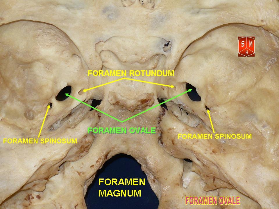
The greater wing of the sphenoid bone, or alisphenoid, is a bony process of the sphenoid bone; there is one on each side, extending from the side of the body of the sphenoid and curving upward, laterally, and backward. Structure The greater wings of the sphenoid are two strong processes of bone, which arise from the sides of the body, and are curved upward, laterally, and backward; the posterior part of each projects as a triangular process that fits into the angle between the squamous and the petrous part of the temporal bone and presents at its apex a downward-directed process, the spine of sphenoid bone. Cerebral surface The superior or cerebral surface of each greater wing ig. 1forms part of the middle cranial fossa; it is deeply concave, and presents depressions for the convolutions of the temporal lobe of the brain. It has a number of foramina (holes) in it: * The foramen rotundum is a circular aperture at its anterior and medial part; it transmits the maxillary nerve. ... [...More Info...] [...Related Items...] OR: [Wikipedia] [Google] [Baidu] |
Sphenoid Bone
The sphenoid bone is an unpaired bone of the neurocranium. It is situated in the middle of the skull towards the front, in front of the basilar part of occipital bone, basilar part of the occipital bone. The sphenoid bone is one of the seven bones that articulate to form the orbit (anatomy), orbit. Its shape somewhat resembles that of a butterfly or bat with its wings extended. Structure It is divided into the following parts: * a median portion, known as the body of sphenoid bone, containing the sella turcica, which houses the pituitary gland as well as the paired paranasal sinuses, the sphenoidal sinuses * two Greater wing of sphenoid bone, greater wings on the lateral side of the body and two Lesser wing of sphenoid bone, lesser wings from the anterior side. * Pterygoid processes of the sphenoides, directed downwards from the junction of the body and the greater wings. Two sphenoidal conchae are situated at the anterior and inferior part of the body. Intrinsic ligaments of ... [...More Info...] [...Related Items...] OR: [Wikipedia] [Google] [Baidu] |
Foramen Petrosum
The lesser petrosal nerve (also known as the small superficial petrosal nerve) is the general visceral efferent (GVE) component of the glossopharyngeal nerve (CN IX), carrying parasympathetic preganglionic fibers from the tympanic plexus to the parotid gland. It synapses in the otic ganglion, from where the postganglionic fibers emerge. Structure After arising in the tympanic plexus, the lesser petrosal nerve passes forward and then through the hiatus for lesser petrosal nerve on the anterior surface of the petrous part of the temporal bone into the middle cranial fossa. It travels across the floor of the middle cranial fossa, then exits the skull via canaliculus innominatus to reach the infratemporal fossa. The fibres synapse in the otic ganglion, and post-ganglionic fibres then travel briefly with the auriculotemporal nerve (a branch of V3) before entering the body of the parotid gland. The lesser petrosal nerve will distribute its parasympathetic post-ganglionic (GVE) fibers ... [...More Info...] [...Related Items...] OR: [Wikipedia] [Google] [Baidu] |
Superior Orbital Fissure
The superior orbital fissure is a foramen or cleft of the skull between the lesser and greater wings of the sphenoid bone. It gives passage to multiple structures, including the oculomotor nerve, trochlear nerve, ophthalmic nerve, abducens nerve, ophthalmic veins, and sympathetic fibres from the cavernous plexus. Structure The superior orbital fissure is usually 22 mm wide in adults, and is much larger medially. Its boundaries are formed by the (caudal surface of the) lesser wing of the sphenoid bone, and (medial border of the) greater wing of the sphenoid bone. Contents The superior orbital fissure is traversed by the following structures: * (superior and inferior divisions of the) oculomotor nerve (CN III) * trochlear nerve (CN IV) * lacrimal, frontal, and nasociliary branches of ophthalmic nerve (CN V1) * abducens nerve (CN VI) * superior ophthalmic vein and superior division of the inferior ophthalmic vein * sympathetic fibres from the cavernous nerve pl ... [...More Info...] [...Related Items...] OR: [Wikipedia] [Google] [Baidu] |
Inferior Orbital Fissure
The inferior orbital fissure is formed by the sphenoid bone and the maxilla. It is located posteriorly along the boundary of the floor and lateral wall of the orbit. It transmits a number of structures, including: * the zygomatic branch of the maxillary nerve * the ascending branches from the pterygopalatine ganglion * the infraorbital vessels, which travel down the infraorbital groove into the infraorbital canal and exit through the infraorbital foramen * the inferior division of the ophthalmic vein Images File:Gray189.png, Left infratemporal fossa. File:Gray191.png, Horizontal section of nasal and orbital cavities. File:Gray787.png, Dissection showing origins of right ocular muscles, and nerves entering by the superior orbital fissure. File:Slide2rome.JPG, Inferior orbital fissure. See also *Foramina of skull *Superior orbital fissure The superior orbital fissure is a foramen or cleft of the skull between the lesser and greater wings of the sphenoid bone. It gives ... [...More Info...] [...Related Items...] OR: [Wikipedia] [Google] [Baidu] |
Frontal Bone
The frontal bone is a bone in the human skull. The bone consists of two portions.''Gray's Anatomy'' (1918) These are the vertically oriented squamous part, and the horizontally oriented orbital part, making up the bony part of the forehead, part of the bony orbital cavity holding the eye, and part of the bony part of the nose respectively. The name comes from the Latin word ''frons'' (meaning " forehead"). Structure of the frontal bone The frontal bone is made up of two main parts. These are the squamous part, and the orbital part. The squamous part marks the vertical, flat, and also the biggest part, and the main region of the forehead. The orbital part is the horizontal and second biggest region of the frontal bone. It enters into the formation of the roofs of the orbital and nasal cavities. Sometimes a third part is included as the nasal part of the frontal bone, and sometimes this is included with the squamous part. The nasal part is between the brow ridges, and ends in ... [...More Info...] [...Related Items...] OR: [Wikipedia] [Google] [Baidu] |
Tensor Veli Palatini
The tensor veli palatini muscle (tensor palati or tensor muscle of the velum palatinum) is a broad, thin, ribbon-like muscle in the head that tenses the soft palate. Structure The tensor veli palatini is found anterior-lateral to the levator veli palatini muscle. It arises by a flat lamella from the scaphoid fossa at the base of the medial pterygoid plate, from the spina angularis of the sphenoid and from the lateral wall of the cartilage of the auditory tube. Descending vertically between the medial pterygoid plate and the medial pterygoid muscle, it ends in a tendon which winds around the pterygoid hamulus, being retained in this situation by some of the fibers of origin of the medial pterygoid muscle. Between the tendon and the hamulus is a small bursa. The tendon then passes medially and is inserted into the palatine aponeurosis and into the surface behind the transverse ridge on the horizontal part of the palatine bone. Nerve supply The tensor veli palatini muscle is ... [...More Info...] [...Related Items...] OR: [Wikipedia] [Google] [Baidu] |
Sphenomandibular Ligament
The sphenomandibular ligament (internal lateral ligament) is one of the three ligaments of the temporomandibular joint. It is situated medially to - and generally separate from - the articular capsule of the joint. Superiorly, it is attached to the spine of the sphenoid bone; inferiorly, it is attached to the lingula of mandible. The SML acts to limit inferior-ward movement of the mandible. The SML is derived from Meckel's cartilage. Anatomy The SML is a flat, thin band. It widens/broadens inferiorlybefore as it reaches its inferior attachment, measuring about 12 mm in width on average at the point of its inferior attachment. Attachments Superiorly, the SML is attached to the spine of the sphenoid bone (spina angularis. Inferiorly, it is attached at to lingula of mandible (which occurs just proximally to the mandibular foramen). Anatomical relations The lateral pterygoid muscle, auriculotemporal nerve, and the maxillary artery and maxillary vein are situated laterally to ... [...More Info...] [...Related Items...] OR: [Wikipedia] [Google] [Baidu] |
Chorda Tympani Nerve
The chorda tympani is a branch of the facial nerve that originates from the taste buds in the front of the tongue, runs through the middle ear, and carries taste messages to the brain. It joins the facial nerve (cranial nerve VII) inside the facial canal, at the level where the facial nerve exits the skull via the stylomastoid foramen, but exits through the petrotympanic fissure and descends in the infratemporal fossa. The chorda tympani is part of one of three cranial nerves that are involved in taste. The taste system involves a complicated feedback loop, with each nerve acting to inhibit the signals of other nerves. Structure The chorda tympani exits the cranial cavity through the internal acoustic meatus along with the facial nerve, then it travels through the middle ear, where it runs from posterior to anterior across the tympanic membrane. It passes between the malleus and the incus, on the medial surface of the neck of the malleus. The nerve continues through the petr ... [...More Info...] [...Related Items...] OR: [Wikipedia] [Google] [Baidu] |
Sphenoidal Spine
The sphenoidal spine (Latin: "''spina angularis''") is a downwardly directed process at the apex of the great wings of the sphenoid bone that serves as the origin of the sphenomandibular ligament. Additional images File:Spine of sphenoid bone.png, Base of skull The base of skull, also known as the cranial base or the cranial floor, is the most inferior area of the skull. It is composed of the endocranium and the lower parts of the calvaria. Structure Structures found at the base of the skull are for .... Inferior surface. Spine of sphenoid bone marked with black circle References External links * - "Schematic view of key landmarks of the infratemporal fossa." * Bones of the head and neck {{musculoskeletal-stub ... [...More Info...] [...Related Items...] OR: [Wikipedia] [Google] [Baidu] |
Foramen Ovale (skull)
The foramen ovale (Latin: oval window) is a hole in the posterior part of the sphenoid bone, posterolateral to the foramen rotundum. It is one of the larger of the several holes (the foramina) in the skull. It transmits the mandibular nerve, a branch of the trigeminal nerve. Structure The foramen ovale is an opening in the greater wing of the sphenoid bone. The foramen ovale is one of two cranial foramina in the greater wing, the other being the foramen spinosum. The foramen ovale is posterolateral to the foramen rotundum and anteromedial to the foramen spinosum. Posterior and medial to the foramen is the opening for the carotid canal. Variation Similar to other foramina, the foramen ovale differs in shape and size throughout the natural life. The earliest perfect ring-shaped formation of the foramen ovale was observed in the 7th fetal month and the latest in 3 years after birth, in a study using over 350 skulls.In a study conducted on 100 skulls, the foramen ovale was d ... [...More Info...] [...Related Items...] OR: [Wikipedia] [Google] [Baidu] |
Lateral Pterygoid Muscle
The lateral pterygoid muscle (or external pterygoid muscle) is a muscle of mastication. It has two heads. It lies superior to the medial pterygoid muscle. It is supplied by pterygoid branches of the maxillary artery, and the lateral pterygoid nerve (from the mandibular nerve, CN V3). It depresses and protrudes the mandible. When each muscle works independently, they can move the mandible side to side. Structure The lateral pterygoid muscle has an upper head and a lower head. * The upper head originates on the infratemporal surface and infratemporal crest of the greater wing of the sphenoid bone. It inserts onto the articular disc and fibrous capsule of the temporomandibular joint. * The lower head originates on the lateral surface of the lateral pterygoid plate. It inserts onto the pterygoid fovea at the neck of the condyloid process of the mandible. It lies superior to the medial pterygoid muscle. Blood supply The lateral pterygoid muscle is supplied by pterygoid branch ... [...More Info...] [...Related Items...] OR: [Wikipedia] [Google] [Baidu] |

.jpg)

