|
Esophageal Varices
Esophageal varices are extremely dilated sub-mucosal veins in the lower third of the esophagus. They are most often a consequence of portal hypertension, commonly due to cirrhosis. People with esophageal varices have a strong tendency to develop severe bleeding which left untreated can be fatal. Esophageal varices are typically diagnosed through an esophagogastroduodenoscopy. Pathogenesis The upper two thirds of the esophagus are drained via the esophageal veins, which carry deoxygenated blood from the esophagus to the azygos vein, which in turn drains directly into the superior vena cava. These veins have no part in the development of esophageal varices. The lower one third of the esophagus is drained into the superficial veins lining the esophageal mucosa, which drain into the left gastric vein, which in turn drains directly into the portal vein. These superficial veins (normally only approximately 1 mm in diameter) become distended up to 1–2 cm in diameter ... [...More Info...] [...Related Items...] OR: [Wikipedia] [Google] [Baidu] |
Gastroenterology
Gastroenterology (from the Greek gastḗr- “belly”, -énteron “intestine”, and -logía "study of") is the branch of medicine focused on the digestive system and its disorders. The digestive system consists of the gastrointestinal tract, sometimes referred to as the ''GI tract,'' which includes the esophagus, stomach, small intestine and large intestine as well as the accessory organs of digestion which includes the pancreas, gallbladder, and liver. The digestive system functions to move material through the GI tract via peristalsis, break down that material via digestion, absorb nutrients for use throughout the body, and remove waste from the body via defecation. Physicians who specialize in the medical specialty of gastroenterology are called gastroenterologists or sometimes ''GI doctors''. Some of the most common conditions managed by gastroenterologists include gastroesophageal reflux disease, gastrointestinal bleeding, irritable bowel syndrome, irritable bow ... [...More Info...] [...Related Items...] OR: [Wikipedia] [Google] [Baidu] |
Esophagogastroduodenoscopy
Esophagogastroduodenoscopy (EGD) or oesophagogastroduodenoscopy (OGD), also called by various other names, is a diagnostic endoscopic procedure that visualizes the upper part of the gastrointestinal tract down to the duodenum. It is considered a minimally invasive procedure since it does not require an incision into one of the major body cavities and does not require any significant recovery after the procedure (unless sedation or anesthesia has been used). However, a sore throat is common. Alternative names The words ''esophagogastroduodenoscopy'' (EGD; American English) and ''oesophagogastroduodenoscopy'' (OGD; British English; see spelling differences) are both pronounced . It is also called ''panendoscopy'' (PES) and ''upper GI endoscopy''. It is also often called just ''upper endoscopy'', ''upper GI'', or even just ''endoscopy''; because EGD is the most commonly performed type of endoscopy, the ambiguous term ''endoscopy'' is sometimes informally used to refer to EGD ... [...More Info...] [...Related Items...] OR: [Wikipedia] [Google] [Baidu] |
Duodenum
The duodenum is the first section of the small intestine in most higher vertebrates, including mammals, reptiles, and birds. In fish, the divisions of the small intestine are not as clear, and the terms anterior intestine or proximal intestine may be used instead of duodenum. In mammals the duodenum may be the principal site for iron absorption. The duodenum precedes the jejunum and ileum and is the shortest part of the small intestine. In humans, the duodenum is a hollow jointed tube about 25–38 cm (10–15 inches) long connecting the stomach to the middle part of the small intestine. It begins with the duodenal bulb and ends at the suspensory muscle of duodenum. Duodenum can be divided into four parts: the first (superior), the second (descending), the third (horizontal) and the fourth (ascending) parts. Structure The duodenum is a C-shaped structure lying adjacent to the stomach. It is divided anatomically into four sections. The first part of the duodenu ... [...More Info...] [...Related Items...] OR: [Wikipedia] [Google] [Baidu] |
Gastric Varices
Gastric varices are dilated submucosal veins in the lining of the stomach, which can be a life-threatening cause of bleeding in the upper gastrointestinal tract. They are most commonly found in patients with portal hypertension, or elevated pressure in the portal vein system, which may be a complication of cirrhosis. Gastric varices may also be found in patients with thrombosis of the splenic vein, into which the short gastric veins that drain the fundus of the stomach flow. The latter may be a complication of acute pancreatitis, pancreatic cancer, or other abdominal tumours, as well as hepatitis C. Gastric varices and associated bleeding are a potential complication of schistosomiasis resulting from portal hypertension. Patients with bleeding gastric varices can present with bloody vomiting (hematemesis), dark, tarry stools (melena), or rectal bleeding. The bleeding may be brisk, and patients may soon develop shock. Treatment of gastric varices can include injection of the v ... [...More Info...] [...Related Items...] OR: [Wikipedia] [Google] [Baidu] |
Splenectomy
A splenectomy is the surgical procedure that partially or completely removes the spleen. The spleen is an important organ in regard to immunological function due to its ability to efficiently destroy encapsulated bacteria. Therefore, removal of the spleen runs the risk of overwhelming post-splenectomy infection, a medical emergency and rapidly fatal disease caused by the inability of the body's immune system to properly fight infection following splenectomy or asplenia. Common indications for splenectomy include trauma, tumors, splenomegaly or for hematological disease such as sickle cell anemia or thalassemia. Indications The spleen is an organ located in the abdomen next to the stomach. It is composed of red pulp which filters the blood, removing foreign material, damaged and worn out red blood cells. It also functions as a storage site for iron, red blood cells and platelets. The rest (~25%) of the spleen is known as the white pulp and functions like a large lymph node ... [...More Info...] [...Related Items...] OR: [Wikipedia] [Google] [Baidu] |
Anastomosis
An anastomosis (, plural anastomoses) is a connection or opening between two things (especially cavities or passages) that are normally diverging or branching, such as between blood vessels, leaf veins, or streams. Such a connection may be normal (such as the foramen ovale in a fetus's heart) or abnormal (such as the patent foramen ovale in an adult's heart); it may be acquired (such as an arteriovenous fistula) or innate (such as the arteriovenous shunt of a metarteriole); and it may be natural (such as the aforementioned examples) or artificial (such as a surgical anastomosis). The reestablishment of an anastomosis that had become blocked is called a reanastomosis. Anastomoses that are abnormal, whether congenital or acquired, are often called fistulas. The term is used in medicine, biology, mycology, geology, and geography. Etymology Anastomosis: medical or Modern Latin, from Greek ἀναστόμωσις, anastomosis, "outlet, opening", Gr ana- "up, on, upon", stoma "mouth", ... [...More Info...] [...Related Items...] OR: [Wikipedia] [Google] [Baidu] |
Varicose Veins
Varicose veins, also known as varicoses, are a medical condition in which superficial veins become enlarged and twisted. These veins typically develop in the legs, just under the skin. Varicose veins usually cause few symptoms. However, some individuals may experience fatigue or pain in the area. Complications can include bleeding or superficial thrombophlebitis. Varices in the scrotum are known as a varicocele, while those around the anus are known as hemorrhoids. Due to the various physical, social, and psychological effects of varicose veins, they can negatively affect one's quality of life. Varicose veins have no specific cause. Risk factors include obesity, lack of exercise, leg trauma, and family history of the condition. They also develop more commonly during pregnancy. Occasionally they result from chronic venous insufficiency. Underlying causes include weak or damaged valves in the veins. They are typically diagnosed by examination, including observation by ultrasound. ... [...More Info...] [...Related Items...] OR: [Wikipedia] [Google] [Baidu] |
Rectum
The rectum is the final straight portion of the large intestine in humans and some other mammals, and the gut in others. The adult human rectum is about long, and begins at the rectosigmoid junction (the end of the sigmoid colon) at the level of the third sacral vertebra or the sacral promontory depending upon what definition is used. Its diameter is similar to that of the sigmoid colon at its commencement, but it is dilated near its termination, forming the rectal ampulla. It terminates at the level of the anorectal ring (the level of the puborectalis sling) or the dentate line, again depending upon which definition is used. In humans, the rectum is followed by the anal canal which is about long, before the gastrointestinal tract terminates at the anal verge. The word rectum comes from the Latin '' rectum intestinum'', meaning ''straight intestine''. Structure The rectum is a part of the lower gastrointestinal tract. The rectum is a continuation of the sigmoi ... [...More Info...] [...Related Items...] OR: [Wikipedia] [Google] [Baidu] |
Stomach
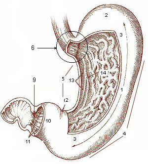
The stomach is a muscular, hollow organ in the gastrointestinal tract of humans and many other animals, including several invertebrates. The stomach has a dilated structure and functions as a vital organ in the digestive system. The stomach is involved in the gastric phase of digestion, following chewing. It performs a chemical breakdown by means of enzymes and hydrochloric acid. In humans and many other animals, the stomach is located between the oesophagus and the small intestine. The stomach secretes digestive enzymes and gastric acid to aid in food digestion. The pyloric sphincter controls the passage of partially digested food ( chyme) from the stomach into the duodenum, where peristalsis takes over to move this through the rest of intestines. Structure In the human digestive system, the stomach lies between the oesophagus and the duodenum (the first part of the small intestine). It is in the left upper quadrant of the abdominal cavity. The top of the stomach lies ... [...More Info...] [...Related Items...] OR: [Wikipedia] [Google] [Baidu] |
Collateral Circulation
Collateral circulation is the alternate circulation around a blocked artery or vein via another path, such as nearby minor vessels. It may occur via preexisting vascular redundancy (analogous to engineered redundancy), as in the circle of Willis in the brain, or it may occur via new branches formed between adjacent blood vessels ( neovascularization), as in the eye after a retinal embolism or in the brain when moyamoya occurs. Its formation may be provoked by pathological conditions such as high vascular resistance or ischaemia. An example of the usefulness of collateral circulation is a systemic thromboembolism in cats. This is when a thrombotic embolus lodges above the external iliac artery (common iliac artery), blocking the external and internal iliac arteries and effectively shutting off all blood supply to the hind leg. Even though the main vessels to the leg are blocked, enough blood can get to the tissues in the leg via the collateral circulation to keep them aliv ... [...More Info...] [...Related Items...] OR: [Wikipedia] [Google] [Baidu] |
Left Gastric Vein
The left gastric vein (or coronary vein) is a vein that derives from tributaries draining the lesser curvature of the stomach. Structure The left gastric vein runs from right to left along the lesser curvature of the stomach. It passes to the esophageal opening of the stomach, where it receives some esophageal veins. It then turns backward and passes from left to right behind the omental bursa. It drains into the portal vein near the superior border of the pancreas. Function The left gastric vein drains deoxygenated blood from the lesser curvature of the stomach. It also acts as collaterals between the portal vein and the systemic venous system of the lower esophagus (azygous vein). Clinical significance Esophageal and paraesophageal varices are supplied primarily by the left gastric vein (due to flow reversal) and typically drain into the azygos/hemiazygos venous system.Siegelman, E.: "Body MRI", page 47. Saunders, 2004 See also * Right gastric vein The right g ... [...More Info...] [...Related Items...] OR: [Wikipedia] [Google] [Baidu] |
Superior Vena Cava
The superior vena cava (SVC) is the anatomical terms of location#Superior and inferior, superior of the two venae cavae, the great vein, venous trunks that return deoxygenated blood from the circulatory system, systemic circulation to the atrium (heart), right atrium of the heart. It is a large-diameter (24 mm) short length vein that receives venous return from the upper half of the body, above the thoracic diaphragm, diaphragm. Venous return from the lower half, below the diaphragm, flows through the inferior vena cava. The SVC is located in the anterior right superior mediastinum. It is the typical site of central venous access via a central venous catheter or a peripherally inserted central catheter. Mentions of "the cava" without further specification usually refer to the SVC. Structure The superior vena cava is formed by the left and right brachiocephalic veins, which receive blood from the upper limbs, head and neck, behind the lower border of the first right costal ca ... [...More Info...] [...Related Items...] OR: [Wikipedia] [Google] [Baidu] |


