|
Dorsal Interossei Of The Foot
In human anatomy, the dorsal interossei of the foot are four muscles situated between the metatarsal bones. Origin The four interossei muscles are bipenniform muscles each originating by two heads from the proximal half of the sides of adjacent metatarsal bones. Insertion The two heads of each muscle form a central tendon which passes forwards deep to the deep transverse metatarsal ligament. The tendons are inserted on the bases of the second, third, and fourth proximal phalanges and into the aponeurosis of the tendons of the extensor digitorum longus Gray's Anatomy, 1918 (see infobox) without attaching to the extensor hoods of the toes. Thus, the first is inserted into the medial side of the second toe; the other three are inserted into the lateral sides of the second, third, and fourth toes. Action The dorsal interossei abduct at the metatarsophalangeal joints of the third and fourth toes. Because there is a pair of dorsal interossei muscles attached on both si ... [...More Info...] [...Related Items...] OR: [Wikipedia] [Google] [Baidu] |
Metatarsals
The metatarsal bones, or metatarsus, are a group of five long bones in the foot, located between the tarsal bones of the hind- and mid-foot and the phalanges of the toes. Lacking individual names, the metatarsal bones are numbered from the medial side (the side of the great toe): the first, second, third, fourth, and fifth metatarsal (often depicted with Roman numerals). The metatarsals are analogous to the metacarpal bones of the hand. The lengths of the metatarsal bones in humans are, in descending order, second, third, fourth, fifth, and first. Structure The five metatarsals are dorsal convex long bones consisting of a shaft or body, a base (proximal Standard anatomical terms of location are used to unambiguously describe the anatomy of animals, including humans. The terms, typically derived from Latin or Greek roots, describe something in its standard anatomical position. This position ...ly), and a head (distally).Platzer 2004, p. 220 The body is prismoid in ... [...More Info...] [...Related Items...] OR: [Wikipedia] [Google] [Baidu] |
Abduction (kinesiology)
Motion, the process of movement, is described using specific anatomical terms. Motion includes movement of organs, joints, limbs, and specific sections of the body. The terminology used describes this motion according to its direction relative to the anatomical position of the body parts involved. Anatomists and others use a unified set of terms to describe most of the movements, although other, more specialized terms are necessary for describing unique movements such as those of the hands, feet, and eyes. In general, motion is classified according to the anatomical plane it occurs in. ''Flexion'' and ''extension'' are examples of ''angular'' motions, in which two axes of a joint are brought closer together or moved further apart. ''Rotational'' motion may occur at other joints, for example the shoulder, and are described as ''internal'' or ''external''. Other terms, such as ''elevation'' and ''depression'', describe movement above or below the horizontal plane. Many anato ... [...More Info...] [...Related Items...] OR: [Wikipedia] [Google] [Baidu] |
Interosseous Muscles Of The Foot
The interosseous muscles of the foot are muscles found near the metatarsal The metatarsal bones, or metatarsus, are a group of five long bones in the foot, located between the tarsal bones of the hind- and mid-foot and the phalanges of the toes. Lacking individual names, the metatarsal bones are numbered from the med ... bones that help to control the toes. They are considered voluntary muscles. They are generally divided into two sets: * 4 Dorsal interossei - Abduct the digits away from the 2nd digit (away from axial line) and are bipennate. * 3 Plantar interossei - Adduct the digits towards the 2nd digit (towards the axial line) and are unipennate. The axial line goes down the middle of the 2nd digit, towards the sole of the foot (it's an imaginary line). Both sets of muscles are innervated by the Lateral plantar nerve. References Muscles of the lower limb Foot {{Muscle-stub ... [...More Info...] [...Related Items...] OR: [Wikipedia] [Google] [Baidu] |
Palmar Interossei Muscles
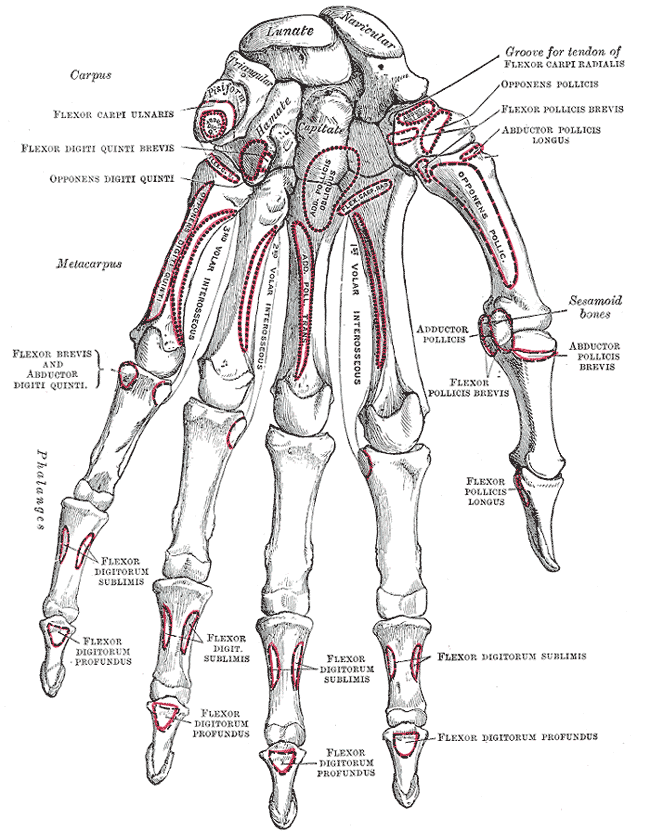
In human anatomy, the palmar or volar interossei (interossei volares in older literature) are three small, unipennate muscles in the hand that lie between the metacarpal bones and are attached to the index, ring, and little fingers. They are smaller than the dorsal interossei of the hand. Structure All palmar interossei originate along the shaft of the metacarpal bone of the digit on which they act. They are inserted into the base of the proximal phalanx and the extensor expansion of the extensor digitorum of the same digit. Pollical palmar interosseous The first palmar interosseous is located at the thumb's medial side. Passing between the first dorsal interosseous and the oblique head of adductor pollicis, it is inserted on the base of the thumb's proximal phalanx together with adductor pollicis. The "pollical" palmar interosseous muscle (PPIM), is present in more than 80% of individuals and was first described by . Its presence has been verified by numerous anatomists sinc ... [...More Info...] [...Related Items...] OR: [Wikipedia] [Google] [Baidu] |
Interosseous Muscles Of The Hand
The interosseous muscles of the hand are muscles found near the metacarpal bones that help to control the fingers. They are considered voluntary muscles. They are generally divided into two sets: * 4 Dorsal interossei - Abduct the digits away from the 3rd digit (away from axial line) and are bipennate. * 3 Palmar interossei In human anatomy, the palmar or volar interossei (interossei volares in older literature) are three small, unipennate muscles in the hand that lie between the metacarpal bones and are attached to the index, ring, and little fingers. They are sm ... - Adduct the digits towards the 3rd digit (towards the axial line) and are unipennate. This is often remembered by the mnemonic PAD-DAB, as the Palmar interosseous muscles ADduct, and the Dorsal interosseous muscles ABduct. The axial line goes down the middle of the 3rd digit, towards the palm of the hand (it's an imaginary line). Both sets of muscles are innervated by the deep branch of the ulnar nerve. Re ... [...More Info...] [...Related Items...] OR: [Wikipedia] [Google] [Baidu] |
Dorsalis Pedis Artery
In human anatomy, the dorsalis pedis artery (dorsal artery of foot) is a blood vessel of the lower limb. It arises from the anterior tibial artery, and ends at the first intermetatarsal space (as the first dorsal metatarsal artery and the deep plantar artery). It carries oxygenated blood to the dorsal side of the foot. It is useful for taking a pulse. It is also at risk during anaesthesia of the deep peroneal nerve. Structure The dorsalis pedis artery is located 1/3 from medial malleolus of the ankle. It arises at the anterior aspect of the ankle joint and is a continuation of the anterior tibial artery. It ends at the proximal part of the first intermetatarsal space. Here, it divides into two branches, the first dorsal metatarsal artery, and the deep plantar artery. It is covered by skin and fascia, but is fairly superficial. The dorsalis pedis communicates with the plantar blood supply of the foot through the deep plantar artery. Along its course, it is accompanied ... [...More Info...] [...Related Items...] OR: [Wikipedia] [Google] [Baidu] |
Deep Peroneal Nerve
The deep fibular nerve (also known as deep peroneal nerve) begins at the bifurcation of the common fibular nerve between the fibula and upper part of the fibularis longus, passes infero-medially, deep to the extensor digitorum longus, to the anterior surface of the interosseous membrane, and comes into relation with the anterior tibial artery above the middle of the leg; it then descends with the artery to the front of the ankle-joint, where it divides into a ''lateral'' and a '' medial terminal branch''. Structure Lateral side of the leg The deep fibular nerve is the nerve of the anterior compartment of the leg and the dorsum of the foot. It is one of the terminal branches of the common fibular nerve. It corresponds to the posterior interosseus nerve of the forearm. It begins at the lateral side of the fibula bone, and then enters the anterior compartment by piercing the anterior intermuscular septum. It then pierces the extensor digitorum longus and lies next to the anterior ti ... [...More Info...] [...Related Items...] OR: [Wikipedia] [Google] [Baidu] |
Arches Of The Foot
The arches of the foot, formed by the tarsal and metatarsal bones, strengthened by ligaments and tendons, allow the foot to support the weight of the body in the erect posture with the least weight. They are categorized as longitudinal and transverse arches. Structure Longitudinal arches The longitudinal arches of the foot can be divided into medial and lateral arches. Medial arch The medial arch is higher than the lateral longitudinal arch. It is made up by the calcaneus, the talus, the navicular, the three cuneiforms (medial, intermediate, and lateral), and the first, second, and third metatarsals. Its summit is at the superior articular surface of the talus, and its two extremities or piers, on which it rests in standing, are the tuberosity on the plantar surface of the calcaneus posteriorly and the heads of the first, second, and third metatarsal bones anteriorly. The chief characteristic of this arch is its elasticity, due to its height and to the number of small j ... [...More Info...] [...Related Items...] OR: [Wikipedia] [Google] [Baidu] |
Dorsal Interossei Of The Hand
In human anatomy, the dorsal interossei (DI) are four muscles in the back of the hand that act to abduct (spread) the index, middle, and ring fingers away from hand's midline (ray of middle finger) and assist in flexion at the metacarpophalangeal joints and extension at the interphalangeal joints of the index, middle and ring fingers. Structure There are four dorsal interossei in each hand. They are specified as 'dorsal' to contrast them with the palmar interossei, which are located on the anterior side of the metacarpals. The dorsal interosseous muscles are bipennate, with each muscle arising by two heads from the adjacent sides of the metacarpal bones, but more extensively from the metacarpal bone of the finger into which the muscle is inserted. They are inserted into the bases of the proximal phalanges and into the extensor expansion of the corresponding extensor digitorum tendon. The middle digit has two dorsal interossei insert onto it while the first digit (thumb) and ... [...More Info...] [...Related Items...] OR: [Wikipedia] [Google] [Baidu] |
Metatarsophalangeal Joints
The metatarsophalangeal joints (MTP joints), also informally known as toe knuckles, are the joints between the metatarsal bones of the foot and the proximal bones ( proximal phalanges) of the toes. They are condyloid joints, meaning that an elliptical or rounded surface (of the metatarsal bones) comes close to a shallow cavity (of the proximal phalanges). The ligaments are the plantar and two collateral. Movements The movements permitted in the metatarsophalangeal joints are flexion, extension, abduction, adduction and circumduction. File:The feet of C. H. Unthan, the armless fiddler Wellcome L0034227.jpg, Left: toes adducted (pulled towards the center) and spread (abducted); right, both feet clenched (plantar flexed) File:Footgym rings1.jpg, The upper foot is clenching (plantarflexing) at the MTP joints and at the joints of the toes; the central foot is lifting the toes (dorsiflexing) at the MTP joints; and the foot flat on the ground off to the side is in a neutral po ... [...More Info...] [...Related Items...] OR: [Wikipedia] [Google] [Baidu] |
Gray's Anatomy
''Gray's Anatomy'' is a reference book of human anatomy written by Henry Gray, illustrated by Henry Vandyke Carter, and first published in London in 1858. It has gone through multiple revised editions and the current edition, the 42nd (October 2020), remains a standard reference, often considered "the doctors' bible". Earlier editions were called ''Anatomy: Descriptive and Surgical'', ''Anatomy of the Human Body'' and ''Gray's Anatomy: Descriptive and Applied'', but the book's name is commonly shortened to, and later editions are titled, ''Gray's Anatomy''. The book is widely regarded as an extremely influential work on the subject. Publication history Origins The English anatomist Henry Gray was born in 1827. He studied the development of the endocrine glands and spleen and in 1853 was appointed Lecturer on Anatomy at St George's Hospital Medical School in London. In 1855, he approached his colleague Henry Vandyke Carter with his idea to produce an inexpensive a ... [...More Info...] [...Related Items...] OR: [Wikipedia] [Google] [Baidu] |
Proximal Phalanges
The phalanges (singular: ''phalanx'' ) are digital bones in the hands and feet of most vertebrates. In primates, the thumbs and big toes have two phalanges while the other digits have three phalanges. The phalanges are classed as long bones. Structure The phalanges are the bones that make up the fingers of the hand and the toes of the foot. There are 56 phalanges in the human body, with fourteen on each hand and foot. Three phalanges are present on each finger and toe, with the exception of the thumb and large toe, which possess only two. The middle and far phalanges of the fifth toes are often fused together (symphalangism). The phalanges of the hand are commonly known as the finger bones. The phalanges of the foot differ from the hand in that they are often shorter and more compressed, especially in the proximal phalanges, those closest to the torso. A phalanx is named according to whether it is proximal, middle, or distal and its associated finger or toe. The proxi ... [...More Info...] [...Related Items...] OR: [Wikipedia] [Google] [Baidu] |


