|
Deep Peroneal Nerve
The deep fibular nerve (also known as deep peroneal nerve) begins at the bifurcation of the common fibular nerve between the fibula and upper part of the fibularis longus, passes infero-medially, deep to the extensor digitorum longus, to the anterior surface of the interosseous membrane, and comes into relation with the anterior tibial artery above the middle of the leg; it then descends with the artery to the front of the ankle-joint, where it divides into a ''lateral'' and a '' medial terminal branch''. Structure Lateral side of the leg The deep fibular nerve is the nerve of the anterior compartment of the leg and the dorsum of the foot. It is one of the terminal branches of the common fibular nerve. It corresponds to the posterior interosseus nerve of the forearm. It begins at the lateral side of the fibula bone, and then enters the anterior compartment by piercing the anterior intermuscular septum. It then pierces the extensor digitorum longus and lies next to the anterior tibia ... [...More Info...] [...Related Items...] OR: [Wikipedia] [Google] [Baidu] |
Anterior Compartment Of Leg
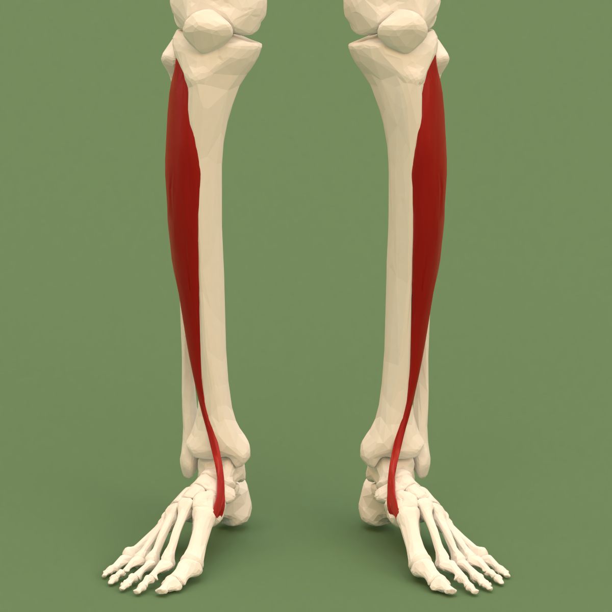
The anterior compartment of the leg is a fascial compartments of leg, fascial compartment of the lower leg. It contains muscles that produce Anatomical terms of motion#Flexion and extension of the foot, dorsiflexion and participate in Anatomical terms of motion#Inversion and eversion, inversion and eversion of the foot, as well as vascular and nervous elements, including the anterior tibial artery and anterior tibial vein, veins and the deep fibular nerve. Muscles The muscles of the compartment are: * tibialis anterior muscle, tibialis anterior * extensor hallucis longus muscle, extensor hallucis longus * extensor digitorum longus muscle, extensor digitorum longus * Fibularis tertius, fibularis (peroneus) tertius Function The compartment contains muscles that are Anatomical terms of motion#Flexion and extension of the foot, dorsiflexors and participate in Anatomical terms of motion#Inversion and eversion, inversion and eversion of the foot. Innervation and blood supply The an ... [...More Info...] [...Related Items...] OR: [Wikipedia] [Google] [Baidu] |
Fibularis Tertius
In human anatomy, the fibularis tertius (also known as the peroneus tertius) is a muscle in the anterior compartment of the leg. It acts to tilt the sole of the foot away from the midline of the body ( eversion) and to pull the foot upward toward the body (dorsiflexion). Structure The fibularis tertius arises from the lower third of the front surface of the fibula, the lower part of the interosseous membrane, and septum, or connective tissue, between it and the fibularis brevis. The septum is sometimes called the intermuscular septum of Otto. The muscle passes downward and ends in a tendon that passes under the superior extensor retinaculum and the inferior extensor retinaculum of the foot in the same canal as the extensor digitorum longus muscle. It may be mistaken as a fifth tendon of the extensor digitorum longus. The tendon inserts into the medial part of the posterior surface of the shaft of the fifth metatarsal bone. The fibularis tertius is supplied by the deep fibula ... [...More Info...] [...Related Items...] OR: [Wikipedia] [Google] [Baidu] |
Extensor Hallucis Brevis
The extensor hallucis brevis is a muscle on the top of the foot that helps to extend the big toe. Structure The extensor hallucis brevis is essentially the medial part of the extensor digitorum brevis muscle. Some anatomists have debated whether these two muscles are distinct entities. The extensor hallucis brevis arises from the calcaneus and inserts on the proximal phalanx of the digit 1 (the big toe). Nerve supply Nerve supplied by lateral terminal branch of Deep Peroneal Nerve (deep fibular nerve) (proximal sciatic branches S1, S2). Same innervation of Extensor Digitorum Brevis Function The extensor hallucis brevis helps to extend the big toe. See also * Extensor digitorum brevis * Extensor hallucis longus The extensor hallucis longus muscle is a thin skeletal muscle, situated between the tibialis anterior and the extensor digitorum longus. It extends the big toe and dorsiflects the foot. It also assists with foot eversion and inversion. Struct ... Additional Ima ... [...More Info...] [...Related Items...] OR: [Wikipedia] [Google] [Baidu] |
Extensor Hallucis Longus (propius)
The extensor hallucis longus muscle is a thin skeletal muscle, situated between the tibialis anterior and the extensor digitorum longus. It extends the big toe and dorsiflects the foot. It also assists with foot eversion and inversion. Structure The extensor hallucis longus muscle arises from the anterior surface of the fibula for about the middle two-fourths of its extent, medial to the origin of the extensor digitorum longus muscle. It also arises from the interosseous membrane of the leg to a similar extent. The anterior tibial vessels and deep fibular nerve lie between it and the tibialis anterior. The fibers pass downward, and end in a tendon, which occupies the anterior border of the muscle, passes through a distinct compartment in the cruciate crural ligament, crosses from the lateral to the medial side of the anterior tibial vessels near the bend of the ankle, and is inserted into the base of the distal phalanx of the great toe. Opposite the metatarsophalangeal ar ... [...More Info...] [...Related Items...] OR: [Wikipedia] [Google] [Baidu] |
Metatarsophalangeal
The metatarsophalangeal joints (MTP joints), also informally known as toe knuckles, are the joints between the metatarsal bones of the foot and the proximal bones (proximal phalanges) of the toes. They are condyloid joints, meaning that an elliptical or rounded surface (of the metatarsal bones) comes close to a shallow cavity (of the proximal phalanges). The ligaments are the plantar and two collateral. Movements The movements permitted in the metatarsophalangeal joints are flexion, extension, abduction, adduction and circumduction. File:The feet of C. H. Unthan, the armless fiddler Wellcome L0034227.jpg, Left: toes adducted (pulled towards the center) and spread (abducted); right, both feet clenched (plantar flexed) File:Footgym rings1.jpg, The upper foot is clenching (plantarflexing) at the MTP joints and at the joints of the toes; the central foot is lifting the toes (dorsiflexing) at the MTP joints; and the foot flat on the ground off to the side is in a neutral position ... [...More Info...] [...Related Items...] OR: [Wikipedia] [Google] [Baidu] |
Extensor Digitorum Brevis
The extensor digitorum brevis muscle (sometimes EDB) is a muscle on the upper surface of the foot that helps extend digits 2 through 4. Structure The muscle originates from the forepart of the upper and lateral surface of the calcaneus (in front of the groove for the peroneus brevis tendon), from the interosseous talocalcaneal ligament and the stem of the inferior extensor retinaculum. The fibres pass obliquely forwards and medially across the dorsum of the foot and end in four tendons. The medial part of the muscle, also known as extensor hallucis brevis, ends in a tendon which crosses the dorsalis pedis artery and inserts into the dorsal surface of the base of the proximal phalanx of the great toe. The other three tendons insert into the lateral sides of the tendons of extensor digitorum longus for the second, third and fourth toes. Nerve supply Nerve supply: lateral terminal branch of Deep Peroneal Nerve (deep fibular nerve) (proximal sciatic branches L4-L5, but most clinicall ... [...More Info...] [...Related Items...] OR: [Wikipedia] [Google] [Baidu] |
Tarsus (skeleton)
In the human body, the tarsus is a cluster of seven articulating bones in each foot situated between the lower end of the tibia and the fibula of the lower leg and the metatarsus. It is made up of the midfoot (Cuboid bone, cuboid, medial, intermediate, and lateral cuneiform bone, cuneiform, and navicular) and hindfoot (Talus bone, talus and calcaneus). The tarsus articulates with the bones of the metatarsus, which in turn articulate with the proximal phalanges of the toes. The joint between the tibia and fibula above and the tarsus below is referred to as the ankle, ankle joint proper. In humans the largest bone in the tarsus is the calcaneus, which is the weight-bearing bone within the heel of the foot. Human anatomy Bones The talus bone or ankle bone is connected superiorly to the two bones of the lower leg, the tibia and fibula, to form the ankle, ankle joint or talocrural joint; inferiorly, at the subtalar joint, to the calcaneus or heel bone. Together, the talus and ... [...More Info...] [...Related Items...] OR: [Wikipedia] [Google] [Baidu] |
Dorsal Interossei Of The Foot
In human anatomy, the dorsal interossei of the foot are four muscles situated between the metatarsal bones. Origin The four interossei muscles are bipenniform muscles each originating by two heads from the proximal half of the sides of adjacent metatarsal bones. Insertion The two heads of each muscle form a central tendon which passes forwards deep to the deep transverse metatarsal ligament. The tendons are inserted on the bases of the second, third, and fourth proximal phalanges and into the aponeurosis of the tendons of the extensor digitorum longus Gray's Anatomy, 1918 (see infobox) without attaching to the extensor hoods of the toes. Thus, the first is inserted into the medial side of the second toe; the other three are inserted into the lateral sides of the second, third, and fourth toes. Action The dorsal interossei abduct at the metatarsophalangeal joints of the third and fourth toes. Because there is a pair of dorsal interossei muscles attached on both sides ... [...More Info...] [...Related Items...] OR: [Wikipedia] [Google] [Baidu] |
Metatarsophalangeal Articulations
The metatarsophalangeal joints (MTP joints), also informally known as toe knuckles, are the joints between the metatarsal bones of the foot and the proximal bones (proximal phalanges) of the toes. They are condyloid joints, meaning that an elliptical or rounded surface (of the metatarsal bones) comes close to a shallow cavity (of the proximal phalanges). The ligaments are the plantar and two collateral. Movements The movements permitted in the metatarsophalangeal joints are flexion, extension, abduction, adduction and circumduction. File:The feet of C. H. Unthan, the armless fiddler Wellcome L0034227.jpg, Left: toes adducted (pulled towards the center) and spread (abducted); right, both feet clenched (plantar flexed) File:Footgym rings1.jpg, The upper foot is clenching (plantarflexing) at the MTP joints and at the joints of the toes; the central foot is lifting the toes (dorsiflexing) at the MTP joints; and the foot flat on the ground off to the side is in a neutral position ... [...More Info...] [...Related Items...] OR: [Wikipedia] [Google] [Baidu] |
Medial Dorsal Cutaneous Branch Of The Superficial Fibular Nerve
Medial may refer to: Mathematics * Medial magma, a mathematical identity in algebra Geometry * Medial axis, in geometry the set of all points having more than one closest point on an object's boundary * Medial graph, another graph that represents the adjacencies between edges in the faces of a plane graph * Medial triangle, the triangle whose vertices lie at the midpoints of an enclosing triangle's sides * Polyhedra: ** Medial deltoidal hexecontahedron ** Medial disdyakis triacontahedron ** Medial hexagonal hexecontahedron ** Medial icosacronic hexecontahedron ** Medial inverted pentagonal hexecontahedron ** Medial pentagonal hexecontahedron ** Medial rhombic triacontahedron Linguistics * A medial sound or letter is one that is found in the middle of a larger unit (like a word) ** Syllable medial, a segment located between the onset and the rime of a syllable * In the older literature, a term for the voiced stops (like ''b'', ''d'', ''g'') * Medial or second person demonstr ... [...More Info...] [...Related Items...] OR: [Wikipedia] [Google] [Baidu] |
Dorsal Digital Nerves Of Foot
Dorsal digital nerves of foot are branches of the intermediate dorsal cutaneous nerve, medial dorsal cutaneous nerve, sural nerve and deep fibular nerve. Structures There are 10 total dorsal digital branches: * The medial terminal branch (internal branch) divides into two dorsal digital nerves (nn. digitales dorsales hallucis lateralis et digiti secundi medialis) which supply the adjacent sides of the great and second toes, * The medial dorsal cutaneous nerve (internal dorsal cutaneous branch) passes in front of the ankle-joint, and divides into three dorsal digital branches, one of which supplies the medial side of the great toe, the other, the adjacent sides of the second and third toes. * The intermediate dorsal cutaneous nerve divides into four dorsal digital branches, which supply the medial and lateral sides of the third and fourth, and of the fourth and fifth toes. * The lateral dorsal cutaneous nerve from the sural nerve turns into a dorsal digital nerve and supplies the ... [...More Info...] [...Related Items...] OR: [Wikipedia] [Google] [Baidu] |
Dorsalis Pedis Artery
In human anatomy, the dorsalis pedis artery (dorsal artery of foot) is a blood vessel of the lower limb. It arises from the anterior tibial artery, and ends at the first intermetatarsal space (as the first dorsal metatarsal artery and the deep plantar artery). It carries oxygenated blood to the dorsal side of the foot. It is useful for taking a pulse. It is also at risk during anaesthesia of the deep peroneal nerve. Structure The dorsalis pedis artery is located 1/3 from medial malleolus of the ankle. It arises at the anterior aspect of the ankle joint and is a continuation of the anterior tibial artery. It ends at the proximal part of the first intermetatarsal space. Here, it divides into two branches, the first dorsal metatarsal artery, and the deep plantar artery. It is covered by skin and fascia, but is fairly superficial. The dorsalis pedis communicates with the plantar blood supply of the foot through the deep plantar artery. Along its course, it is accompanied by a deep ... [...More Info...] [...Related Items...] OR: [Wikipedia] [Google] [Baidu] |

