|
Palmar Interossei Muscles
In human anatomy, the palmar or volar interossei (interossei volares in older literature) are four muscles, one on the thumb that is occasionally missing, and three small, unipennate, central muscles in the hand that lie between the Metacarpus, metacarpal bones and are attached to the Index finger, index, Ring finger, ring, and Little finger, little fingers. They are smaller than the dorsal interossei of the hand. Structure All palmar interossei originate along the shaft of the metacarpal bone of the digit on which they act. They are inserted into the base of the Phalanx bone, proximal phalanx and the extensor expansion of the Extensor digitorum muscle, extensor digitorum of the same digit. Pollical palmar interosseous The first palmar interosseous is located at the thumb's medial side. Passing between the first dorsal interosseous and the oblique head of Adductor pollicis muscle, adductor pollicis, it is inserted on the base of the thumb's proximal phalanx together with Adductor ... [...More Info...] [...Related Items...] OR: [Wikipedia] [Google] [Baidu] |
Metacarpals
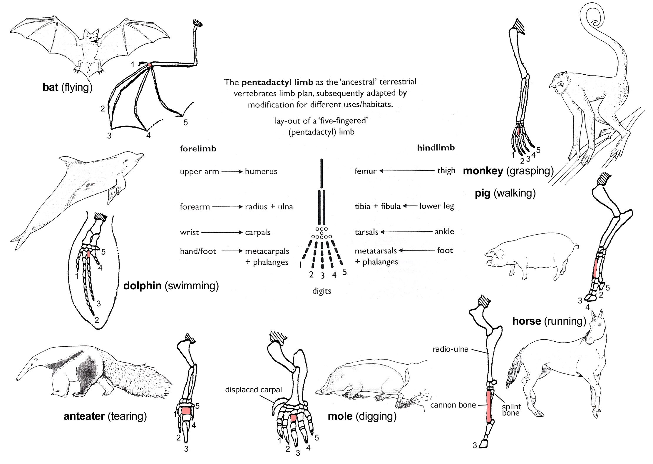
In human anatomy, the metacarpal bones or metacarpus, also known as the "palm bones", are the appendicular skeleton, appendicular bones that form the intermediate part of the hand between the phalanges (fingers) and the carpal bones (wrist, wrist bones), which joint, articulate with the forearm. The metacarpal bones are homologous to the metatarsal bones in the foot. Structure The metacarpals form a transverse arch to which the rigid row of distal carpal bones are fixed. The peripheral metacarpals (those of the thumb and little finger) form the sides of the cup of the palmar gutter and as they are brought together they deepen this concavity. The index metacarpal is the most firmly fixed, while the thumb metacarpal articulates with the trapezium and acts independently from the others. The middle metacarpals are tightly united to the carpus by intrinsic interlocking bone elements at their bases. The ring metacarpal is somewhat more mobile while the fifth metacarpal is semi-indepen ... [...More Info...] [...Related Items...] OR: [Wikipedia] [Google] [Baidu] |
Flexor Pollicis Brevis Muscle
The flexor pollicis brevis is a muscle in the hand that flexes the thumb. It is one of three thenar muscles. It has both a superficial part and a deep part. Origin and insertion The muscle's superficial head arises from the distal edge of the flexor retinaculum and the tubercle of the trapezium, the most lateral bone in the distal row of carpal bones. It passes along the radial side of the tendon of the flexor pollicis longus. The deeper (and medial) head "varies in size and may be absent."Gray's 37th British Edition, p. 630" It arises from the trapezoid and capitate bones on the floor of the carpal tunnel, as well as the ligaments of the distal carpal row. Both heads become tendinous and insert together into the radial side of the base of the proximal phalanx of the thumb; at the junction between the tendinous heads there is a sesamoid bone.''Gray's Anatomy'' 1918, see infobox Innervation The superficial head is usually innervated by the lateral terminal branch of the med ... [...More Info...] [...Related Items...] OR: [Wikipedia] [Google] [Baidu] |
Interphalangeal Articulations Of Hand
The interphalangeal joints of the hand are the hinge joints between the phalanges of the fingers that provide flexion towards the palm of the hand. There are two sets in each finger (except in the thumb, which has only one joint): * "proximal interphalangeal joints" (PIJ or PIP), those between the first (also called proximal) and second (intermediate) phalanges * "distal interphalangeal joints" (DIJ or DIP), those between the second (intermediate) and third (distal) phalanges Anatomically, the proximal and distal interphalangeal joints are very similar. There are some minor differences in how the palmar plates are attached proximally and in the segmentation of the flexor tendon sheath, but the major differences are the smaller dimension and reduced mobility of the distal joint. Joint structure The PIP joint exhibits great lateral stability. Its transverse diameter is greater than its antero-posterior diameter and its thick collateral ligaments are tight in all positions dur ... [...More Info...] [...Related Items...] OR: [Wikipedia] [Google] [Baidu] |
Tendon
A tendon or sinew is a tough band of fibrous connective tissue, dense fibrous connective tissue that connects skeletal muscle, muscle to bone. It sends the mechanical forces of muscle contraction to the skeletal system, while withstanding tension (physics), tension. Tendons, like ligaments, are made of collagen. The difference is that ligaments connect bone to bone, while tendons connect muscle to bone. There are about 4,000 tendons in the adult human body. Structure A tendon is made of dense regular connective tissue, whose main cellular components are special fibroblasts called tendon cells (tenocytes). Tendon cells synthesize the tendon's extracellular matrix, which abounds with densely-packed collagen fibers. The collagen fibers run parallel to each other and are grouped into fascicles. Each fascicle is bound by an endotendineum, which is a delicate loose connective tissue containing thin collagen fibrils and elastic fibers. A set of fascicles is bound by an epitenon, whi ... [...More Info...] [...Related Items...] OR: [Wikipedia] [Google] [Baidu] |
Myocyte
A muscle cell, also known as a myocyte, is a mature contractile Cell (biology), cell in the muscle of an animal. In humans and other vertebrates there are three types: skeletal muscle, skeletal, smooth muscle, smooth, and Cardiac muscle, cardiac (cardiomyocytes). A skeletal muscle cell is long and threadlike with multinucleated, many nuclei and is called a ''muscle fiber''. Muscle cells develop from embryonic precursor cells called myoblasts. Skeletal muscle cells form by cell fusion, fusion of myoblasts to produce multinucleated cells (syncytium, syncytia) in a process known as myogenesis. Skeletal muscle cells and cardiac muscle cells both contain myofibrils and sarcomeres and form a striated muscle tissue. Cardiac muscle cells form the cardiac muscle in the walls of the heart chambers, and have a single central Cell nucleus, nucleus. Cardiac muscle cells are joined to neighboring cells by intercalated discs, and when joined in a visible unit they are described as a ''cardiac m ... [...More Info...] [...Related Items...] OR: [Wikipedia] [Google] [Baidu] |
Anatomical Terms Of Motion
Motion, the process of movement, is described using specific anatomical terms. Motion includes movement of organs, joints, limbs, and specific sections of the body. The terminology used describes this motion according to its direction relative to the anatomical position of the body parts involved. Anatomists and others use a unified set of terms to describe most of the movements, although other, more specialized terms are necessary for describing unique movements such as those of the hands, feet, and eyes. In general, motion is classified according to the anatomical plane it occurs in. ''Flexion'' and ''extension'' are examples of ''angular'' motions, in which two axes of a joint are brought closer together or moved further apart. ''Rotational'' motion may occur at other joints, for example the shoulder, and are described as ''internal'' or ''external''. Other terms, such as ''elevation'' and ''depression'', describe movement above or below the horizontal plane. Many anatom ... [...More Info...] [...Related Items...] OR: [Wikipedia] [Google] [Baidu] |
Lumbricals Of The Hand
The lumbricals are intrinsic muscles of the hand that flexion, flex the metacarpophalangeal joints, and extension (kinesiology), extend the Interphalangeal articulations of hand, interphalangeal joints. p. 97 The lumbrical muscle of the foot, lumbrical muscles of the foot also have a similar action, though they are of less clinical concern. Structure The lumbricals are four, small, worm-like muscles on each hand. These muscles are unusual in that they do not attach to bone. Instead, they attach proximally to the tendons of flexor digitorum profundus, and distally to the extensor expansions. The first and second lumbricals are Anatomical terms of muscle, unipennate, while the third and fourth lumbricals are Anatomical terms of muscle, bipennate. Nerve supply The first and second lumbricals (the most radial two) are Nerve, innervated by the median nerve. The third and fourth lumbricals (most ulnar two) are innervated by the deep branch of ulnar nerve. This is the usual inne ... [...More Info...] [...Related Items...] OR: [Wikipedia] [Google] [Baidu] |
Lumbrical Muscle (hand)
The lumbricals are intrinsic muscles of the hand that flex the metacarpophalangeal joints, and extend the interphalangeal joints. p. 97 The lumbrical muscles of the foot also have a similar action, though they are of less clinical concern. Structure The lumbricals are four, small, worm-like muscles on each hand. These muscles are unusual in that they do not attach to bone. Instead, they attach proximally to the tendons of flexor digitorum profundus, and distally to the extensor expansions. The first and second lumbricals are unipennate, while the third and fourth lumbricals are bipennate. Nerve supply The first and second lumbricals (the most radial two) are innervated by the median nerve. The third and fourth lumbricals (most ulnar two) are innervated by the deep branch of ulnar nerve. This is the usual innervation of the lumbricals (occurring in 60% of individuals). However 1:3 (median:ulnar - 20% of individuals) and 3:1 (median:ulnar - 20% of individuals) also ... [...More Info...] [...Related Items...] OR: [Wikipedia] [Google] [Baidu] |
Deep Palmar Arch
The deep palmar arch (deep volar arch) is an arterial network found in the palm. It is usually primarily formed from the terminal part of the radial artery. The ulnar artery also contributes through an anastomosis. This is in contrast to the superficial palmar arch, which is formed predominantly by the ulnar artery. Structure The deep palmar arch is usually primarily formed from the radial artery. The ulnar artery also contributes through an anastomosis. The deep palmar arch lies upon the bases of the metacarpal bones and on the interossei of the hand. It is deep to the oblique head of the adductor pollicis muscle, the flexor tendons of the fingers, and the lumbricals of the hand. Alongside of it, but running in the opposite direction—toward the radial side of the hand—is the deep branch of the ulnar nerve. The superficial palmar arch is more distally located than the deep palmar arch. If one were to fully extend the thumb and draw a line from the distal border of the thu ... [...More Info...] [...Related Items...] OR: [Wikipedia] [Google] [Baidu] |
Palmar Metacarpal Arteries
The palmar metacarpal arteries (volar metacarpal arteries, palmar interosseous arteries) are three or four arteries that arise from the convexity of the deep palmar arch. Structure The palmar metacarpal arteries arise from the convexity of the deep palmar arch. They run distally upon the palmar interossei muscles. They anastomose at the clefts of the fingers with the common palmar digital arteries which arise from the superficial palmar arch The superficial palmar arch is formed predominantly by the ulnar artery, with a contribution from the superficial palmar branch of the radial artery. However, in some individuals the contribution from the radial artery might be absent, and instea .... References External links * ("Palm of the hand, deep dissection, anterior view") Arteries of the upper limb {{circulatory-stub ... [...More Info...] [...Related Items...] OR: [Wikipedia] [Google] [Baidu] |
Deep Branch Of The Ulnar Nerve
The deep branch of the ulnar nerve is a terminal, primarily motor branch of the ulnar nerve. It is accompanied by the deep palmar branch of ulnar artery. Structure It passes between the abductor digiti minimi and the flexor digiti minimi brevis. It then perforates the opponens digiti minimi and follows the course of the deep palmar arch beneath the flexor tendons. As the deep ulnar nerve passes across the palm, it lies in a fibrous tunnel formed between the hook of the hamate and the pisiform ( Guyon's canal). Function At its origin it innervates the hypothenar muscles. As it crosses the deep part of the hand, it innervates all the interosseous muscles and the third and fourth lumbricals. It ends by innervating the adductor pollicis and the medial (deep) head of the flexor pollicis brevis The flexor pollicis brevis is a muscle in the hand that flexes the thumb. It is one of three thenar muscles. It has both a superficial part and a deep part. Origin and insertion The mus ... [...More Info...] [...Related Items...] OR: [Wikipedia] [Google] [Baidu] |
Deep Transverse Metacarpal Ligament
The deep transverse metacarpal ligament (also called the deep transverse palmar ligament) connects the palmar surfaces of metacarpophalangeal joints The metacarpophalangeal joints (MCP) are situated between the metacarpal bones and the proximal phalanges of the fingers. These joints are of the condyloid kind, formed by the reception of the rounded heads of the metacarpal bones into shallow ... of all the fingers of the hand except the thumb. Structure The deep transverse metacarpal ligament is a narrow fibrous band. It blends with the palmar metacarpophalangeal ligaments. Its palmar surface is concave where the flexor tendons pass over it. it, the tendons of the interosseous muscles of the hand pass to their insertions. Clinical significance Rarely, the deep transverse metacarpal ligament may rupture. Additional images File:Dissection of the human hand - 02.jpg, Wrist joint. Deep dissection. Posterior view. File:Dissection of the human hand - 01.jpg, Wrist joint. D ... [...More Info...] [...Related Items...] OR: [Wikipedia] [Google] [Baidu] |


