|
Crystallographic Electron Microscopy
Electron crystallography is a method to determine the arrangement of atoms in solids using a transmission electron microscope (TEM). Comparison with X-ray crystallography It can complement X-ray crystallography for studies of very small crystals ( 1 micrometer) crystals impervious to electrons, which only penetrate short distances. One of the main difficulties in X-ray crystallography is determining phases in the diffraction pattern. Because of the complexity of X-ray lenses, it is difficult to form an image of the crystal being diffracted, and hence phase information is lost. Fortunately, electron microscopes can resolve atomic structure in real space and the crystallographic structure factor phase information can be experimentally determined from an image's Fourier transform. The Fourier transform of an atomic resolution image is similar, but different, to a diffraction pattern—with reciprocal lattice spots reflecting the symmetry and spacing of a crystal. Aaron Klug was th ... [...More Info...] [...Related Items...] OR: [Wikipedia] [Google] [Baidu] |
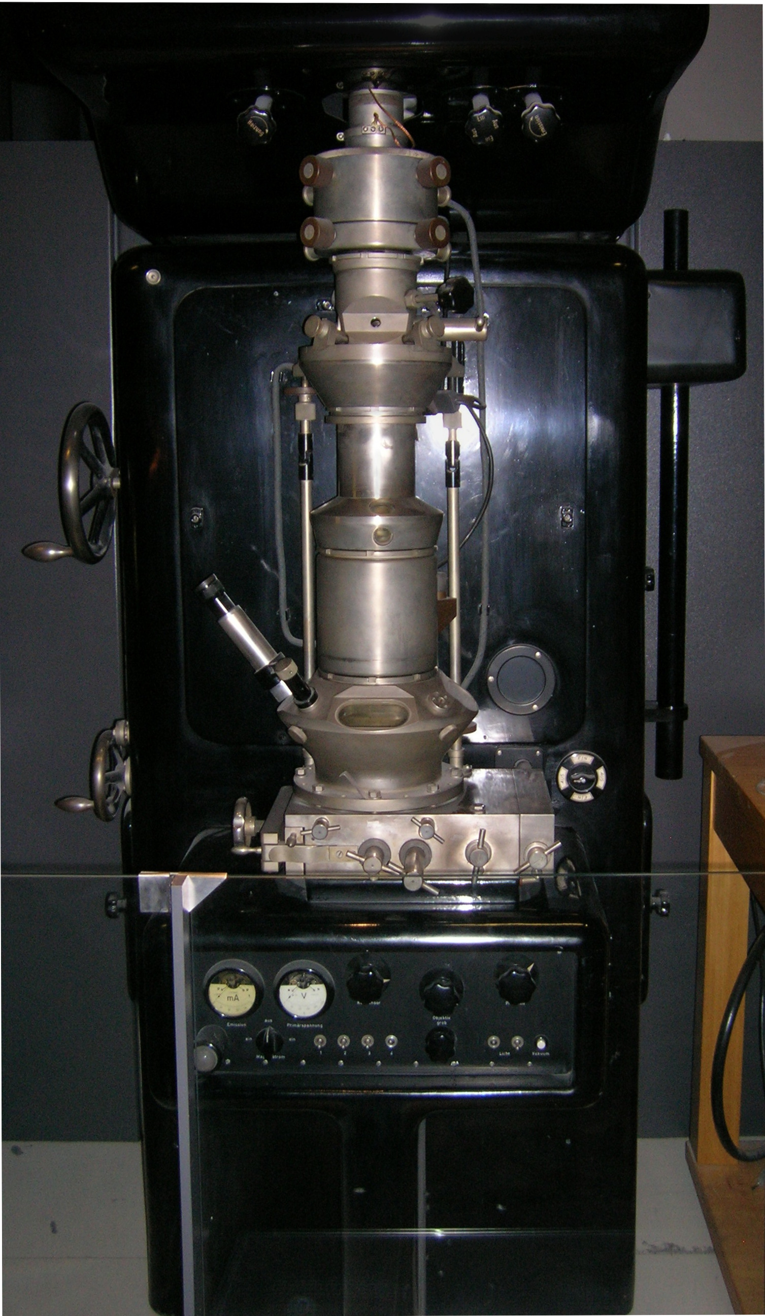
Transmission Electron Microscope
Transmission electron microscopy (TEM) is a microscopy technique in which a beam of electrons is transmitted through a specimen to form an image. The specimen is most often an ultrathin section less than 100 nm thick or a suspension on a grid. An image is formed from the interaction of the electrons with the sample as the beam is transmitted through the specimen. The image is then magnified and focused onto an imaging device, such as a fluorescent screen, a layer of photographic film, or a sensor such as a scintillator attached to a charge-coupled device. Transmission electron microscopes are capable of imaging at a significantly higher resolution than light microscopes, owing to the smaller de Broglie wavelength of electrons. This enables the instrument to capture fine detail—even as small as a single column of atoms, which is thousands of times smaller than a resolvable object seen in a light microscope. Transmission electron microscopy is a major analytical method in ... [...More Info...] [...Related Items...] OR: [Wikipedia] [Google] [Baidu] |
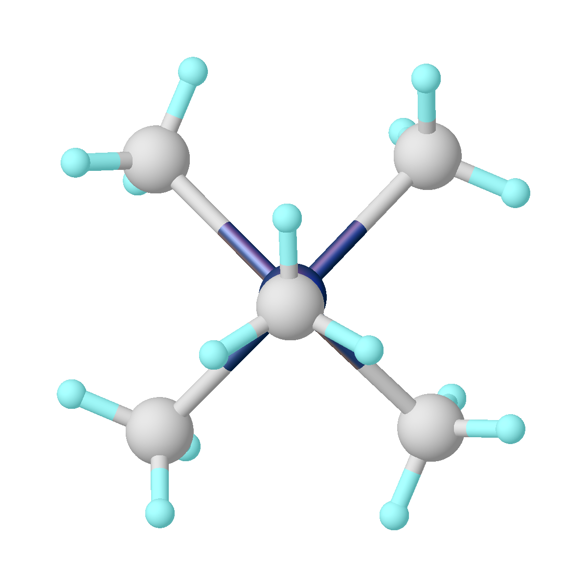
Liquid Nitrogen
Liquid nitrogen—LN2—is nitrogen in a liquid state at low temperature. Liquid nitrogen has a boiling point of about . It is produced industrially by fractional distillation of liquid air. It is a colorless, low viscosity liquid that is widely used as a coolant. Physical properties The diatomic character of the N2 molecule is retained after liquefaction. The weak van der Waals interaction between the N2 molecules results in little interatomic interaction, manifested in its very low boiling point. The temperature of liquid nitrogen can readily be reduced to its freezing point by placing it in a vacuum chamber pumped by a vacuum pump. Liquid nitrogen's efficiency as a coolant is limited by the fact that it boils immediately on contact with a warmer object, enveloping the object in an insulating layer of nitrogen gas bubbles. This effect, known as the Leidenfrost effect, occurs when any liquid comes in contact with a surface which is significantly hotter than its boiling ... [...More Info...] [...Related Items...] OR: [Wikipedia] [Google] [Baidu] |
Tantalum Oxide EM Image
Tantalum is a chemical element with the symbol Ta and atomic number 73. Previously known as ''tantalium'', it is named after Tantalus, a villain in Greek mythology. Tantalum is a very hard, ductile, lustrous, blue-gray transition metal that is highly corrosion-resistant. It is part of the refractory metals group, which are widely used as components of strong high-melting-point alloys. It is a group 5 element, along with vanadium and niobium, and it always occurs in geologic sources together with the chemically similar niobium, mainly in the mineral groups tantalite, columbite and coltan. The chemical inertness and very high melting point of tantalum make it valuable for laboratory and industrial equipment such as reaction vessels and vacuum furnaces. It is used in tantalum capacitors for electronic equipment such as computers. Tantalum is considered a technology-critical element by the European Commission. History Tantalum was discovered in Sweden in 1802 by Anders E ... [...More Info...] [...Related Items...] OR: [Wikipedia] [Google] [Baidu] |
Microcrystal Electron Diffraction
Microcrystal electron diffraction, or MicroED, is a CryoEM method that was developed by the Gonen laboratory in late 2013 at the Janelia Research Campus of the Howard Hughes Medical Institute. MicroED is a form of electron crystallography where thin 3D crystals are used for structure determination by electron diffraction. The method was developed for structure determination of proteins from nanocrystals that are typically not suitable for X-ray diffraction because of their size. Crystals that are one billionth the size needed for X-ray crystallography can yield high quality data. The samples are frozen hydrated as for all other CryoEM modalities but instead of using the transmission electron microscope (TEM) in imaging mode one uses it in diffraction mode with an extremely low electron exposure (typically < 0.01 e−/Å2/s). The nano crystal is exposed to the diffracting beam and continuously rotated while diffraction is collected on a fast ... [...More Info...] [...Related Items...] OR: [Wikipedia] [Google] [Baidu] |
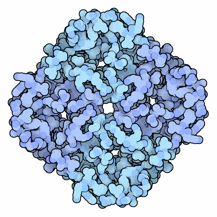
Aquaporin
Aquaporins, also called water channels, are channel proteins from a larger family of major intrinsic proteins that form pores in the membrane of biological cells, mainly facilitating transport of water between cells. The cell membranes of a variety of different bacteria, fungi, animal and plant cells contain aquaporins through which water can flow more rapidly into and out of the cell than by diffusing through the phospholipid bilayer. Aquaporins have six membrane-spanning alpha helical domains with both carboxylic and amino terminals on the cytoplasmic side. Two hydrophobic loops contain conserved asparagine- proline-alanine ("NPA motif") which form a barrel surrounding a central pore-like region that contains additional protein density. Because aquaporins are usually always open and are prevalent in just about every cell type, this leads to a misconception that water readily passes through the cell membrane down its concentration gradient. Water can pass through the cell mem ... [...More Info...] [...Related Items...] OR: [Wikipedia] [Google] [Baidu] |
Flagellum
A flagellum (; ) is a hairlike appendage that protrudes from certain plant and animal sperm cells, and from a wide range of microorganisms to provide motility. Many protists with flagella are termed as flagellates. A microorganism may have from one to many flagella. A gram-negative bacterium ''Helicobacter pylori'' for example uses its multiple flagella to propel itself through the mucus lining to reach the stomach epithelium, where it may cause a gastric ulcer to develop. In some bacteria the flagellum can also function as a sensory organelle, being sensitive to wetness outside the cell. Across the three domains of Bacteria, Archaea, and Eukaryota the flagellum has a different structure, protein composition, and mechanism of propulsion but shares the same function of providing motility. The Latin word means " whip" to describe its lash-like swimming motion. The flagellum in archaea is called the archaellum to note its difference from the bacterial flagellum. Eukaryotic ... [...More Info...] [...Related Items...] OR: [Wikipedia] [Google] [Baidu] |
Nicotinic Acetylcholine Receptor
Nicotinic acetylcholine receptors, or nAChRs, are receptor polypeptides that respond to the neurotransmitter acetylcholine. Nicotinic receptors also respond to drugs such as the agonist nicotine. They are found in the central and peripheral nervous system, muscle, and many other tissues of many organisms. At the neuromuscular junction they are the primary receptor in muscle for motor nerve-muscle communication that controls muscle contraction. In the peripheral nervous system: (1) they transmit outgoing signals from the presynaptic to the postsynaptic cells within the sympathetic and parasympathetic nervous system, and (2) they are the receptors found on skeletal muscle that receive acetylcholine released to signal for muscular contraction. In the immune system, nAChRs regulate inflammatory processes and signal through distinct intracellular pathways. In insects, the cholinergic system is limited to the central nervous system. The nicotinic receptors are considered cholinergi ... [...More Info...] [...Related Items...] OR: [Wikipedia] [Google] [Baidu] |
Light-harvesting Complex
A light-harvesting complex consists of a number of chromophores which are complex subunit proteins that may be part of a larger super complex of a photosystem, the functional unit in photosynthesis. It is used by plants and photosynthetic bacteria to collect more of the incoming light than would be captured by the photosynthetic reaction center alone. The light which is captured by the chromophores is capable of exciting molecules from their ground state to a higher energy state, known as the excited state. This excited state does not last very long and is known to be short-lived. Light-harvesting complexes are found in a wide variety among the different photosynthetic species, with no homology among the major groups. The complexes consist of proteins and photosynthetic pigments and surround a photosynthetic reaction center to focus energy, attained from photons absorbed by the pigment, toward the reaction center using Förster resonance energy transfer. Function Photosynthesis i ... [...More Info...] [...Related Items...] OR: [Wikipedia] [Google] [Baidu] |
Laboratory Of Molecular Biology
The Medical Research Council (MRC) Laboratory of Molecular Biology (LMB) is a research institute in Cambridge, England, involved in the revolution in molecular biology which occurred in the 1950–60s. Since then it has remained a major medical research laboratory at the forefront of scientific discovery, dedicated to improving the understanding of key biological processes at atomic, molecular and cellular levels using multidisciplinary methods, with a focus on using this knowledge to address key issues in human health. A new replacement building constructed close by to the original site on the Cambridge Biomedical Campus was opened by Her Majesty the Queen in May 2013. The road outside the new building is named Francis Crick Avenue after the 1962 joint Nobel Prize winner and LMB alumnus, who co-discovered the helical structure of DNA in 1953. History Origins: 1947-61 Max Perutz, following undergraduate training in organic chemistry, left Austria in 1936 and came to the Universi ... [...More Info...] [...Related Items...] OR: [Wikipedia] [Google] [Baidu] |
Medical Research Council (UK)
The Medical Research Council (MRC) is responsible for co-coordinating and funding medical research in the United Kingdom. It is part of United Kingdom Research and Innovation (UKRI), which came into operation 1 April 2018, and brings together the UK's seven research councils, Innovate UK and Research England. UK Research and Innovation is answerable to, although politically independent from, the Department for Business, Energy and Industrial Strategy. The MRC focuses on high-impact research and has provided the financial support and scientific expertise behind a number of medical breakthroughs, including the development of penicillin and the discovery of the structure of DNA. Research funded by the MRC has produced 32 Nobel Prize winners to date. History The MRC was founded as the Medical Research Committee and Advisory Council in 1913, with its prime role being the distribution of medical research funds under the terms of the National Insurance Act 1911. This was a consequen ... [...More Info...] [...Related Items...] OR: [Wikipedia] [Google] [Baidu] |
Richard Henderson (molecular Biologist)
Richard Henderson (born 19 July 1945) is a Scottish molecular biologist and biophysicist and pioneer in the field of electron microscopy of biological molecules. Henderson shared the Nobel Prize in Chemistry in 2017 with Jacques Dubochet and Joachim Frank. Education Henderson was educated at Newcastleton primary school, Hawick High School and Boroughmuir High School. He went on to study Physics at the University of Edinburgh graduating with a BSc degree in Physics, 1st Class honours in 1966. He then commenced postgraduate study at Corpus Christi College, Cambridge, and obtained his PhD degree from the University of Cambridge in 1969. Career and research Research Henderson worked on the structure and mechanism of chymotrypsin for his doctorate under the supervision of David Mervyn Blow at the MRC Laboratory of Molecular Biology. [...More Info...] [...Related Items...] OR: [Wikipedia] [Google] [Baidu] |


