|
Microcrystal Electron Diffraction
Microcrystal electron diffraction, or MicroED, is a CryoEM method that was developed by the Gonen laboratory in late 2013 at the Janelia Research Campus of the Howard Hughes Medical Institute. MicroED is a form of electron crystallography where thin 3D crystals are used for structure determination by electron diffraction. The method was developed for structure determination of proteins from nanocrystals that are typically not suitable for X-ray diffraction because of their size. Crystals that are one billionth the size needed for X-ray crystallography can yield high quality data. The samples are frozen hydrated as for all other CryoEM modalities but instead of using the transmission electron microscope ( TEM) in imaging mode one uses it in diffraction mode with an extremely low electron exposure (typically < 0.01 e−/Å2/s). The nano crystal is exposed to the diffracting beam and continuously rotated while diffraction is collected on a fas ... [...More Info...] [...Related Items...] OR: [Wikipedia] [Google] [Baidu] |
Cryogenic Electron Microscopy
Cryogenic electron microscopy (cryo-EM) is a cryomicroscopy technique applied on samples cooled to cryogenic temperatures. For biological specimens, the structure is preserved by embedding in an environment of vitreous ice. An aqueous sample solution is applied to a grid-mesh and plunge-frozen in liquid ethane or a mixture of liquid ethane and propane. While development of the technique began in the 1970s, recent advances in detector technology and software algorithms have allowed for the determination of biomolecular structures at near-atomic resolution. This has attracted wide attention to the approach as an alternative to X-ray crystallography or NMR spectroscopy for macromolecular structure determination without the need for crystallization. In 2017, the Nobel Prize in Chemistry was awarded to Jacques Dubochet, Joachim Frank, and Richard Henderson "for developing cryo-electron microscopy for the high-resolution structure determination of biomolecules in solution." ''Natur ... [...More Info...] [...Related Items...] OR: [Wikipedia] [Google] [Baidu] |
X-ray Crystallography
X-ray crystallography is the experimental science determining the atomic and molecular structure of a crystal, in which the crystalline structure causes a beam of incident X-rays to diffract into many specific directions. By measuring the angles and intensities of these diffracted beams, a crystallographer can produce a three-dimensional picture of the density of electrons within the crystal. From this electron density, the mean positions of the atoms in the crystal can be determined, as well as their chemical bonds, their crystallographic disorder, and various other information. Since many materials can form crystals—such as salts, metals, minerals, semiconductors, as well as various inorganic, organic, and biological molecules—X-ray crystallography has been fundamental in the development of many scientific fields. In its first decades of use, this method determined the size of atoms, the lengths and types of chemical bonds, and the atomic-scale differences among vari ... [...More Info...] [...Related Items...] OR: [Wikipedia] [Google] [Baidu] |
Alpha-synuclein
Alpha-synuclein is a protein that, in humans, is encoded by the ''SNCA'' gene. Alpha-synuclein is a neuronal protein that regulates synaptic vesicle trafficking and subsequent neurotransmitter release. It is abundant in the brain, while smaller amounts are found in the heart, muscle and other tissues. In the brain, alpha-synuclein is found mainly in the axon terminals of presynaptic neurons. Within these terminals, alpha-synuclein interacts with phospholipids and proteins. Presynaptic terminals release chemical messengers, called neurotransmitters, from compartments known as synaptic vesicles. The release of neurotransmitters relays signals between neurons and is critical for normal brain function. The human alpha-synuclein protein is made of 140 amino acids. An alpha-synuclein fragment, known as the non- Abeta component (NAC) of Alzheimer's disease amyloid, originally found in an amyloid-enriched fraction, was shown to be a fragment of its precursor protein, NACP. It was late ... [...More Info...] [...Related Items...] OR: [Wikipedia] [Google] [Baidu] |
Goniometer
A goniometer is an instrument that either measures an angle or allows an object to be rotated to a precise angular position. The term goniometry derives from two Greek words, Wikt:γωνία, γωνία (''gōnía'') 'angle' and Wikt:μέτρον#Ancient Greek, μέτρον (''métron'') 'Measurement, measure'. The first known description of a goniometer, based on the astrolabe, was by Gemma Frisius in 1538. Applications Surveying Prior to the invention of the theodolite, the goniometer was used in surveying. The application of triangulation to geodesy was described in the second (1533) edition of ''Cosmograficus liber'' by Petri Appiani as a 16-page appendix by Frisius entitled ''Libellus de locorum describendorum ratione''. Communications The Bellini–Tosi direction finder was a type of radio direction finder that was widely used from World War I to World War II. It used the signals from two crossed antennas, or four individual antennas simulating two crossed ones, to r ... [...More Info...] [...Related Items...] OR: [Wikipedia] [Google] [Baidu] |
CMOS
Complementary metal–oxide–semiconductor (CMOS, pronounced "sea-moss", ) is a type of metal–oxide–semiconductor field-effect transistor (MOSFET) fabrication process that uses complementary and symmetrical pairs of p-type and n-type MOSFETs for logic functions. CMOS technology is used for constructing integrated circuit (IC) chips, including microprocessors, microcontrollers, memory chips (including CMOS BIOS), and other digital logic circuits. CMOS technology is also used for analog circuits such as image sensors ( CMOS sensors), data converters, RF circuits ( RF CMOS), and highly integrated transceivers for many types of communication. The CMOS process was originally conceived by Frank Wanlass at Fairchild Semiconductor and presented by Wanlass and Chih-Tang Sah at the International Solid-State Circuits Conference in 1963. Wanlass later filed US patent 3,356,858 for CMOS circuitry and it was granted in 1967. commercialized the technology with the trademar ... [...More Info...] [...Related Items...] OR: [Wikipedia] [Google] [Baidu] |
Charge-coupled Device
A charge-coupled device (CCD) is an integrated circuit containing an array of linked, or coupled, capacitors. Under the control of an external circuit, each capacitor can transfer its electric charge to a neighboring capacitor. CCD sensors are a major technology used in digital imaging. In a CCD image sensor, pixels are represented by p-doped metal–oxide–semiconductor (MOS) capacitors. These MOS capacitors, the basic building blocks of a CCD, are biased above the threshold for inversion when image acquisition begins, allowing the conversion of incoming photons into electron charges at the semiconductor-oxide interface; the CCD is then used to read out these charges. Although CCDs are not the only technology to allow for light detection, CCD image sensors are widely used in professional, medical, and scientific applications where high-quality image data are required. In applications with less exacting quality demands, such as consumer and professional digital camera ... [...More Info...] [...Related Items...] OR: [Wikipedia] [Google] [Baidu] |
Selected Area Diffraction
Selected area (electron) diffraction (abbreviated as SAD or SAED), is a crystallography, crystallographic experimental technique typically performed using a transmission electron microscope (TEM). It is a specific case of electron diffraction used primarily in material science and solid state physics as one of the most common experimental techniques. Especially with appropriate analytical software, SAD patterns (SADP) can be used to determine crystal Orientation (geometry) , orientation, measure lattice constants or examine its Crystallographic defects , defects. Principle In transmission electron microscope, a thin crystalline sample is illuminated by parallel beam of electrons accelerated to kinetic energy , energy of hundreds of electron volt, kiloelectron volts. At these energies, even metallic samples are Transparency (optics), transparent for the electrons if the sample is thinned enough (typically less than 100 nanometer, nm). Due to the wave–particle duality, t ... [...More Info...] [...Related Items...] OR: [Wikipedia] [Google] [Baidu] |
Medipix
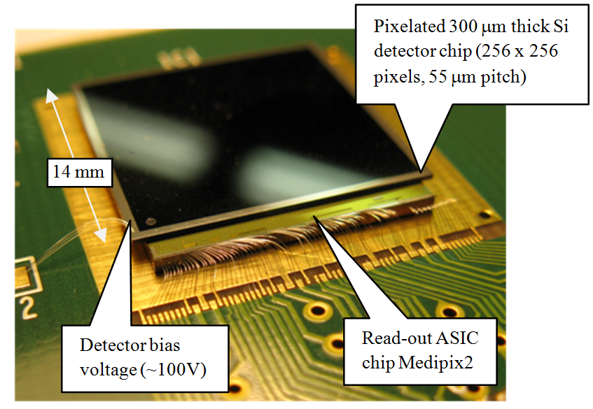
Medipix is a family of photon counting and particle tracking pixel detectors developed by an international collaboration, hosted by CERN. Design These are hybrid detectors as a semiconductor sensor layer is bonded to a processing electronics layer. The sensor layer is a semiconductor, such as silicon, GaAs, or CdTe in which the incident radiation makes an electron hole/cloud. The charge is then collected to pixel electrodes and, via bump bonds, conducted to the CMOS electronics layer. The pixel electronics first amplifies the signal and then compares the signal amplitude with a pre-set discrimination level (an energy threshold). The subsequent signal processing depends on the type of device. A standard Medipix detector increases the counter in the appropriate pixel if the signal is above the discrimination level. The Medipix device also contains an upper discrimination level and hence only signals within a range of amplitude could be accepted (within an energy window). Time ... [...More Info...] [...Related Items...] OR: [Wikipedia] [Google] [Baidu] |
Lysozyme
Lysozyme (EC 3.2.1.17, muramidase, ''N''-acetylmuramide glycanhydrolase; systematic name peptidoglycan ''N''-acetylmuramoylhydrolase) is an antimicrobial enzyme produced by animals that forms part of the innate immune system. It is a glycoside hydrolase that catalyzes the following process: : Hydrolysis of (1→4)-β-linkages between ''N''-acetylmuramic acid and ''N''-acetyl-D-glucosamine residues in a peptidoglycan and between ''N''-acetyl-D-glucosamine residues in chitodextrins Peptidoglycan is the major component of gram-positive bacterial cell wall. This hydrolysis in turn compromises the integrity of bacterial cell walls causing lysis of the bacteria. Lysozyme is abundant in secretions including tears, saliva, human milk, and mucus. It is also present in cytoplasmic granules of the macrophages and the polymorphonuclear neutrophils (PMNs). Large amounts of lysozyme can be found in egg white. C-type lysozymes are closely related to α-lactalbumin in sequence and s ... [...More Info...] [...Related Items...] OR: [Wikipedia] [Google] [Baidu] |
Diffraction
Diffraction is defined as the interference or bending of waves around the corners of an obstacle or through an aperture into the region of geometrical shadow of the obstacle/aperture. The diffracting object or aperture effectively becomes a secondary source of the propagating wave. Italian scientist Francesco Maria Grimaldi coined the word ''diffraction'' and was the first to record accurate observations of the phenomenon in 1660. In classical physics, the diffraction phenomenon is described by the Huygens–Fresnel principle that treats each point in a propagating wavefront as a collection of individual spherical wavelets. The characteristic bending pattern is most pronounced when a wave from a coherent source (such as a laser) encounters a slit/aperture that is comparable in size to its wavelength, as shown in the inserted image. This is due to the addition, or interference, of different points on the wavefront (or, equivalently, each wavelet) that travel by paths of ... [...More Info...] [...Related Items...] OR: [Wikipedia] [Google] [Baidu] |
Tamir Gonen
Tamir Gonen (born 1975) is an American structural biochemist and membrane biophysicist best known for his contributions to structural biology of membrane proteins, membrane biochemistry and electron cryo-microscopy (cryoEM) particularly in electron crystallography of 2D crystals and for the development of 3D electron crystallography from microscopic crystals known as MicroED. Gonen is an Investigator of the Howard Hughes Medical Institute, a professor at the University of California, Los Angeles, the founding director of the MicroED Imaging Center at UCLA and a Member of the Royal Society of New Zealand. Education Gonen attended the University of Auckland in New Zealand and graduated with a Bachelor of Science double major in Inorganic Chemistry and Biological Sciences, followed by First Class Honors in Biological Sciences in 1998. He then obtained a Doctor of Philosophy in Biological Science in 2002 from the University of Auckland for research with by Edward N. Baker and Joerg ... [...More Info...] [...Related Items...] OR: [Wikipedia] [Google] [Baidu] |


