|
Cryomicroscopy
Cryomicroscopy is a technique in which a microscope is equipped in such a fashion that the object intended to be inspected can be cooled to below room temperature. Technically, cryomicroscopy implies compatibility between a cryostat and a microscope. Most cryostats make use of a cryogenic fluid such as liquid helium or liquid nitrogen. There exists two common motivations for performing a cryomicroscopy. One is to improve upon the process of performing a standard microscopy. Cryogenic electron microscopy, for example, enables the studying of proteins with limited radiation damage. In this case, the protein structure may not change with temperature, but the cryogenic environment enables the improvement of the electron microscopy process. Another motivation for performing a cryomicroscopy is to apply the microscopy to a low-temperature phenomenon. A scanning tunnelling microscopy under a cryogenic environment, for example, allows for the studying of superconductivity, which does not ... [...More Info...] [...Related Items...] OR: [Wikipedia] [Google] [Baidu] |
Microscope
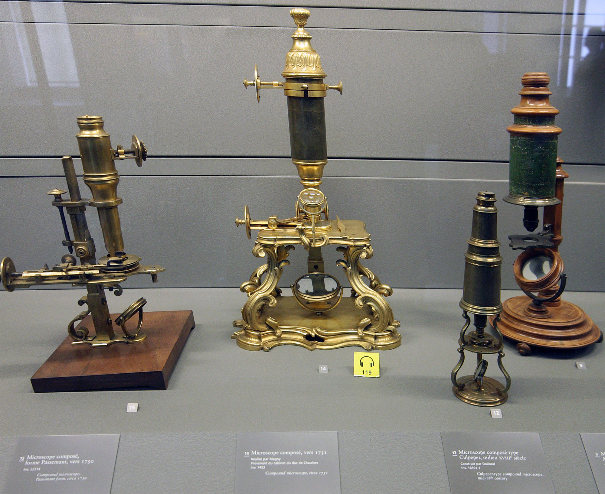
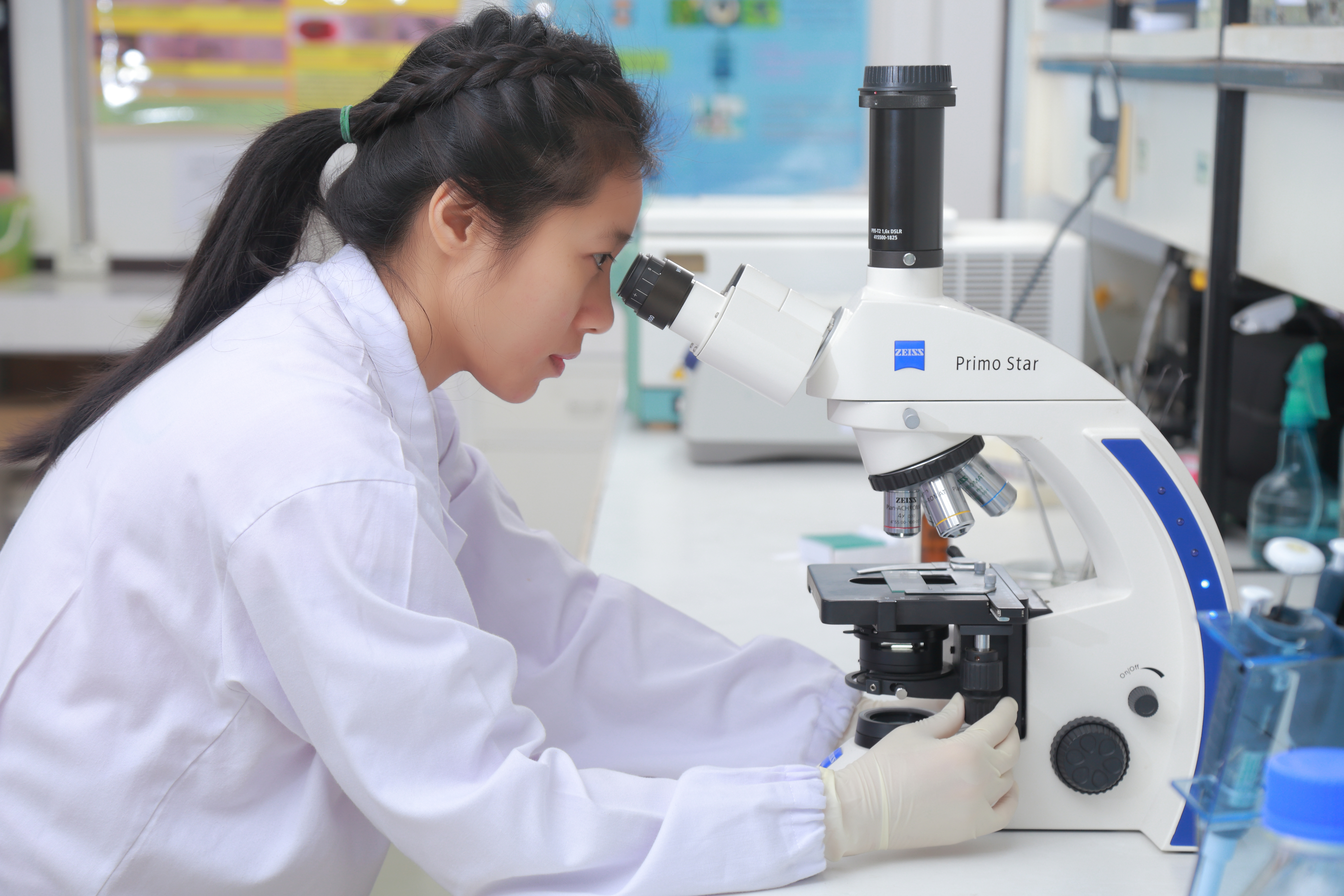
A microscope () is a laboratory instrument used to examine objects that are too small to be seen by the naked eye. Microscopy is the science of investigating small objects and structures using a microscope. Microscopic means being invisible to the eye unless aided by a microscope. There are many types of microscopes, and they may be grouped in different ways. One way is to describe the method an instrument uses to interact with a sample and produce images, either by sending a beam of light or electrons through a sample in its optical path, by detecting photon emissions from a sample, or by scanning across and a short distance from the surface of a sample using a probe. The most common microscope (and the first to be invented) is the optical microscope, which uses lenses to refract visible light that passed through a thinly sectioned sample to produce an observable image. Other major types of microscopes are the fluorescence microscope, electron microscope (both the transmi ... [...More Info...] [...Related Items...] OR: [Wikipedia] [Google] [Baidu] |
Richard Henderson (biologist)
Richard Henderson (born 19 July 1945) is a Scottish molecular biologist and biophysicist and pioneer in the field of electron microscopy of biological molecules. Henderson shared the Nobel Prize in Chemistry in 2017 with Jacques Dubochet and Joachim Frank. Education Henderson was educated at Newcastleton primary school, Hawick High School and Boroughmuir High School. He went on to study Physics at the University of Edinburgh graduating with a BSc degree in Physics, 1st Class honours in 1966. He then commenced postgraduate study at Corpus Christi College, Cambridge, and obtained his PhD degree from the University of Cambridge in 1969. Career and research Research Henderson worked on the structure and mechanism of chymotrypsin for his doctorate under the supervision of David Mervyn Blow at the MRC Laboratory of Molecular Biology. [...More Info...] [...Related Items...] OR: [Wikipedia] [Google] [Baidu] |
Stefan Hell
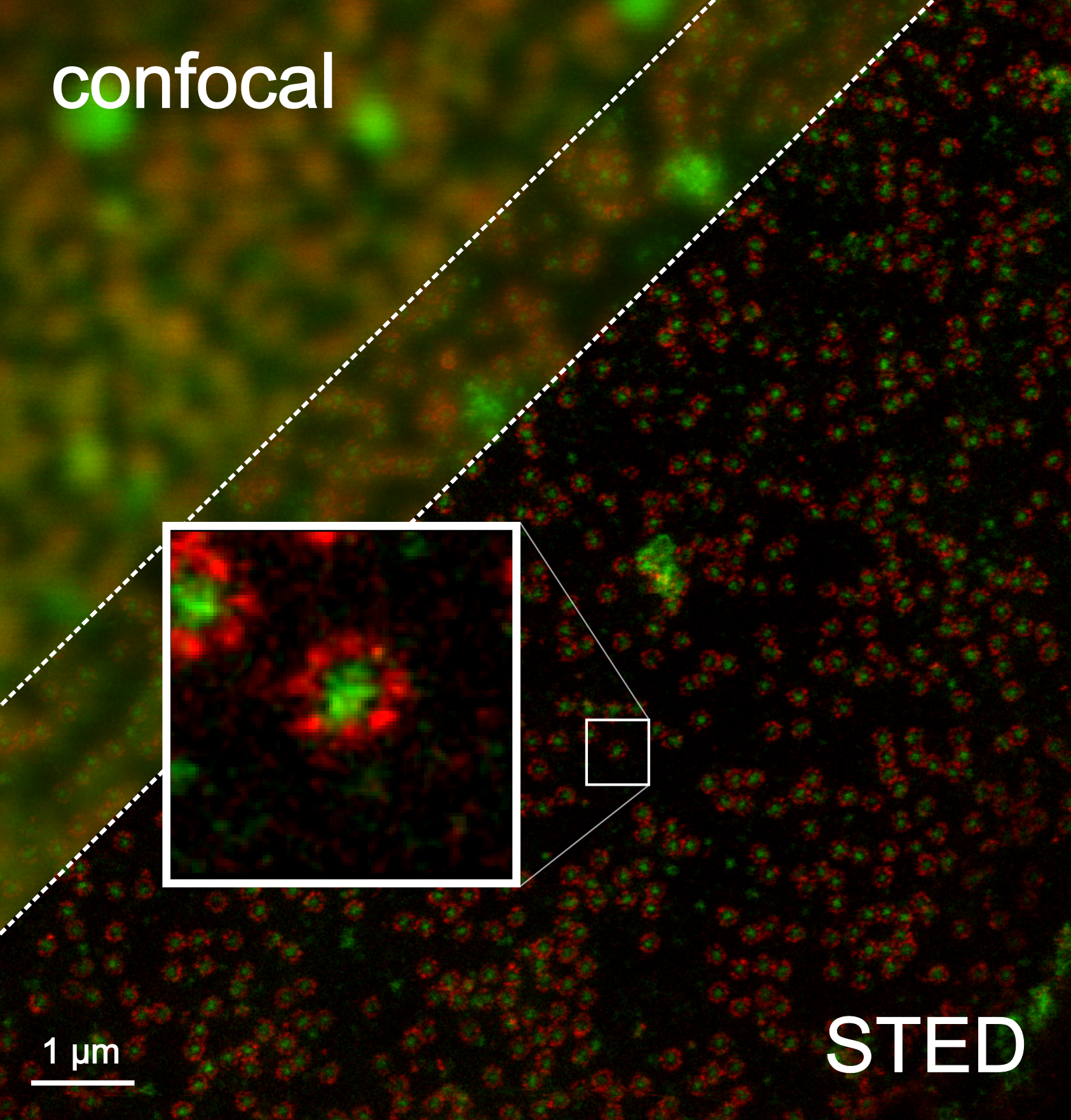
Stefan Walter Hell HonFRMS (: born 23 December 1962) is a Romanian-German physicist and one of the directors of the Max Planck Institute for Biophysical Chemistry in Göttingen, Germany. He received the Nobel Prize in Chemistry in 2014 "for the development of super-resolved fluorescence microscopy", together with Eric Betzig and William Moerner. Life Born into a Roman Catholic Banat Swabian family in Arad, Romania, he grew up at his parents' home in nearby Sântana. Hell attended primary school there between 1969 and 1977. Andreea Pocotila"Fizicianul premiat cu Nobelul pentru chimie vorbește românește și ține legătura cu mediul științific din țara noastră" ''România Liberă'', October 8, 2014 Subsequently, he attended one year of secondary education at the Nikolaus Lenau High School in Timișoara before leaving with his parents to West Germany in 1978. His father was an engineer and his mother a teacher; the family settled in Ludwigshafen after emigrating. He ... [...More Info...] [...Related Items...] OR: [Wikipedia] [Google] [Baidu] |
Eric Betzig
Robert Eric Betzig (born January 13, 1960) is an American physicist who works as a professor of physics and professor of molecular and cell biology at the University of California, Berkeley. He is also a senior fellow at the Janelia Farm Research Campus in Ashburn, Virginia. Betzig has worked to develop the field of fluorescence microscopy and photoactivated localization microscopy. He was awarded the 2014 Nobel Prize in Chemistry for "the development of super-resolved fluorescence microscopy" along with Stefan Hell and fellow Cornell alumnus William E. Moerner. Early life and education Betzig was born in Ann Arbor, Michigan, in 1960, the son of Helen Betzig and engineer Robert Betzig. Aspiring to work in the aerospace industry, Betzig studied physics at the California Institute of Technology and graduated with a BS degree in 1983. He then went on to study at Cornell University where he was advised by Aaron Lewis and Michael Isaacson. There he obtained an MS degree and a ... [...More Info...] [...Related Items...] OR: [Wikipedia] [Google] [Baidu] |
Diffraction-limited System
The resolution of an optical imaging system a microscope, telescope, or camera can be limited by factors such as imperfections in the lenses or misalignment. However, there is a principal limit to the resolution of any optical system, due to the physics of diffraction. An optical system with resolution performance at the instrument's theoretical limit is said to be diffraction-limited. The diffraction-limited angular resolution of a telescopic instrument is inversely proportional to the wavelength of the light being observed, and proportional to the diameter of its objective's entrance aperture. For telescopes with circular apertures, the size of the smallest feature in an image that is diffraction limited is the size of the Airy disk. As one decreases the size of the aperture of a telescopic lens, diffraction proportionately increases. At small apertures, such as f/22, most modern lenses are limited only by diffraction and not by aberrations or other imperfections in the cons ... [...More Info...] [...Related Items...] OR: [Wikipedia] [Google] [Baidu] |
Fluorescence Microscope
A fluorescence microscope is an optical microscope that uses fluorescence instead of, or in addition to, scattering, reflection, and attenuation or absorption, to study the properties of organic or inorganic substances. "Fluorescence microscope" refers to any microscope that uses fluorescence to generate an image, whether it is a simple set up like an epifluorescence microscope or a more complicated design such as a confocal microscope, which uses optical sectioning to get better resolution of the fluorescence image. Principle The specimen is illuminated with light of a specific wavelength (or wavelengths) which is absorbed by the fluorophores, causing them to emit light of longer wavelengths (i.e., of a different color than the absorbed light). The illumination light is separated from the much weaker emitted fluorescence through the use of a spectral emission filter. Typical components of a fluorescence microscope are a light source (xenon arc lamp or mercury-vapor lamp are ... [...More Info...] [...Related Items...] OR: [Wikipedia] [Google] [Baidu] |
Birefringence
Birefringence is the optical property of a material having a refractive index that depends on the polarization and propagation direction of light. These optically anisotropic materials are said to be birefringent (or birefractive). The birefringence is often quantified as the maximum difference between refractive indices exhibited by the material. Crystals with non-cubic crystal structures are often birefringent, as are plastics under mechanical stress. Birefringence is responsible for the phenomenon of double refraction whereby a ray of light, when incident upon a birefringent material, is split by polarization into two rays taking slightly different paths. This effect was first described by Danish scientist Rasmus Bartholin in 1669, who observed it in calcite, a crystal having one of the strongest birefringences. In the 19th century Augustin-Jean Fresnel described the phenomenon in terms of polarization, understanding light as a wave with field components in transverse polariz ... [...More Info...] [...Related Items...] OR: [Wikipedia] [Google] [Baidu] |
Polarized Light Microscopy
Polarized light microscopy can mean any of a number of optical microscopy techniques involving polarized light. Simple techniques include illumination of the sample with polarized light. Directly transmitted light can, optionally, be blocked with a polariser orientated at 90 degrees to the illumination. More complex microscopy techniques which take advantage of polarized light include differential interference contrast microscopy and interference reflection microscopy. Scientists will often use a device called a polarizing plate to convert natural light into polarized light. These illumination techniques are most commonly used on birefringent samples where the polarized light interacts strongly with the sample and so generating contrast with the background. Polarized light microscopy is used extensively in optical mineralogy. History Although the invention of the polarizing microscope is typically attributed to David Brewster around 1815, Brewster clearly acknowledges the prio ... [...More Info...] [...Related Items...] OR: [Wikipedia] [Google] [Baidu] |
Optical Microscope
The optical microscope, also referred to as a light microscope, is a type of microscope that commonly uses visible light and a system of lenses to generate magnified images of small objects. Optical microscopes are the oldest design of microscope and were possibly invented in their present compound form in the 17th century. Basic optical microscopes can be very simple, although many complex designs aim to improve resolution and sample contrast. The object is placed on a stage and may be directly viewed through one or two eyepieces on the microscope. In high-power microscopes, both eyepieces typically show the same image, but with a stereo microscope, slightly different images are used to create a 3-D effect. A camera is typically used to capture the image (micrograph). The sample can be lit in a variety of ways. Transparent objects can be lit from below and solid objects can be lit with light coming through ( bright field) or around (dark field) the objective lens. Polarised ... [...More Info...] [...Related Items...] OR: [Wikipedia] [Google] [Baidu] |
Transmission Electron Microscopy
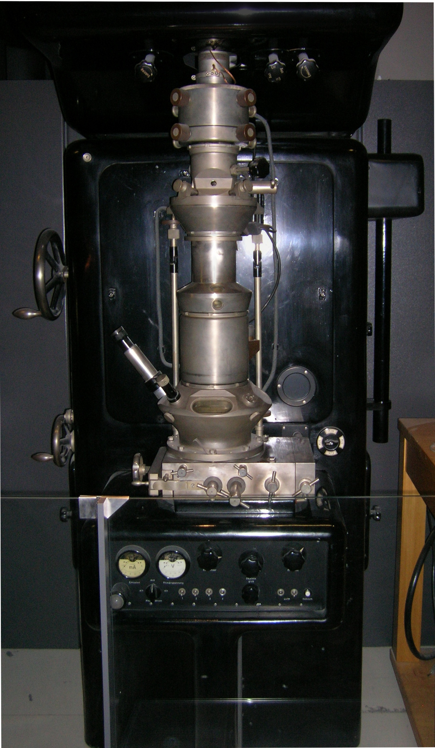
Transmission electron microscopy (TEM) is a microscopy technique in which a beam of electrons is transmitted through a specimen to form an image. The specimen is most often an ultrathin section less than 100 nm thick or a suspension on a grid. An image is formed from the interaction of the electrons with the sample as the beam is transmitted through the specimen. The image is then magnified and focused onto an imaging device, such as a fluorescent screen, a layer of photographic film, or a sensor such as a scintillator attached to a charge-coupled device. Transmission electron microscopes are capable of imaging at a significantly higher resolution than light microscopes, owing to the smaller de Broglie wavelength of electrons. This enables the instrument to capture fine detail—even as small as a single column of atoms, which is thousands of times smaller than a resolvable object seen in a light microscope. Transmission electron microscopy is a major analytical method i ... [...More Info...] [...Related Items...] OR: [Wikipedia] [Google] [Baidu] |
Joachim Frank
Joachim Frank () (born September 12, 1940) is a German-American biophysicist at Columbia University and a Nobel laureate. He is regarded as the founder of single-particle cryo-electron microscopy (cryo-EM), for which he shared the Nobel Prize in Chemistry in 2017 with Jacques Dubochet and Richard Henderson. He also made significant contributions to structure and function of the ribosome from bacteria and eukaryotes. Life and career Frank was born in Siegen in the borough of Weidenau. After completing his Vordiplom (B.S.) degree in physics at the University of Freiburg (1963) and his Diplom under Walter Rollwagen's mentorship at the Ludwig Maximilian University of Munich with the thesis "Untersuchung der Sekundärelektronen-Emission von Gold am Schmelzpunkt" (Investigation of secondary electron emission of gold at its melting point) (1967), Frank obtained his Ph.D. from the Technical University of Munich for graduate studies in Walter Hoppe's lab at the Max Planck Instit ... [...More Info...] [...Related Items...] OR: [Wikipedia] [Google] [Baidu] |
Cryostat
A cryostat (from ''cryo'' meaning cold and ''stat'' meaning stable) is a device used to maintain low cryogenic temperatures of samples or devices mounted within the cryostat. Low temperatures may be maintained within a cryostat by using various refrigeration methods, most commonly using cryogenic fluid bath such as liquid helium. Hence it is usually assembled into a vessel, similar in construction to a vacuum flask or Dewar. Cryostats have numerous applications within science, engineering, and medicine. Types Closed-cycle cryostats Closed-cycle cryostats consist of a chamber through which cold helium vapour is pumped. An external mechanical refrigerator extracts the warmer helium exhaust vapour, which is cooled and recycled. Closed-cycle cryostats consume a relatively large amount of electrical power, but need not be refilled with helium and can run continuously for an indefinite period. Objects may be cooled by attaching them to a metallic coldplate inside a vacuum ch ... [...More Info...] [...Related Items...] OR: [Wikipedia] [Google] [Baidu] |


.jpg)


