|
Chronic Relapsing Inflammatory Optic Neuropathy
Chronic relapsing inflammatory optic neuropathy (CRION) is a form of recurrent optic neuritis that is steroid responsive and dependent. Patients typically present with pain associated with visual loss. CRION is a clinical diagnosis of exclusion, and other demyelinating, autoimmune, and systemic causes should be ruled out. An accurate antibody test which became available commercially in 2017 has allowed most patients previously diagnosed with CRION to be re-identified as having MOG antibody disease, which is not a diagnosis of exclusion. Early recognition is crucial given risks for severe visual loss and because it is treatable with immunosuppressive treatment such as steroids or B-cell depleting therapy. Relapse that occurs after reducing or stopping steroids is a characteristic feature. Signs and symptoms Pain, visual loss, relapse, and steroid response are typical of CRION. Ocular pain is typical, although there are some cases with no reported pain. Bilateral severe visual l ... [...More Info...] [...Related Items...] OR: [Wikipedia] [Google] [Baidu] |
Ophthalmology
Ophthalmology ( ) is a surgical subspecialty within medicine that deals with the diagnosis and treatment of eye disorders. An ophthalmologist is a physician who undergoes subspecialty training in medical and surgical eye care. Following a medical degree, a doctor specialising in ophthalmology must pursue additional postgraduate residency training specific to that field. This may include a one-year integrated internship that involves more general medical training in other fields such as internal medicine or general surgery. Following residency, additional specialty training (or fellowship) may be sought in a particular aspect of eye pathology. Ophthalmologists prescribe medications to treat eye diseases, implement laser therapy, and perform surgery when needed. Ophthalmologists provide both primary and specialty eye care - medical and surgical. Most ophthalmologists participate in academic research on eye diseases at some point in their training and many include research as part ... [...More Info...] [...Related Items...] OR: [Wikipedia] [Google] [Baidu] |
Neuromyelitis Optica
Neuromyelitis optica spectrum disorders (NMOSD), including neuromyelitis optica (NMO), are autoimmune diseases characterized by acute inflammation of the optic nerve (optic neuritis, ON) and the spinal cord (myelitis). Episodes of ON and myelitis can be simultaneous or successive. A relapsing disease course is common, especially in untreated patients. In more than 80% of cases, NMO is caused by immunoglobulin G Autoantibody, autoantibodies to aquaporin 4 (anti-AQP4 diseases, anti-AQP4), the most abundant Aquaporin, water channel protein in the central nervous system. A subset of anti-AQP4-negative cases is associated with antibodies against myelin oligodendrocyte glycoprotein (Anti-MOG associated encephalomyelitis, anti-MOG). Rarely, NMO may occur in the context of other autoimmune diseases (e.g. Connective tissue disease, connective tissue disorders, paraneoplastic syndromes) or infectious diseases. In some cases, the etiology remains unknown (Idiopathic disease, idiopathic NMO). ... [...More Info...] [...Related Items...] OR: [Wikipedia] [Google] [Baidu] |
Oligoclonal Band
Oligoclonal bands (OCBs) are bands of immunoglobulins that are seen when a patient's blood serum, or cerebrospinal fluid (CSF) is analyzed. They are used in the diagnosis of various neurological and blood diseases, especially in multiple sclerosis. Two methods of analysis are possible: (a) protein electrophoresis, a method of analyzing the composition of fluids, also known as "SDS-PAGE (sodium dodecyl sulphate polyacrylamide gel electrophoresis)/Coomassie blue staining", and (b) the combination of isoelectric focusing/silver staining. The latter is more sensitive. For the analysis of cerebrospinal fluid, a sample is first collected via lumbar puncture (LP). Normally it is assumed that all the proteins that appear in the CSF, but are not present in the serum, are produced intrathecally (inside the central nervous system). Therefore, it is normal to subtract bands in serum from bands in CSF when investigating CNS diseases. A sample of blood serum is usually obtained from a clotted bl ... [...More Info...] [...Related Items...] OR: [Wikipedia] [Google] [Baidu] |
Autoimmune Optic Neuropathy
Autoimmune optic neuropathy (AON), sometimes called autoimmune optic neuritis, may be a forme fruste of systemic lupus erythematosus (SLE) associated optic neuropathy. AON is more than the presence of any optic neuritis Optic neuritis describes any condition that causes inflammation of the optic nerve; it may be associated with demyelinating diseases, or infectious or inflammatory processes. It is also known as optic papillitis (when the head of the optic nerv ... in a patient with an autoimmune process, as it describes a relatively specific clinical syndrome. AON is characterized by chronically progressive or recurrent vision loss associated with serological evidence of autoimmunity. Specifically, this term has been suggested for cases of optic neuritis with serological evidence of vasculitis by positive ANA, despite the lack of meeting criteria for SLE. The clinical manifestations include progressive vision loss that tends to be steroid-responsive and steroid dependent. ... [...More Info...] [...Related Items...] OR: [Wikipedia] [Google] [Baidu] |
Serostatus
Serostatus refers to the presence or absence of a serological marker in the blood. The presence of detectable levels of a specific marker within the serum is considered seropositivity, while the absence of such levels is considered seronegativity. HIV/AIDS The term serostatus is commonly used in HIV/AIDS prevention efforts. In the late 20th and early 21st centuries, social advocacy has emphasized the importance of learning one's HIV/AIDS serostatus in an effort to curtail the spread of the disease. Autoimmune disease Researchers have investigated the effects of autoantibody serostatus on autoimmune disease presentation. Study of seronegative patient populations has led to the identification of additional autoantibodies that could potentially help with diagnosis. See also * Seroconversion * Correlates of immunity Correlates of immunity or correlates of protection to a virus or other infectious pathogen are measurable signs that a person (or other potential host) is immune, in th ... [...More Info...] [...Related Items...] OR: [Wikipedia] [Google] [Baidu] |
Glial Fibrillary Acidic Protein
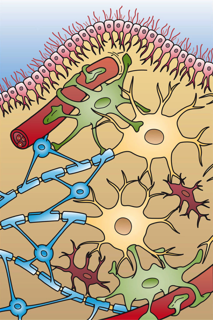
Glial fibrillary acidic protein (GFAP) is a protein that is encoded by the ''GFAP'' gene in humans. It is a type III intermediate filament (IF) protein that is expressed by numerous cell types of the central nervous system (CNS), including astrocytes and ependymal cells during development. GFAP has also been found to be expressed in glomeruli and peritubular fibroblasts taken from rat kidneys, Leydig cells of the testis in both hamsters and humans, human keratinocytes, human osteocytes and chondrocytes and stellate cells of the pancreas and liver in rats. GFAP is closely related to the other three non-epithelial type III IF family members, vimentin, desmin and peripherin, which are all involved in the structure and function of the cell’s cytoskeleton. GFAP is thought to help to maintain astrocyte mechanical strength as well as the shape of cells, but its exact function remains poorly understood, despite the number of studies using it as a cell marker. The protein was named and ... [...More Info...] [...Related Items...] OR: [Wikipedia] [Google] [Baidu] |
Aquaporin 4
Aquaporin-4, also known as AQP-4, is a water channel protein encoded by the ''AQP4'' gene in humans. AQP-4 belongs to the aquaporin family of integral membrane proteins that conduct water through the cell membrane. A limited number of aquaporins are found within the central nervous system (CNS): AQP1, 3, 4, 5, 8, 9, and 11, but more exclusive representation of AQP1, 4, and 9 are found in the brain and spinal cord. AQP4 shows the largest presence in the cerebellum and spinal cord grey matter. In the CNS, AQP4 is the most prevalent aquaporin channel, specifically located at the perimicrovessel astrocyte foot processes, glia limitans, and ependyma. In addition, this channel is commonly found facilitating water movement near cerebrospinal fluid and vasculature. Aquaporin-4 was first identified in 1986. It was the first evidence of the existence of water transport channels. The method that was used to discover the existence of the transport channels was through knockout experiments. Wit ... [...More Info...] [...Related Items...] OR: [Wikipedia] [Google] [Baidu] |
Thyroid Function Tests
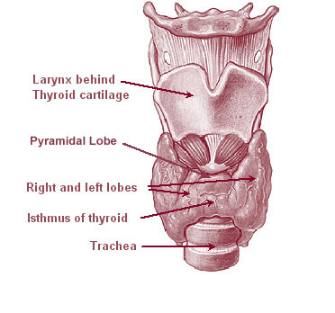
The thyroid, or thyroid gland, is an endocrine gland in vertebrates. In humans it is in the neck and consists of two connected lobes. The lower two thirds of the lobes are connected by a thin band of tissue called the thyroid isthmus. The thyroid is located at the front of the neck, below the Adam's apple. Microscopically, the functional unit of the thyroid gland is the spherical thyroid follicle, lined with follicular cells (thyrocytes), and occasional parafollicular cells that surround a lumen containing colloid. The thyroid gland secretes three hormones: the two thyroid hormones triiodothyronine (T3) and thyroxine (T4)and a peptide hormone, calcitonin. The thyroid hormones influence the metabolic rate and protein synthesis, and in children, growth and development. Calcitonin plays a role in calcium homeostasis. Secretion of the two thyroid hormones is regulated by thyroid-stimulating hormone (TSH), which is secreted from the anterior pituitary gland. TSH is regulated ... [...More Info...] [...Related Items...] OR: [Wikipedia] [Google] [Baidu] |
Folate Deficiency
Folate deficiency, also known as vitamin B9 deficiency, is a low level of folate and derivatives in the body. Signs of folate deficiency are often subtle. A low number of red blood cells (anemia) is a late finding in folate deficiency and folate deficiency anemia is the term given for this medical condition. It is characterized by the appearance of large-sized, abnormal red blood cells (megaloblasts), which form when there are inadequate stores of folic acid within the body. Signs and symptoms Loss of appetite and weight loss can occur. Additional signs are weakness, sore tongue, headaches, heart palpitations, irritability, and behavioral disorders. In adults, anemia (macrocytic, megaloblastic anemia) can be a sign of advanced folate deficiency. Women with folate deficiency who become pregnant are more likely to give birth to low birth weight premature infants, and infants with neural tube defects and even spina bifida. In infants and children, folate deficiency can lead to f ... [...More Info...] [...Related Items...] OR: [Wikipedia] [Google] [Baidu] |
B12 Deficiency
Vitamin B12 deficiency, also known as cobalamin deficiency, is the medical condition in which the blood and tissue have a lower than normal level of vitamin B12. Symptoms can vary from none to severe. Mild deficiency may have few or absent symptoms. In moderate deficiency, feeling tired, anemia, soreness of the tongue, mouth ulcers, breathlessness, feeling faint, rapid heartbeat, low blood pressure, pallor, hair loss, decreased ability to think and severe joint pain and the beginning of neurological symptoms, including abnormal sensations such as ''pins and needles'', numbness and tinnitus may occur. Severe deficiency may include symptoms of reduced heart function as well as more severe neurological symptoms, including changes in reflexes, poor muscle function, memory problems, blurred vision, irritability, ataxia, decreased smell and taste, decreased level of consciousness, depression, anxiety, guilt and psychosis. If left untreated, some of these changes can become perm ... [...More Info...] [...Related Items...] OR: [Wikipedia] [Google] [Baidu] |
Anti-nuclear Antibody
Antinuclear antibodies (ANAs, also known as antinuclear factor or ANF) are autoantibodies that bind to contents of the cell nucleus. In normal individuals, the immune system produces antibodies to foreign proteins (antigens) but not to human proteins (autoantigens). In some cases, antibodies to human antigens are produced. There are many subtypes of ANAs such as anti-Ro antibodies, anti-La antibodies, anti-Sm antibodies, anti-nRNP antibodies, anti-Scl-70 antibodies, anti-dsDNA antibodies, anti-histone antibodies, antibodies to nuclear pore complexes, anti-centromere antibodies and anti-sp100 antibodies. Each of these antibody subtypes binds to different proteins or protein complexes within the nucleus. They are found in many disorders including autoimmunity, cancer and infection, with different prevalences of antibodies depending on the condition. This allows the use of ANAs in the diagnosis of some autoimmune disorders, including systemic lupus erythematosus, Sjögren synd ... [...More Info...] [...Related Items...] OR: [Wikipedia] [Google] [Baidu] |
Leber's Hereditary Optic Neuropathy
Leber's hereditary optic neuropathy (LHON) is a mitochondrially inherited (transmitted from mother to offspring) degeneration of retinal ganglion cells (RGCs) and their axons that leads to an acute or subacute loss of central vision; it predominantly affects young adult males. LHON is transmitted only through the mother, as it is primarily due to mutations in the mitochondrial (not nuclear) genome, and only the egg contributes mitochondria to the embryo. LHON is usually due to one of three pathogenic mitochondrial DNA (mtDNA) point mutations. These mutations are at nucleotide positions 11778 G to A, 3460 G to A and 14484 T to C, respectively in the ND4, ND1 and ND6 subunit genes of complex I of the oxidative phosphorylation chain in mitochondria. Men cannot pass on the disease to their offspring. Signs and symptoms Clinically, there is an acute onset of visual loss, first in one eye, and then a few weeks to months later in the other. Onset is usually young adulthood, b ... [...More Info...] [...Related Items...] OR: [Wikipedia] [Google] [Baidu] |


