|
Cardiac Aberration
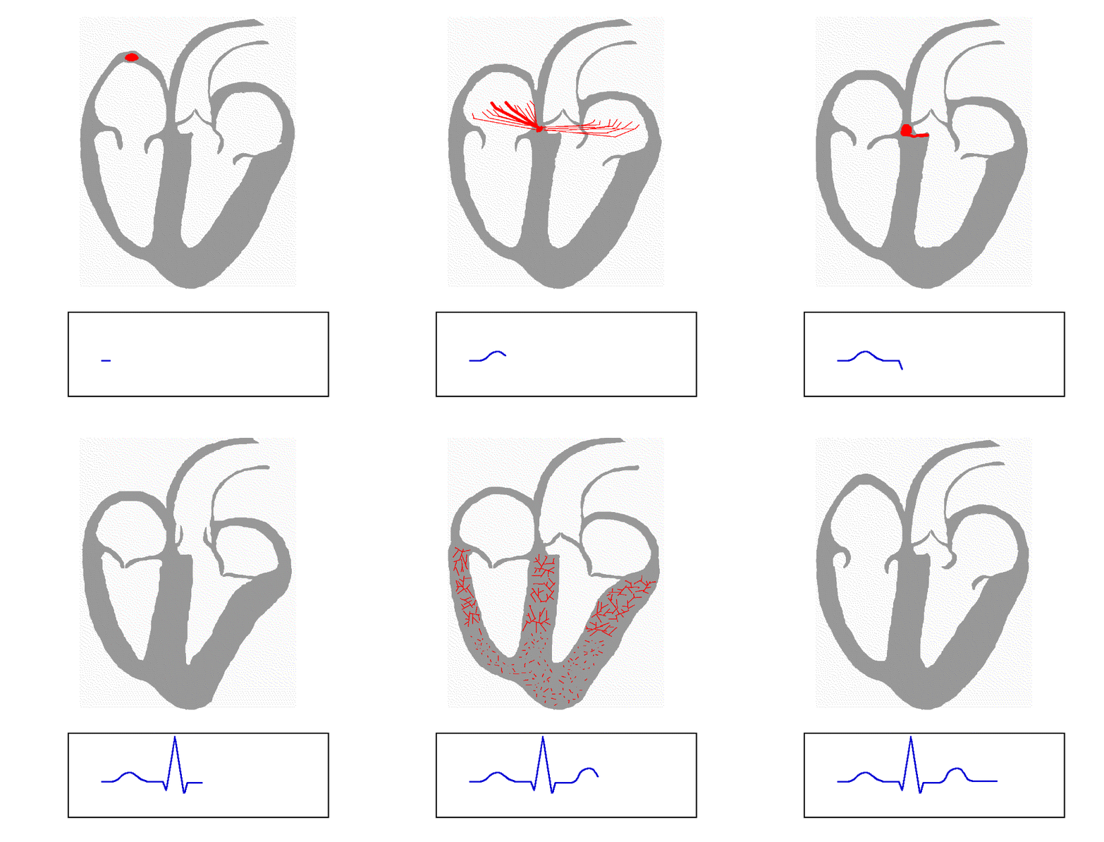
Cardiac aberrancy is a type of aberration in the shape of the EKG signal, representing activation of the heart muscle via the electrical conduction system of the heart. Aberration occurs when the electrical activation of the heart, which is caused by a series of action potentials is conducting improperly, which can be due to: * left anterior fascicular block * left posterior fascicular block * bundle branch block * Wolff–Parkinson–White syndrome Wolff–Parkinson–White syndrome (WPWS) is a disorder due to a specific type of problem with the electrical system of the heart. About 60% of people with the electrical problem developed symptoms, which may include an abnormally fast heartbeat, ... See also * Electrocardiography {{circulatory-stub Cardiac electrophysiology ... [...More Info...] [...Related Items...] OR: [Wikipedia] [Google] [Baidu] |
EKG
Electrocardiography is the process of producing an electrocardiogram (ECG or EKG), a recording of the heart's electrical activity. It is an electrogram of the heart which is a graph of voltage versus time of the electrical activity of the heart using electrodes placed on the skin. These electrodes detect the small electrical changes that are a consequence of cardiac muscle depolarization followed by repolarization during each cardiac cycle (heartbeat). Changes in the normal ECG pattern occur in numerous cardiac abnormalities, including cardiac rhythm disturbances (such as atrial fibrillation and ventricular tachycardia), inadequate coronary artery blood flow (such as myocardial ischemia and myocardial infarction), and electrolyte disturbances (such as hypokalemia and hyperkalemia). Traditionally, "ECG" usually means a 12-lead ECG taken while lying down as discussed below. However, other devices can record the electrical activity of the heart such as a Holter monito ... [...More Info...] [...Related Items...] OR: [Wikipedia] [Google] [Baidu] |
Electrical Conduction System Of The Heart
The cardiac conduction system (CCS) (also called the electrical conduction system of the heart) transmits the signals generated by the sinoatrial node – the heart's pacemaker, to cause the heart muscle to contract, and pump blood through the body's circulatory system. The pacemaking signal travels through the right atrium to the atrioventricular node, along the bundle of His, and through the bundle branches to Purkinje fibers in the walls of the ventricles. The Purkinje fibers transmit the signals more rapidly to stimulate contraction of the ventricles. The conduction system consists of specialized heart muscle cells, situated within the myocardium. There is a skeleton of fibrous tissue that surrounds the conduction system which can be seen on an ECG. Dysfunction of the conduction system can cause irregular heart rhythms including rhythms that are too fast or too slow. Structure Electrical signals arising in the SA node (located in the right atrium) stimulate the atr ... [...More Info...] [...Related Items...] OR: [Wikipedia] [Google] [Baidu] |
Action Potentials
An action potential occurs when the membrane potential of a specific cell location rapidly rises and falls. This depolarization then causes adjacent locations to similarly depolarize. Action potentials occur in several types of animal cells, called excitable cells, which include neurons, muscle cells, and in some plant cells. Certain endocrine cells such as pancreatic beta cells, and certain cells of the anterior pituitary gland are also excitable cells. In neurons, action potentials play a central role in cell-cell communication by providing for—or with regard to saltatory conduction, assisting—the propagation of signals along the neuron's axon toward synaptic boutons situated at the ends of an axon; these signals can then connect with other neurons at synapses, or to motor cells or glands. In other types of cells, their main function is to activate intracellular processes. In muscle cells, for example, an action potential is the first step in the chain of events leadi ... [...More Info...] [...Related Items...] OR: [Wikipedia] [Google] [Baidu] |
Left Anterior Fascicular Block
Left anterior fascicular block (LAFB) is an abnormal condition of the left ventricle of the heart, related to, but distinguished from, left bundle branch block (LBBB). It is caused by only the left anterior fascicle – one half of the left bundle branch being defective. It is manifested on the ECG by left axis deviation. It is much more common than left posterior fascicular block. Mechanism Normal activation of the left ventricle (LV) proceeds down the left bundle branch, which consist of three fascicles, the left anterior fascicle, the left posterior fascicle, and the septal fascicle. The posterior fascicle supplies the posterior and inferoposterior walls of the LV, the anterior fascicle supplies the upper and anterior parts of the LV and the septal fascicle supplies the septal wall with innervation. LAFB — which is also known as left anterior hemiblock (LAHB) — occurs when a cardiac impulse spreads first through the left posterior fascicle, causing a delay in activation of ... [...More Info...] [...Related Items...] OR: [Wikipedia] [Google] [Baidu] |
Left Posterior Fascicular Block
A left posterior fascicular block (LPFB), also known as left posterior hemiblock (LPH), is a condition where the left posterior fascicle, which travels to the inferior and posterior portion of the left ventricle, does not conduct the electrical impulses from the atrioventricular node. The wave-front instead moves more quickly through the left anterior fascicle and right bundle branch, leading to a right axis deviation seen on the ECG. Definition The American Heart Association has defined a LPFB as: * Frontal plane axis between 90° and 180° in adults * rS pattern in leads I and aVL * qR pattern in leads III and aVF * QRS duration less than 120 ms The broad nature of the posterior bundle as well as its dual blood supply makes isolated LPFB rare. See also * Left bundle branch block * Left anterior fascicular block Left anterior fascicular block (LAFB) is an abnormal condition of the left ventricle of the heart, related to, but distinguished from, left bundle branch block (LBBB) ... [...More Info...] [...Related Items...] OR: [Wikipedia] [Google] [Baidu] |
Bundle Branch Block
A bundle branch block is a defect in one the bundle branches in the electrical conduction system of the heart. Anatomy and physiology The heart's electrical activity begins in the sinoatrial node (the heart's natural pacemaker), which is situated on the upper right atrium. The impulse travels next through the left and right atria and summates at the atrioventricular node. From the AV node the electrical impulse travels down the bundle of His and divides into the right and left bundle branches. The right bundle branch contains one fascicle. The left bundle branch subdivides into two fascicles: the left anterior fascicle, and the left posterior fascicle. Other sources divide the left bundle branch into three fascicles: the left anterior, the left posterior, and the left septal fascicle. The thicker left posterior fascicle bifurcates, with one fascicle being in the septal aspect. Ultimately, the fascicles divide into millions of Purkinje fibres, which in turn interdigitate with ind ... [...More Info...] [...Related Items...] OR: [Wikipedia] [Google] [Baidu] |
Wolff–Parkinson–White Syndrome
Wolff–Parkinson–White syndrome (WPWS) is a disorder due to a specific type of problem with the electrical system of the heart. About 60% of people with the electrical problem developed symptoms, which may include an abnormally fast heartbeat, palpitations, shortness of breath, lightheadedness, or syncope. Rarely, cardiac arrest may occur. The most common type of irregular heartbeat that occurs is known as paroxysmal supraventricular tachycardia. The cause of WPW is typically unknown and is likely due to a combination of chance and genetic factors. A small number of cases are due to a mutation of the PRKAG2 gene which may be inherited from a person's parents in an autosomal dominant fashion. The underlying mechanism involves an accessory electrical conduction pathway between the atria and the ventricles. It is associated with other conditions such as Ebstein anomaly and hypokalemic periodic paralysis. The diagnosis of WPW occurs with a combination of palpitations and when ... [...More Info...] [...Related Items...] OR: [Wikipedia] [Google] [Baidu] |
Electrocardiography
Electrocardiography is the process of producing an electrocardiogram (ECG or EKG), a recording of the heart's electrical activity. It is an electrogram of the heart which is a graph of voltage versus time of the electrical activity of the heart using electrodes placed on the skin. These electrodes detect the small electrical changes that are a consequence of cardiac muscle depolarization followed by repolarization during each cardiac cycle (heartbeat). Changes in the normal ECG pattern occur in numerous cardiac abnormalities, including cardiac rhythm disturbances (such as atrial fibrillation and ventricular tachycardia), inadequate coronary artery blood flow (such as myocardial ischemia and myocardial infarction), and electrolyte disturbances (such as hypokalemia and hyperkalemia). Traditionally, "ECG" usually means a 12-lead ECG taken while lying down as discussed below. However, other devices can record the electrical activity of the heart such as a Holter monitor but also s ... [...More Info...] [...Related Items...] OR: [Wikipedia] [Google] [Baidu] |


