|
Anterior Cerebral Artery
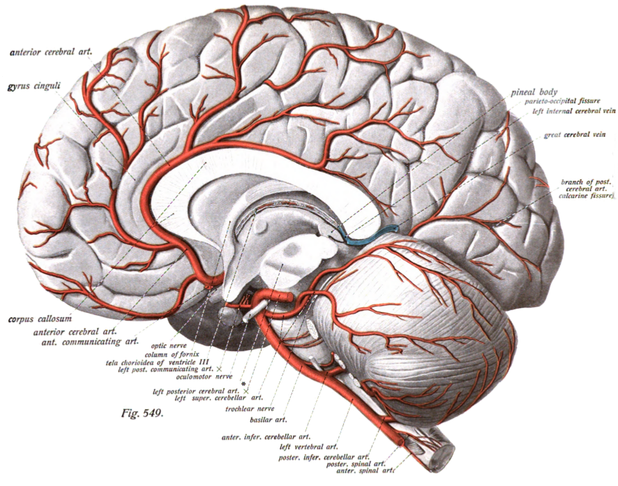
The anterior cerebral artery (ACA) is one of a pair of cerebral arteries that supplies oxygenated blood to most midline portions of the frontal lobes and superior medial parietal lobes of the brain. The two anterior cerebral arteries arise from the internal carotid artery and are part of the circle of Willis. The left and right anterior cerebral arteries are connected by the anterior communicating artery. Anterior cerebral artery syndrome refers to symptoms that follow a stroke occurring in the area normally supplied by one of the arteries. It is characterized by weakness and sensory loss in the lower leg and foot opposite to the lesion and behavioral changes. Structure The anterior cerebral artery is divided into 5 segments. Its smaller branches: the callosal (supracallosal) arteries are considered to be the A4 and A5 segments. *A1 originates from the internal carotid artery and extends to the ''anterior communicating artery'' (AComm). The ''anteromedial central'' (medial lent ... [...More Info...] [...Related Items...] OR: [Wikipedia] [Google] [Baidu] |
Internal Carotid Artery
The internal carotid artery (Latin: arteria carotis interna) is an artery in the neck which supplies the anterior circulation of the brain. In human anatomy, the internal and external carotids arise from the common carotid arteries, where these bifurcate at cervical vertebrae C3 or C4. The internal carotid artery supplies the brain, including the eyes, while the external carotid nourishes other portions of the head, such as the face, scalp, skull, and meninges. Classification Terminologia Anatomica in 1998 subdivided the artery into four parts: "cervical", "petrous", "cavernous", and "cerebral". However, in clinical settings, the classification system of the internal carotid artery usually follows the 1996 recommendations by Bouthillier, describing seven anatomical segments of the internal carotid artery, each with a corresponding alphanumeric identifier—C1 cervical, C2 petrous, C3 lacerum, C4 cavernous, C5 clinoid, C6 ophthalmic, and C7 communicating. The Bouthillier nomenclat ... [...More Info...] [...Related Items...] OR: [Wikipedia] [Google] [Baidu] |
Anastomosis
An anastomosis (, plural anastomoses) is a connection or opening between two things (especially cavities or passages) that are normally diverging or branching, such as between blood vessels, leaf#Veins, leaf veins, or streams. Such a connection may be normal (such as the foramen ovale (heart), foramen ovale in a fetus's heart) or abnormal (such as the atrial septal defect#Patent foramen ovale, patent foramen ovale in an adult's heart); it may be acquired (such as an arteriovenous fistula) or innate (such as the arteriovenous shunt of a metarteriole); and it may be natural (such as the aforementioned examples) or artificial (such as a surgical anastomosis). The reestablishment of an anastomosis that had become blocked is called a reanastomosis. Anastomoses that are abnormal, whether congenital disorder, congenital or acquired, are often called fistulas. The term is used in medicine, biology, mycology, geology, and geography. Etymology Anastomosis: medical or Modern Latin, from Gre ... [...More Info...] [...Related Items...] OR: [Wikipedia] [Google] [Baidu] |
Motor Cortex
The motor cortex is the region of the cerebral cortex believed to be involved in the planning, control, and execution of voluntary movements. The motor cortex is an area of the frontal lobe located in the posterior precentral gyrus immediately anterior to the central sulcus. Components of the motor cortex The motor cortex can be divided into three areas: 1. The primary motor cortex is the main contributor to generating neural impulses that pass down to the spinal cord and control the execution of movement. However, some of the other motor areas in the brain also play a role in this function. It is located on the anterior paracentral lobule on the medial surface. 2. The premotor cortex is responsible for some aspects of motor control, possibly including the preparation for movement, the sensory guidance of movement, the spatial guidance of reaching, or the direct control of some movements with an emphasis on control of proximal and trunk muscles of the body. Located anterior ... [...More Info...] [...Related Items...] OR: [Wikipedia] [Google] [Baidu] |
Paralysis
Paralysis (also known as plegia) is a loss of motor function in one or more muscles. Paralysis can also be accompanied by a loss of feeling (sensory loss) in the affected area if there is sensory damage. In the United States, roughly 1 in 50 people have been diagnosed with some form of permanent or transient paralysis. The word "paralysis" derives from the Greek παράλυσις, meaning "disabling of the nerves" from παρά (''para'') meaning "beside, by" and λύσις (''lysis'') meaning "making loose". A paralysis accompanied by involuntary tremors is usually called "palsy". Causes Paralysis is most often caused by damage in the nervous system, especially the spinal cord. Other major causes are stroke, trauma with nerve injury, poliomyelitis, cerebral palsy, peripheral neuropathy, Parkinson's disease, ALS, botulism, spina bifida, multiple sclerosis, and Guillain–Barré syndrome. Temporary paralysis occurs during REM sleep, and dysregulation of this system can lead ... [...More Info...] [...Related Items...] OR: [Wikipedia] [Google] [Baidu] |
Collateral Circulation
Collateral circulation is the alternate circulation around a blocked artery or vein via another path, such as nearby minor vessels. It may occur via preexisting vascular redundancy (analogous to engineered redundancy), as in the circle of Willis in the brain, or it may occur via new branches formed between adjacent blood vessels (neovascularization), as in the eye after a retinal embolism or in the brain when moyamoya occurs. Its formation may be provoked by pathological conditions such as high vascular resistance or ischaemia. An example of the usefulness of collateral circulation is a systemic thromboembolism in cats. This is when a thrombotic embolus lodges above the external iliac artery (common iliac artery), blocking the external and internal iliac arteries and effectively shutting off all blood supply to the hind leg. Even though the main vessels to the leg are blocked, enough blood can get to the tissues in the leg via the collateral circulation to keep them alive. Br ... [...More Info...] [...Related Items...] OR: [Wikipedia] [Google] [Baidu] |
Stroke
A stroke is a medical condition in which poor blood flow to the brain causes cell death. There are two main types of stroke: ischemic, due to lack of blood flow, and hemorrhagic, due to bleeding. Both cause parts of the brain to stop functioning properly. Signs and symptoms of a stroke may include an inability to move or feel on one side of the body, problems understanding or speaking, dizziness, or loss of vision to one side. Signs and symptoms often appear soon after the stroke has occurred. If symptoms last less than one or two hours, the stroke is a transient ischemic attack (TIA), also called a mini-stroke. A hemorrhagic stroke may also be associated with a severe headache. The symptoms of a stroke can be permanent. Long-term complications may include pneumonia and loss of bladder control. The main risk factor for stroke is high blood pressure. Other risk factors include high blood cholesterol, tobacco smoking, obesity, diabetes mellitus, a previous TIA, end-st ... [...More Info...] [...Related Items...] OR: [Wikipedia] [Google] [Baidu] |
Brain Areas Supplied By Anterior Cerebral Artery
A brain is an organ that serves as the center of the nervous system in all vertebrate and most invertebrate animals. It is located in the head, usually close to the sensory organs for senses such as vision. It is the most complex organ in a vertebrate's body. In a human, the cerebral cortex contains approximately 14–16 billion neurons, and the estimated number of neurons in the cerebellum is 55–70 billion. Each neuron is connected by synapses to several thousand other neurons. These neurons typically communicate with one another by means of long fibers called axons, which carry trains of signal pulses called action potentials to distant parts of the brain or body targeting specific recipient cells. Physiologically, brains exert centralized control over a body's other organs. They act on the rest of the body both by generating patterns of muscle activity and by driving the secretion of chemicals called hormones. This centralized control allows rapid and coordinated responses ... [...More Info...] [...Related Items...] OR: [Wikipedia] [Google] [Baidu] |
Globus Pallidus
The globus pallidus (GP), also known as paleostriatum or dorsal pallidum, is a subcortical structure of the brain. It consists of two adjacent segments, one external, known in rodents simply as the globus pallidus, and one internal, known in rodents as the entopeduncular nucleus. It is part of the telencephalon, but retains close functional ties with the subthalamus in the diencephalon – both of which are part of the extrapyramidal motor system. The globus pallidus is a major component of the basal ganglia, with principal inputs from the striatum, and principal direct outputs to the thalamus and the substantia nigra. The latter is made up of similar neuronal elements, has similar afferents from the striatum, similar projections to the thalamus, and has a similar synaptology. Neither receives direct cortical afferents, and both receive substantial additional inputs from the intralaminar thalamus. Globus pallidus is Latin for "pale globe". Structure Pallidal nuclei are ma ... [...More Info...] [...Related Items...] OR: [Wikipedia] [Google] [Baidu] |
Magnetic Resonance Angiography
Magnetic resonance angiography (MRA) is a group of techniques based on magnetic resonance imaging (MRI) to image blood vessels. Magnetic resonance angiography is used to generate images of arteries (and less commonly veins) in order to evaluate them for stenosis (abnormal narrowing), Vascular occlusion, occlusions, aneurysms (vessel wall dilatations, at risk of rupture) or other abnormalities. MRA is often used to evaluate the arteries of the neck and brain, the thoracic and abdominal aorta, the renal arteries, and the legs (the latter exam is often referred to as a "run-off"). Acquisition A variety of techniques can be used to generate the pictures of blood vessels, both artery, arteries and veins, based on flow effects or on contrast (inherent or pharmacologically generated). The most frequently applied MRA methods involve the use intravenous MRI contrast agent, contrast agents, particularly those containing gadolinium to shorten the Spin–lattice relaxation, ''T''1 of blood ... [...More Info...] [...Related Items...] OR: [Wikipedia] [Google] [Baidu] |
Embryo
An embryo is an initial stage of development of a multicellular organism. In organisms that reproduce sexually, embryonic development is the part of the life cycle that begins just after fertilization of the female egg cell by the male sperm cell. The resulting fusion of these two cells produces a single-celled zygote that undergoes many cell divisions that produce cells known as blastomeres. The blastomeres are arranged as a solid ball that when reaching a certain size, called a morula, takes in fluid to create a cavity called a blastocoel. The structure is then termed a blastula, or a blastocyst in mammals. The mammalian blastocyst hatches before implantating into the endometrial lining of the womb. Once implanted the embryo will continue its development through the next stages of gastrulation, neurulation, and organogenesis. Gastrulation is the formation of the three germ layers that will form all of the different parts of the body. Neurulation forms the nervous ... [...More Info...] [...Related Items...] OR: [Wikipedia] [Google] [Baidu] |
Anterior Choroidal Artery
The anterior choroidal artery originates from the internal carotid artery. However, it may (rarely) arise from the middle cerebral artery. Structure The anterior choroidal artery originates from the distal carotid artery 5 mm after the origin of the posterior communicating artery and just before the carotid terminus. It serves structures in the prosencephalon, diencephalon, and mesencephalon: * choroid plexus of the lateral ventricle and third ventricle * optic chiasm and optic tract * internal capsule * lateral geniculate body * globus pallidus * tail of the caudate nucleus * hippocampus * amygdala * substantia nigra * red nucleus * crus cerebri Clinical significance The full extent of the damage caused by occlusion of the anterior choroidal artery is not known. However, studies show that the interruption of blood flow from this vessel can result in hemiplegia on the contralateral (opposite) side of the body, contralateral hemi-hypoesthesia, and homonymous hemianopsia. These sym ... [...More Info...] [...Related Items...] OR: [Wikipedia] [Google] [Baidu] |
Middle Cerebral Artery
The middle cerebral artery (MCA) is one of the three major paired cerebral artery, cerebral arteries that supply blood to the cerebrum. The MCA arises from the internal carotid artery and continues into the lateral sulcus where it then branches and projects to many parts of the lateral cerebral cortex. It also supplies blood to the anterior temporal lobes and the insular cortex, insular cortices. The left and right MCAs rise from trifurcations of the internal carotid artery, internal carotid arteries and thus are connected to the anterior cerebral artery, anterior cerebral arteries and the posterior communicating artery, posterior communicating arteries, which connect to the posterior cerebral artery, posterior cerebral arteries. The MCAs are not considered a part of the Circle of Willis. Structure The middle cerebral artery divides into four segments, named by the region they supply as opposed to order of branching as the latter can be somewhat variable: *M1: The ''sphenoidal' ... [...More Info...] [...Related Items...] OR: [Wikipedia] [Google] [Baidu] |


_Phase_Contrast_(PC)_sequence_MRI_of_arterial_dissections.jpg)

