|
Yim Ebbin Syndrome
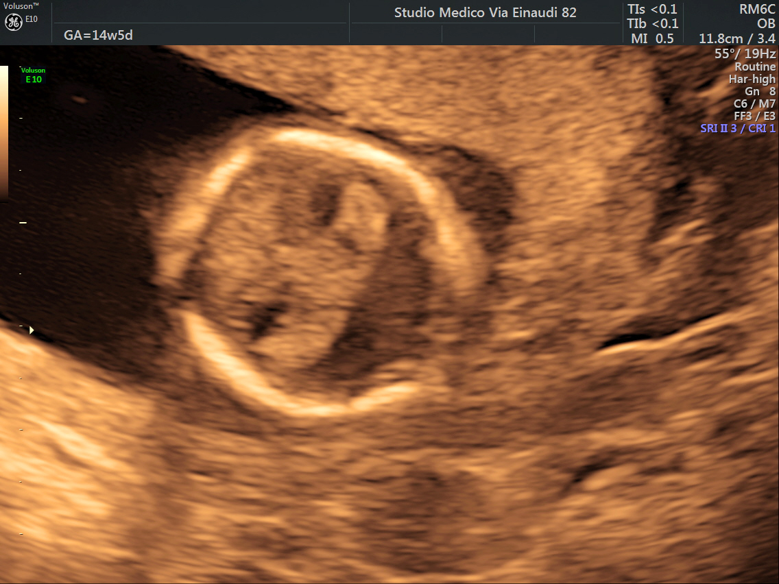
Brachial amelia, cleft lip, and holoprosencephaly, or Yim–Ebbin syndrome, is a very rare multi-systemic genetic disorder which is characterized by brachial amelia (mainly that affecting the upper limbs) cleft lip, and forebrain defects such as holoprosencephaly. Approximately five cases of this disorder have been described in medical literature. 'Other signs include hydrocephalus and an iris coloboma. It was first described by Yim and Ebbin in 1982, and later by Thomas and Donnai in 1994. In 1996, a third case was reported by Froster et al. who suggested that the three cases were related and represented a distinct syndrome. In 2000, a similar case was reported by Pierri et al. References External linksYim–Ebbin syndromeat Online Mendelian Inheritance in Man {{Medical resources , DiseasesDB = , ICD10 = none , ICD9 = none , ICDO = , OMIM = 601357 , MedlinePlus = , eMedicineSubj = , eMedicineTo ... [...More Info...] [...Related Items...] OR: [Wikipedia] [Google] [Baidu] |
Medical Genetics
Medical genetics is the branch tics in that human genetics is a field of scientific research that may or may not apply to medicine, while medical genetics refers to the application of genetics to medical care. For example, research on the causes and inheritance of genetic disorders would be considered within both human genetics and medical genetics, while the diagnosis, management, and counselling people with genetic disorders would be considered part of medical genetics. In contrast, the study of typically non-medical phenotypes such as the genetics of eye color would be considered part of human genetics, but not necessarily relevant to medical genetics (except in situations such as albinism). ''Genetic medicine'' is a newer term for medical genetics and incorporates areas such as gene therapy, personalized medicine, and the rapidly emerging new medical specialty, predictive medicine. Scope Medical genetics encompasses many different areas, including clinical practice of ... [...More Info...] [...Related Items...] OR: [Wikipedia] [Google] [Baidu] |
Genetic Disorder
A genetic disorder is a health problem caused by one or more abnormalities in the genome. It can be caused by a mutation in a single gene (monogenic) or multiple genes (polygenic) or by a chromosomal abnormality. Although polygenic disorders are the most common, the term is mostly used when discussing disorders with a single genetic cause, either in a gene or chromosome. The mutation responsible can occur spontaneously before embryonic development (a ''de novo'' mutation), or it can be Heredity, inherited from two parents who are carriers of a faulty gene (autosomal recessive inheritance) or from a parent with the disorder (autosomal dominant inheritance). When the genetic disorder is inherited from one or both parents, it is also classified as a hereditary disease. Some disorders are caused by a mutation on the X chromosome and have X-linked inheritance. Very few disorders are inherited on the Y linkage, Y chromosome or Mitochondrial disease#Causes, mitochondrial DNA (due to t ... [...More Info...] [...Related Items...] OR: [Wikipedia] [Google] [Baidu] |
Amelia (birth Defect)
Amelia is the birth defect of lacking one or more limbs. It can also result in a shrunken or deformed limb. The term may be modified to indicate the number of legs or arms missing at birth, such as tetra-amelia for the absence of all four limbs. A related term is meromelia, which is the partial absence of a limb or limbs. The term is from Greek ἀ- "lack of" plus μέλος (plural: μέλεα or μέλη) "limb" Symptoms The diagnosis of tetra-amelia syndrome is established clinically and can be made on routine prenatal ultrasonography. WNT3 is the only gene known to be associated with tetra-amelia syndrome. Molecular genetic testing on a clinical basis can be used to diagnose the incidence of the syndrome. The mutation detection frequency is unknown as only a limited number of families have been studied. Affected infants are often stillborn or die shortly after birth. Description Amelia may be present as an isolated defect, but it is often associated with major malformations ... [...More Info...] [...Related Items...] OR: [Wikipedia] [Google] [Baidu] |
Cleft Lip
A cleft lip contains an opening in the upper lip that may extend into the nose. The opening may be on one side, both sides, or in the middle. A cleft palate occurs when the palate (the roof of the mouth) contains an opening into the nose. The term orofacial cleft refers to either condition or to both occurring together. These disorders can result in feeding problems, speech problems, hearing problems, and frequent ear infections. Less than half the time the condition is associated with other disorders. Cleft lip and palate are the result of tissues of the face not joining properly during development. As such, they are a type of birth defect. The cause is unknown in most cases. Risk factors include smoking during pregnancy, diabetes, obesity, an older mother, and certain medications (such as some used to treat seizures). Cleft lip and cleft palate can often be diagnosed during pregnancy with an ultrasound exam. A cleft lip or palate can be successfully treated with surgery. ... [...More Info...] [...Related Items...] OR: [Wikipedia] [Google] [Baidu] |
Holoprosencephaly
Holoprosencephaly (HPE) is a cephalic disorder in which the prosencephalon (the forebrain of the embryo) fails to develop into two hemispheres, typically occurring between the 18th and 28th day of gestation. Normally, the forebrain is formed and the face begins to develop in the fifth and sixth weeks of human pregnancy. The condition also occurs in other species. Holoprosencephaly is estimated to occur in approximately 1 in every 250 conceptions and most cases are not compatible with life and result in fetal death in utero due to deformities to the skull and brain. However, holoprosencephaly is still estimated to occur in approximately 1 in every 8,000 live births. When the embryo's forebrain does not divide to form bilateral cerebral hemispheres (the left and right halves of the brain), it causes defects in the development of the face and in brain structure and function. The severity of holoprosencephaly is highly variable. In less severe cases, babies are born with normal or ... [...More Info...] [...Related Items...] OR: [Wikipedia] [Google] [Baidu] |
Hydrocephalus
Hydrocephalus is a condition in which an accumulation of cerebrospinal fluid (CSF) occurs within the brain. This typically causes increased intracranial pressure, pressure inside the skull. Older people may have headaches, double vision, poor balance, urinary incontinence, personality changes, or mental impairment. In babies, it may be seen as a rapid increase in head size. Other symptoms may include vomiting, sleepiness, seizures, and Parinaud's syndrome, downward pointing of the eyes. Hydrocephalus can occur due to birth defects or be acquired later in life. Associated birth defects include neural tube defects and those that result in aqueductal stenosis. Other causes include meningitis, brain tumors, traumatic brain injury, intraventricular hemorrhage, and subarachnoid hemorrhage. The four types of hydrocephalus are communicating, noncommunicating, ''ex vacuo'', and normal pressure hydrocephalus, normal pressure. Diagnosis is typically made by physical examination and medic ... [...More Info...] [...Related Items...] OR: [Wikipedia] [Google] [Baidu] |
Iris (anatomy)
In humans and most mammals and birds, the iris (plural: ''irides'' or ''irises'') is a thin, annular structure in the eye, responsible for controlling the diameter and size of the pupil, and thus the amount of light reaching the retina. Eye color is defined by the iris. In optical terms, the pupil is the eye's aperture, while the iris is the diaphragm. Structure The iris consists of two layers: the front pigmented fibrovascular layer known as a stroma and, beneath the stroma, pigmented epithelial cells. The stroma is connected to a sphincter muscle (sphincter pupillae), which contracts the pupil in a circular motion, and a set of dilator muscles ( dilator pupillae), which pull the iris radially to enlarge the pupil, pulling it in folds. The sphincter pupillae is the opposing muscle of the dilator pupillae. The pupil's diameter, and thus the inner border of the iris, changes size when constricting or dilating. The outer border of the iris does not change size. The constricti ... [...More Info...] [...Related Items...] OR: [Wikipedia] [Google] [Baidu] |
Coloboma
A coloboma (from the Greek , meaning defect) is a hole in one of the structures of the eye, such as the iris, retina, choroid, or optic disc. The hole is present from birth and can be caused when a gap called the choroid fissure, which is present during early stages of prenatal development, fails to close up completely before a child is born. Ocular coloboma is relatively uncommon, affecting less than one in every 10,000 births. The classical description in medical literature is of a keyhole-shaped defect. A coloboma can occur in one eye (unilateral) or both eyes (bilateral). Most cases of coloboma affect only the iris. The level of vision impairment of those with a coloboma can range from having no vision problems to being able to see only light or dark, depending on the position and extent of the coloboma (or colobomata if more than one is present). Signs and symptoms Visual effects may be mild to more severe depending on the size and location of the coloboma. If, for exam ... [...More Info...] [...Related Items...] OR: [Wikipedia] [Google] [Baidu] |
Online Mendelian Inheritance In Man
Online Mendelian Inheritance in Man (OMIM) is a continuously updated catalog of human genes and genetic disorders and traits, with a particular focus on the gene-phenotype relationship. , approximately 9,000 of the over 25,000 entries in OMIM represented phenotypes; the rest represented genes, many of which were related to known phenotypes. Versions and history OMIM is the online continuation of Dr. Victor A. McKusick's ''Mendelian Inheritance in Man'' (MIM), which was published in 12 editions between 1966 and 1998.McKusick, V. A. ''Mendelian Inheritance in Man. Catalogs of Autosomal Dominant, Autosomal Recessive and X-Linked Phenotypes.'' Baltimore, MD: Johns Hopkins University Press, 1st ed, 1996; 2nd ed, 1969; 3rd ed, 1971; 4th ed, 1975; 5th ed, 1978; 6th ed, 1983; 7th ed, 1986; 8th ed, 1988; 9th ed, 1990; 10th ed, 1992. Nearly all of the 1,486 entries in the first edition of MIM discussed phenotypes. MIM/OMIM is produced and curated at the Johns Hopkins School of Medicine ... [...More Info...] [...Related Items...] OR: [Wikipedia] [Google] [Baidu] |


