|
Yasui Procedure
The Yasui procedure is a pediatric heart operation used to bypass the Ventricular outflow tract, left ventricular outflow tract (LVOT) that combines the aortic repair of the Norwood procedure and a shunt similar to that used in the Rastelli procedure in a single operation. It is used to repair defects that result in the physiology of hypoplastic left heart syndrome even though both ventricles are functioning normally. These defects are common in DiGeorge syndrome and include interrupted aortic arch and Ventricular outflow tract obstruction, LVOT obstruction (IAA/LVOTO); aortic atresia-Aortic stenosis, severe stenosis with ventricular septal defect (AA/VSD); and aortic atresia with interrupted aortic arch and aortopulmonary window. This procedure allows the surgeon to keep the Ventricle (heart), left ventricle connected to the Circulatory system, systemic circulation while using the pulmonary valve as its outflow valve, by connecting them through the ventricular septal defect. The Yas ... [...More Info...] [...Related Items...] OR: [Wikipedia] [Google] [Baidu] |
Ventricular Outflow Tract
A ventricular outflow tract is a portion of either the left ventricle or right ventricle of the heart through which blood passes in order to enter the great arteries. The right ventricular outflow tract (RVOT) is an infundibular extension of the ventricular cavity that connects to the pulmonary artery. The left ventricular outflow tract (LVOT), which connects to the aorta, is nearly indistinguishable from the rest of the ventricle. The outflow tract is derived from the secondary heart field, during cardiogenesis. Both the left and right outflow tract have their own term. The right outflow tract is called "conus arteriosus" from the outside, and infundibulum from the inside. In the left ventricle the outflow tract is the "aortic vestibule". They both possess smooth walls, and are derived from the embryonic bulbus cordis In both left and right ventricle there are specific structures separating the inflow and outflow of blood. In the right ventricle, the inflow and outflow is separ ... [...More Info...] [...Related Items...] OR: [Wikipedia] [Google] [Baidu] |
Ascending Aorta
The ascending aorta (AAo) is a portion of the aorta commencing at the upper part of the base of the left ventricle, on a level with the lower border of the third costal cartilage behind the left half of the sternum. Structure It passes obliquely upward, forward, and to the right, in the direction of the heart's axis, as high as the upper border of the second right costal cartilage, describing a slight curve in its course, and being situated, about behind the posterior surface of the sternum. The total length is about . Components The aortic root is the portion of the aorta beginning at the aortic annulus and extending to the sinotubular junction. It is sometimes regarded as a part of the ascending aorta, and sometimes regarded as a separate entity from the rest of the ascending aorta. Between each commissure of the aortic valve and opposite the cusps of the aortic valve, three small dilatations called the aortic sinuses. The sinotubular junction is the point in the ascendi ... [...More Info...] [...Related Items...] OR: [Wikipedia] [Google] [Baidu] |
Pericardium
The pericardium, also called pericardial sac, is a double-walled sac containing the heart and the roots of the great vessels. It has two layers, an outer layer made of strong connective tissue (fibrous pericardium), and an inner layer made of serous membrane (serous pericardium). It encloses the pericardial cavity, which contains pericardial fluid, and defines the middle mediastinum. It separates the heart from interference of other structures, protects it against infection and blunt trauma, and lubricates the heart's movements. The English name originates from the Ancient Greek prefix "''peri-''" (περί; "around") and the suffix "''-cardion''" (κάρδιον; "heart"). Anatomy The pericardium is a tough fibroelastic sac which covers the heart from all sides except at the cardiac root (where the great vessels join the heart) and the bottom (where only the serous pericardium exists to cover the upper surface of the central tendon of diaphragm). The fibrous pericardiu ... [...More Info...] [...Related Items...] OR: [Wikipedia] [Google] [Baidu] |
Right Ventricular Hypertrophy
Right ventricular hypertrophy (RVH) is a condition defined by an abnormal enlargement of the cardiac muscle surrounding the right ventricle. The right ventricle is one of the four chambers of the heart. It is located towards the lower-end of the heart and it receives blood from the right atrium and pumps blood into the lungs. Since RVH is an enlargement of muscle it arises when the muscle is required to work harder. Therefore, the main causes of RVH are pathologies of systems related to the right ventricle such as the pulmonary artery, the tricuspid valve or the airways. RVH can be benign and have little impact on day-to-day life or it can lead to conditions such as heart failure, which has a poor prognosis. Signs and symptoms Symptoms Although presentations vary, individuals with right ventricular hypertrophy can experience symptoms that are associated with pulmonary hypertension, heart failure and/or a reduced cardiac output. These include: * Difficulty breathing on exertio ... [...More Info...] [...Related Items...] OR: [Wikipedia] [Google] [Baidu] |
Aorta
The aorta ( ) is the main and largest artery in the human body, originating from the left ventricle of the heart and extending down to the abdomen, where it splits into two smaller arteries (the common iliac arteries). The aorta distributes oxygenated blood to all parts of the body through the systemic circulation. Structure Sections In anatomical sources, the aorta is usually divided into sections. One way of classifying a part of the aorta is by anatomical compartment, where the thoracic aorta (or thoracic portion of the aorta) runs from the heart to the diaphragm. The aorta then continues downward as the abdominal aorta (or abdominal portion of the aorta) from the diaphragm to the aortic bifurcation. Another system divides the aorta with respect to its course and the direction of blood flow. In this system, the aorta starts as the ascending aorta, travels superiorly from the heart, and then makes a hairpin turn known as the aortic arch. Following the aortic arch ... [...More Info...] [...Related Items...] OR: [Wikipedia] [Google] [Baidu] |
Pulmonary Artery
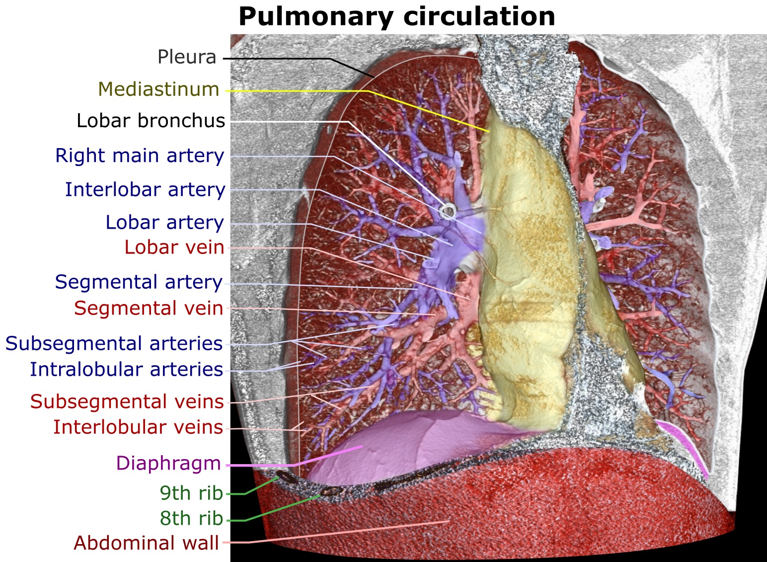
A pulmonary artery is an artery in the pulmonary circulation that carries deoxygenated blood from the right side of the heart to the lungs. The largest pulmonary artery is the ''main pulmonary artery'' or ''pulmonary trunk'' from the heart, and the smallest ones are the arterioles, which lead to the capillaries that surround the pulmonary alveoli. Structure The pulmonary arteries are blood vessels that carry systemic venous blood from the right ventricle of the heart to the microcirculation of the lungs. Unlike in other organs where arteries supply oxygenated blood, the blood carried by the pulmonary arteries is deoxygenated, as it is venous blood returning to the heart. The main pulmonary arteries emerge from the right side of the heart, and then split into smaller arteries that progressively divide and become arterioles, eventually narrowing into the capillary microcirculation of the lungs where gas exchange occurs. Pulmonary trunk In order of blood flow, the pulmonary art ... [...More Info...] [...Related Items...] OR: [Wikipedia] [Google] [Baidu] |
Patent Ductus Arteriosus
''Patent ductus arteriosus'' (PDA) is a medical condition in which the ''ductus arteriosus'' fails to close after birth: this allows a portion of oxygenated blood from the left heart to flow back to the lungs by flowing from the aorta, which has a higher pressure, to the pulmonary artery. Symptoms are uncommon at birth and shortly thereafter, but later in the first year of life there is often the onset of an increased work of breathing and failure to gain weight at a normal rate. With time, an uncorrected PDA usually leads to pulmonary hypertension followed by right-sided heart failure. The ''ductus arteriosus'' is a fetal blood vessel that normally closes soon after birth. In a PDA, the vessel does not close, but remains ''patent'' (open), resulting in an abnormal transmission of blood from the aorta to the pulmonary artery. PDA is common in newborns with persistent respiratory problems such as hypoxia, and has a high occurrence in premature newborns. Premature newborns are ... [...More Info...] [...Related Items...] OR: [Wikipedia] [Google] [Baidu] |
Cardiopulmonary Bypass
Cardiopulmonary bypass (CPB) is a technique in which a machine temporarily takes over the function of the heart and lungs during surgery, maintaining the circulation of blood and oxygen to the body. The CPB pump itself is often referred to as a heart–lung machine or "the pump". Cardiopulmonary bypass pumps are operated by perfusionists. CPB is a form of extracorporeal circulation. Extracorporeal membrane oxygenation is generally used for longer-term treatment. CPB mechanically circulates and oxygenates blood for the body while bypassing the heart and lungs. It uses a heart–lung machine to maintain perfusion to other body organs and tissues while the surgeon works in a bloodless surgical field. The surgeon places a cannula in the right atrium, vena cava, or femoral vein to withdraw blood from the body. Venous blood is removed from the body by the cannula and then filtered, cooled or warmed, and oxygenated before it is returned to the body by a mechanical pump. The cannula used ... [...More Info...] [...Related Items...] OR: [Wikipedia] [Google] [Baidu] |
Median Sternotomy
Median sternotomy is a type of surgical procedure in which a vertical inline incision is made along the sternum, after which the sternum itself is divided using a sternal saw. This procedure provides access to the heart and lungs for surgical procedures such as heart transplant, lung transplant, corrective surgery for congenital heart defects, or coronary artery bypass surgery Coronary artery bypass surgery, also known as coronary artery bypass graft (CABG, pronounced "cabbage") is a surgical procedure to treat coronary artery disease (CAD), the buildup of plaques in the arteries of the heart. It can relieve chest pai .... References {{Cardiac surgery Thoracic surgical procedures Surgical incisions ... [...More Info...] [...Related Items...] OR: [Wikipedia] [Google] [Baidu] |
Coarctation Of The Aorta
Coarctation of the aorta (CoA or CoAo), also called aortic narrowing, is a congenital condition whereby the aorta is narrow, usually in the area where the ductus arteriosus (ligamentum arteriosum after regression) inserts. The word ''coarctation'' means "pressing or drawing together; narrowing". Coarctations are most common in the aortic arch. The arch may be small in babies with coarctations. Other heart defects may also occur when coarctation is present, typically occurring on the left side of the heart. When a patient has a coarctation, the left ventricle has to work harder. Since the aorta is narrowed, the left ventricle must generate a much higher pressure than normal in order to force enough blood through the aorta to deliver blood to the lower part of the body. If the narrowing is severe enough, the left ventricle may not be strong enough to push blood through the coarctation, thus resulting in a lack of blood to the lower half of the body. Physiologically its complete form ... [...More Info...] [...Related Items...] OR: [Wikipedia] [Google] [Baidu] |
Konno Procedure
Konno (written: 金野, 今野 or 紺野) is a Japanese surname. Notable people with the surname include: *, Japanese footballer *, Japanese ski jumper *, Japanese idol and singer *, Japanese politician *Ford Konno (born 1933), American swimmer *, Japanese video game designer *, Japanese voice actress *, Japanese basketball player *, Japanese judoka *, Japanese voice actor *, Japanese actress * Mari Konno (born 1980), Japanese basketball player *, Japanese actress and essayist *, Japanese rugby union player *, Japanese table tennis player *, Japanese footballer *, Japanese women's footballer Fictional characters *, the main character of the anime film ''The Girl Who Leapt Through Time'' *, one of the main characters of the anime series ''Zombie Land Saga'' See also *Konno Station is a railway station in the city of Gujō, Gifu Prefecture, Japan, operated by the third sector railway operator Nagaragawa Railway. Lines Konno Station is a station of the Etsumi-Nan Line, and is ... [...More Info...] [...Related Items...] OR: [Wikipedia] [Google] [Baidu] |
Ross Procedure
The Ross procedure, also known as pulmonary autograft, is a heart valve replacement operation to treat severe aortic valve disease, such as in children and young adults with a bicuspid aortic valve. It involves removing the diseased aortic valve, situated at the exit of the left side of the heart (where the aorta begins), and replacing it with the person's own healthy pulmonary valve (autograft), removed from the exit of the heart's right side (where the pulmonary artery begins). To reconstruct the right sided exit, a pulmonary valve from a cadaver (homograft), or a stentless xenograft, is used to replace the removed pulmonary valve. Compared to a mechanical valve replacement, it avoids the requirement for thinning the blood, has favourable blood flow dynamics, allows growth of the valve with growth of the child and has less risk of endocarditis. It is not performed in Marfan syndrome, if pulmonary valve disease, or if immune problems like lupus. Other contraindications in ... [...More Info...] [...Related Items...] OR: [Wikipedia] [Google] [Baidu] |


