|
X-chromosome Reactivation
X chromosome reactivation (XCR) is the process by which the inactive X chromosome (the Xi) is re-activated in the cells of eutherian female mammals. Therian female mammalian cells have two X chromosomes, while males have only one, requiring X-chromosome inactivation (XCI) for sex-chromosome dosage compensation. In eutherians, XCI is the random inactivation of one of the X chromosomes, silencing its expression. Much of the scientific knowledge currently known about XCR comes from research limited to mouse models or stem cells. Partial XCR may derepress one or more genes on the Xi, and the level of restored gene expression may not be as high as it would normally be on the active X chromosome (the Xa). Complete XCR restores the Xi to Xa and erases the epigenetic memory of XCI, meaning that inducing X-inactivation again will randomly select an X chromosome to silence, rather than deterministically silencing the original Xi. XCR is an emerging topic of interest for multiple reasons: ... [...More Info...] [...Related Items...] OR: [Wikipedia] [Google] [Baidu] |
Eutherian
Eutheria (; from Greek , 'good, right' and , 'beast'; ) is the clade consisting of all therian mammals that are more closely related to placentals than to marsupials. Eutherians are distinguished from noneutherians by various phenotypic traits of the feet, ankles, jaws and teeth. All extant eutherians lack epipubic bones, which are present in all other living mammals (marsupials and monotremes). This allows for expansion of the abdomen during pregnancy. The oldest-known eutherian species is '' Juramaia sinensis'', dated at from the early Late Jurassic ( Oxfordian) of China. Eutheria was named in 1872 by Theodore Gill; in 1880 Thomas Henry Huxley defined it to encompass a more broadly defined group than Placentalia. Characteristics Distinguishing features are: *an enlarged malleolus ("little hammer") at the bottom of the tibia, the larger of the two shin bones *the joint between the first metatarsal bone and the entocuneiform bone (the innermost of the three cuneiform ... [...More Info...] [...Related Items...] OR: [Wikipedia] [Google] [Baidu] |
Heterochromatin
Heterochromatin is a tightly packed form of DNA or '' condensed DNA'', which comes in multiple varieties. These varieties lie on a continue between the two extremes of constitutive heterochromatin and facultative heterochromatin. Both play a role in the expression of genes. Because it is tightly packed, it was thought to be inaccessible to polymerases and therefore not transcribed; however, according to Volpe et al. (2002), and many other papers since, much of this DNA is in fact transcribed, but it is continuously turned over via RNA-induced transcriptional silencing (RITS). Recent studies with electron microscopy and OsO4 staining reveal that the dense packing is not due to the chromatin. Constitutive heterochromatin can affect the genes near itself (e.g. position-effect variegation). It is usually repetitive and forms structural functions such as centromeres or telomeres, in addition to acting as an attractor for other gene-expression or repression signals. Facultative hete ... [...More Info...] [...Related Items...] OR: [Wikipedia] [Google] [Baidu] |
Autosome
An autosome is any chromosome that is not a sex chromosome. The members of an autosome pair in a diploid cell have the same morphology, unlike those in allosome, allosomal (sex chromosome) pairs, which may have different structures. The DNA in autosomes is collectively known as atDNA or auDNA. For example, humans have a diploid human genome, genome that usually contains 22 pairs of autosomes and one allosome pair (46 chromosomes total). The autosome pairs are labeled with numbers (1–22 in humans) roughly in order of their sizes in base pairs, while allosomes are labelled with their letters. By contrast, the allosome pair consists of two X chromosomes in females or one X and one Y chromosome in males. Unusual combinations of XYY syndrome, XYY, Klinefelter syndrome, XXY, Triple X syndrome, XXX, XXXX syndrome, XXXX, XXXXX syndrome, XXXXX or XXYY syndrome, XXYY, among Aneuploidy, other Salome combinations, are known to occur and usually cause developmental abnormalities. Autosomes ... [...More Info...] [...Related Items...] OR: [Wikipedia] [Google] [Baidu] |
Oncogenesis
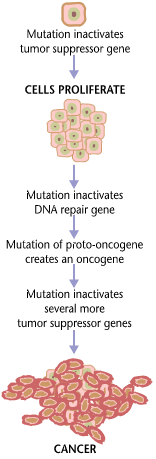
Carcinogenesis, also called oncogenesis or tumorigenesis, is the formation of a cancer, whereby normal cells are transformed into cancer cells. The process is characterized by changes at the cellular, genetic, and epigenetic levels and abnormal cell division. Cell division is a physiological process that occurs in almost all tissues and under a variety of circumstances. Normally, the balance between proliferation and programmed cell death, in the form of apoptosis, is maintained to ensure the integrity of tissues and organs. According to the prevailing accepted theory of carcinogenesis, the somatic mutation theory, mutations in DNA and epimutations that lead to cancer disrupt these orderly processes by interfering with the programming regulating the processes, upsetting the normal balance between proliferation and cell death. This results in uncontrolled cell division and the evolution of those cells by natural selection in the body. Only certain mutations lead to cancer w ... [...More Info...] [...Related Items...] OR: [Wikipedia] [Google] [Baidu] |
Dominance (genetics)
In genetics, dominance is the phenomenon of one variant (allele) of a gene on a chromosome masking or overriding the effect of a different variant of the same gene on the other copy of the chromosome. The first variant is termed dominant and the second recessive. This state of having two different variants of the same gene on each chromosome is originally caused by a mutation in one of the genes, either new (''de novo'') or inherited. The terms autosomal dominant or autosomal recessive are used to describe gene variants on non-sex chromosomes ( autosomes) and their associated traits, while those on sex chromosomes (allosomes) are termed X-linked dominant, X-linked recessive or Y-linked; these have an inheritance and presentation pattern that depends on the sex of both the parent and the child (see Sex linkage). Since there is only one copy of the Y chromosome, Y-linked traits cannot be dominant or recessive. Additionally, there are other forms of dominance such as incomplete d ... [...More Info...] [...Related Items...] OR: [Wikipedia] [Google] [Baidu] |
Oogonium
An oogonium (plural oogonia) is a small diploid cell which, upon maturation, forms a primordial follicle in a female fetus or the female (haploid or diploid) gametangium of certain thallophytes. In the mammalian fetus Oogonia are formed in large numbers by mitosis early in fetal development from primordial germ cells. In humans they start to develop between weeks 4 and 8 and are present in the fetus between weeks 5 and 30. Structure Normal oogonia in human ovaries are spherical or ovoid in shape and are found amongst neighboring somatic cells and oocytes at different phases of development. Oogonia can be distinguished from neighboring somatic cells, under an electron microscope, by observing their nuclei. Oogonial nuclei contain randomly dispersed fibrillar and granular material whereas the somatic cells have a more condensed nucleus that creates a darker outline under the microscope. Oogonial nuclei also contain dense prominent nucleoli. The chromosomal material in the nucleu ... [...More Info...] [...Related Items...] OR: [Wikipedia] [Google] [Baidu] |
Zygote
A zygote (, ) is a eukaryotic cell formed by a fertilization event between two gametes. The zygote's genome is a combination of the DNA in each gamete, and contains all of the genetic information of a new individual organism. In multicellular organisms, the zygote is the earliest developmental stage. In humans and most other anisogamous organisms, a zygote is formed when an egg cell and sperm cell come together to create a new unique organism. In single-celled organisms, the zygote can divide asexually by mitosis to produce identical offspring. German zoologists Oscar and Richard Hertwig made some of the first discoveries on animal zygote formation in the late 19th century. Humans In human fertilization, a released ovum (a haploid secondary oocyte with replicate chromosome copies) and a haploid sperm cell (male gamete) combine to form a single diploid cell called the zygote. Once the single sperm fuses with the oocyte, the latter completes the division of the second ... [...More Info...] [...Related Items...] OR: [Wikipedia] [Google] [Baidu] |
Extraembryonic Membrane
The extraembryonic membranes are four membranes which assist in the development of an animal's embryo. Such membranes occur in a range of animals from humans to insects. They originate from the embryo, but are not considered part of it. They typically perform roles in nutrition, gas exchange, and waste removal. There are four standard extraembryonic membranes in birds, reptiles, and mammals: the yolk sac which surrounds the yolk, the amnion which surrounds and cushions the embryo, the allantois which among avians stores embryonic waste and assists with the exchange of carbon dioxide with oxygen as well as the resorption of calcium from the shell, and the chorion which surrounds all of these and in avians successively merges with the allantois in the later stages of egg development to form a combined respiratory and excretory organ called the chorioallantois. The extraembryonic membranes in insects include a serous membrane originating from blastoderm cells, an amnion or amniot ... [...More Info...] [...Related Items...] OR: [Wikipedia] [Google] [Baidu] |
Oogenesis
Oogenesis, ovogenesis, or oögenesis is the differentiation of the ovum (egg cell) into a cell competent to further develop when fertilized. It is developed from the primary oocyte by maturation. Oogenesis is initiated in the embryonic stage. Oogenesis in non-human mammals In mammals, the first part of oogenesis starts in the germinal epithelium, which gives rise to the development of ovarian follicles, the functional unit of the ovary. Oogenesis consists of several sub-processes: oocytogenesis, ootidogenesis, and finally maturation to form an ovum (oogenesis proper). Folliculogenesis is a separate sub-process that accompanies and supports all three oogenetic sub-processes. Oogonium —(Oocytogenesis)—> Primary Oocyte —(Meiosis I)—> First Polar body (Discarded afterward) + Secondary oocyte —(Meiosis II)—> Second Polar Body (Discarded afterward) + Ovum Oocyte meiosis, important to all animal life cycles yet unlike all other instances of animal cell ... [...More Info...] [...Related Items...] OR: [Wikipedia] [Google] [Baidu] |
Primordial Germ Cell Migration
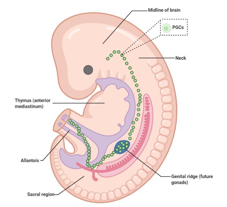
Primordial germ cell (PGC) migration is the process of distribution of primordial germ cells throughout the embryo during embryogenesis. Process Primordial germ cells are among the first lineages that are established in development and they are the precursors for gametes. It is thought that the process of primordial germ cell migration itself has been conserved rather than the specific mechanisms within it, as chemoattraction and repulsion seem to have been borrowed from blood cells, neurones, and the mesoderm. For most organisms, PGC migration starts in the posterior (back end) of the embryo. This process is in most cases distinct from PGC proliferation, with the exception of mammals in which both processes occur at the same time. In most mammals, specification occurs first, followed by migration, and then the proliferation process begins in the gonads. PGCs interact with a wide range of cell types as they move from the epiblast to the gonads. The PGCs move passively (with ... [...More Info...] [...Related Items...] OR: [Wikipedia] [Google] [Baidu] |
Epiblast
In amniote embryonic development, the epiblast (also known as the primitive ectoderm) is one of two distinct cell layers arising from the inner cell mass in the mammalian blastocyst, or from the blastula in reptiles and birds, the other layer is the hypoblast. It derives the embryo proper through its differentiation into the three primary germ layers, ectoderm, mesoderm and endoderm, during gastrulation. The amnionic ectoderm and extraembryonic mesoderm also originate from the epiblast. The other layer of the inner cell mass, the hypoblast, gives rise to the yolk sac, which in turn gives rise to the chorion. Discovery of the epiblast The epiblast was first discovered by Christian Heinrich Pander (1794-1865), a Baltic German biologist and embryologist. With the help of anatomist Ignaz Döllinger (1770–1841) and draftsman Eduard Joseph d'Alton (1772-1840), Pander observed thousands of chicken eggs under a microscope, and ultimately discovered and described the chicken blastoderm ... [...More Info...] [...Related Items...] OR: [Wikipedia] [Google] [Baidu] |


.png)


