|
Williams Flexion Exercises
Williams flexion exercises (WFE) – also called Williams lumbar flexion exercises – are a set or system of related physical exercises intended to enhance lumbar flexion, avoid lumbar extension, and strengthen the abdominal and gluteal musculature in an effort to manage low back pain non-surgically. The system was first devised in 1937 by Dallas orthopedic surgeon Dr. Paul C. Williams. WFEs have been a cornerstone in the management of lower back pain for many years for treating a wide variety of back problems, regardless of diagnosis or chief complaint. In many cases they are used when the disorder’s cause or characteristics were not fully understood by the physician, athletic trainer or physical therapist. Also, physical therapists and athletic trainers often teach these exercises with their own modifications. History The WFEs were developed out of the Regen exercise (also called “squat exercise”), advocated in the 1930s by Eugene M. Regen, a Tennessee orthopedic surg ... [...More Info...] [...Related Items...] OR: [Wikipedia] [Google] [Baidu] |
Lumbar Flexion
In tetrapod anatomy, lumbar is an adjective that means ''of or pertaining to the abdominal segment of the torso, between the diaphragm (anatomy), diaphragm and the sacrum.'' The lumbar region is sometimes referred to as the lower vertebral column, spine, or as an area of the back in its proximity. In human anatomy the five lumbar vertebrae (vertebrae in the lumbar region of the back) are the largest and strongest in the movable part of the spinal column, and can be distinguished by the absence of a foramen transversarium, foramen in the transverse process, and by the absence of facets on the sides of the body. In most mammals, the lumbar region of the spine curves outward. The actual spinal cord terminates between vertebrae one and two of this series, called L1 and L2. The central nervous system, nervous tissue that extends below this point are individual strands that collectively form the cauda equina. In between each lumbar vertebra a nerve root exits, and these nerve roots co ... [...More Info...] [...Related Items...] OR: [Wikipedia] [Google] [Baidu] |
JOSPT
The ''Journal of Orthopaedic & Sports Physical Therapy'' is a peer-reviewed medical journal covering research about musculoskeletal rehabilitation, orthopaedics, physical therapy, and sports medicine. It was established in 1979, following the founding of the Orthopedic and Sports Medicine sections of the American Physical Therapy Association and resulted from a merger of the ''Bulletin of the Orthopaedic Section'' and the ''Bulletin of the Sports Medicine Section''. Initially published quarterly, the journal is now monthly. It is abstracted and indexed by PubMed/MEDLINE MEDLINE (Medical Literature Analysis and Retrieval System Online, or MEDLARS Online) is a bibliographic database of life sciences and biomedical information. It includes bibliographic information for articles from academic journals covering medic ... and CINAHL. External links * Publications established in 1979 Orthopedics journals Physical therapy journals English-language journals Delayed open acces ... [...More Info...] [...Related Items...] OR: [Wikipedia] [Google] [Baidu] |
List Of Eponymous Medical Treatments
Eponymous medical treatments are generally named after the physician or surgeon In modern medicine, a surgeon is a medical professional who performs surgery. Although there are different traditions in different times and places, a modern surgeon usually is also a licensed physician or received the same medical training as ... who described the treatment. References {{Reflist treatments ... [...More Info...] [...Related Items...] OR: [Wikipedia] [Google] [Baidu] |
Nucleus Pulposus
An intervertebral disc (or intervertebral fibrocartilage) lies between adjacent vertebrae in the vertebral column. Each disc forms a fibrocartilaginous joint (a symphysis), to allow slight movement of the vertebrae, to act as a ligament to hold the vertebrae together, and to function as a shock absorber for the spine. Structure Intervertebral discs consist of an outer fibrous ring, the anulus fibrosus disci intervertebralis, which surrounds an inner gel-like center, the nucleus pulposus. The ''anulus fibrosus'' consists of several layers (laminae) of fibrocartilage made up of both type I and type II collagen. Type I is concentrated toward the edge of the ring, where it provides greater strength. The stiff laminae can withstand compressive forces. The fibrous intervertebral disc contains the ''nucleus pulposus'' and this helps to distribute pressure evenly across the disc. This prevents the development of stress concentrations which could cause damage to the underlying vertebrae ... [...More Info...] [...Related Items...] OR: [Wikipedia] [Google] [Baidu] |
McKenzie Method
The McKenzie method (full name: McKenzie method of mechanical diagnosis and therapy (MDT)) is a technique primarily used in physical therapy. It was developed in the late 1950s by New Zealand physiotherapist Robin McKenzie (1931–2013). In 1981 he launched the concept which he called ''Mechanical Diagnosis and Therapy (MDT)'' – a system encompassing assessment, diagnosis and treatment for the spine and extremities. MDT categorises patients' complaints not on an anatomical basis, but subgroups them by the clinical presentation of patients. McKenzie exercises involve spinal extension exercises, as opposed to William flexion exercises, which involve lumbar flexion exercises. Effectiveness There is only weak evidence for the effectiveness of the method's use for treating lower back pain Low back pain (LBP) or lumbago is a common disorder involving the muscles, nerves, and bones of the back, in between the lower edge of the ribs and the lower fold of the buttocks. Pain ... [...More Info...] [...Related Items...] OR: [Wikipedia] [Google] [Baidu] |
Spine (journal)
''Spine'' is a biweekly peer-reviewed medical journal covering research in the field of orthopaedics, especially concerning the spine. It was established in 1976 and is published by Lippincott Williams & Wilkins. The current editor-in-chief An editor-in-chief (EIC), also known as lead editor or chief editor, is a publication's editorial leader who has final responsibility for its operations and policies. The highest-ranking editor of a publication may also be titled editor, managing ... is Andrew J. Schoenfeld, M.D.. Spine is considered the leading orthopaedic journal covering cutting-edge spine research. Spine is available in print and online. Spine is considered the most cited journal in orthopaedics. Affiliated societies The following societies are affiliated with ''Spine'': References External links * {{Official website, http://www.spinejournal.com Biweekly journals Lippincott Williams & Wilkins academic journals Publications established in 1976 English-lan ... [...More Info...] [...Related Items...] OR: [Wikipedia] [Google] [Baidu] |
Phys Ther
The ''Physical Therapy & Rehabilitation Journal'' is a monthly peer-reviewed medical journal covering research about physical therapy. It is published by Oxford University Press on behalf of the American Physical Therapy Association and was established in 1921. According to the ''Journal Citation Reports'', the journal has a 2021 impact factor The impact factor (IF) or journal impact factor (JIF) of an academic journal is a scientometric index calculated by Clarivate that reflects the yearly mean number of citations of articles published in the last two years in a given journal, as i ... of 3.671. The journal obtained its current title in 2021. Previous names include ''P.T. Review'' (1921-1926), ''Physiotherapy Review'' (1926-1948), ''Physical Therapy Review'' (1948-1961), ''Journal of the American Physical Therapy Association'' (1962-1963), and ''Physical Therapy'' (1964-2020). References External links * Physical therapy journals Academic journals established in 1921 ... [...More Info...] [...Related Items...] OR: [Wikipedia] [Google] [Baidu] |
Sacrospinalis
The erector spinae ( ) or spinal erectors is a set of muscles that straighten and rotate the back. The spinal erectors work together with the glutes (gluteus maximus, gluteus medius and gluteus minimus) to maintain stable posture standing or sitting. Structure The erector spinae is not just one muscle, but a group of muscles and tendons which run more or less the length of the spine on the left and the right, from the sacrum, or sacral region, and hips to the base of the skull. They are also known as the sacrospinalis group of muscles. These muscles lie on either side of the spinous processes of the vertebrae and extend throughout the lumbar, thoracic, and cervical regions. The erector spinae is covered in the lumbar and thoracic regions by the thoracolumbar fascia, and in the cervical region by the nuchal ligament. This large muscular and tendinous mass varies in size and structure at different parts of the vertebral column. In the sacral region, it is narrow and pointed, and at ... [...More Info...] [...Related Items...] OR: [Wikipedia] [Google] [Baidu] |
J Bone Joint Surg
''The Journal of Bone and Joint Surgery'' is a biweekly peer reviewed medical journal in the field of orthopedic surgery. It is published by the non-profit corporation The Journal of Bone and Joint Surgery, Inc. It was established as the ''Transactions of the American Orthopedic Association'' in 1889, published by the American Orthopedic Association. In 1903, volume 16 of the ''Transactions'' became the first volume of the ''American Journal of Orthopedic Surgery'', which was renamed ''Journal of Orthopaedic Surgery'' in 1919 and also became the official journal of the British Orthopaedic Association. The journal obtained its current name in 1921. As of 2016, it had a ''Journal Citation Reports'' impact factor of 4.8 and ranking of 10/197 (surgery), 2/76 (orthopedics). The journal became the organ of the newly founded American Academy of Orthopaedic Surgeons in 1933. A British volume was established in 1948, using the name under license from the American volume. In 1954, the Ameri ... [...More Info...] [...Related Items...] OR: [Wikipedia] [Google] [Baidu] |
Zygapophysial Joint
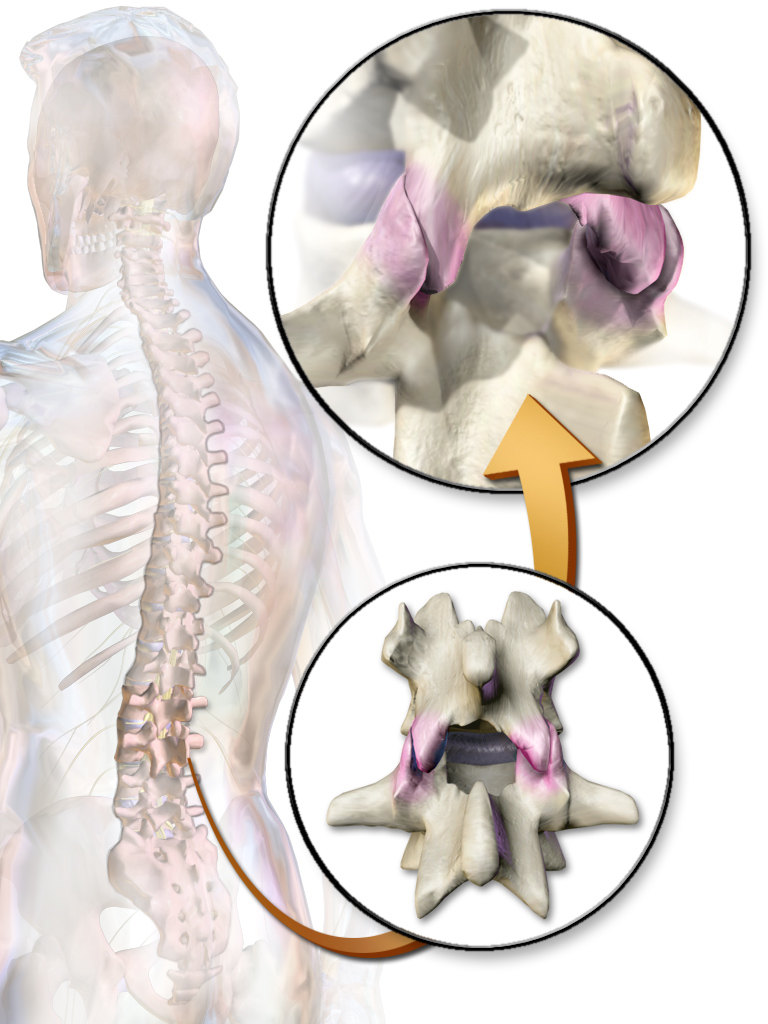
The facet joints (or zygapophysial joints, zygapophyseal, apophyseal, or Z-joints) are a set of synovial joint, synovial, plane joints between the articular processes of two adjacent vertebrae. There are two facet joints in each functional spinal unit, spinal motion segment and each facet joint is innervated by the Meningeal branches of spinal nerve, recurrent meningeal nerves. Innervation Innervation to the facet joints vary between segments of the spinal, but they are generally innervated by medial branch nerves that come off the dorsal rami. It is thought that these nerves are for primary sensory input, though there is some evidence that they have some motor input local musculature. Within the cervical spine, most joints are innervated by the medial branch nerve (a branch of the dorsal rami) from the same levels. In other words, the facet joint between C4 and C5 vertebral segments is innervated by the C4 and C5 medial branch nerves. However, there are two exceptions: # The ... [...More Info...] [...Related Items...] OR: [Wikipedia] [Google] [Baidu] |
Ligament
A ligament is the fibrous connective tissue that connects bones to other bones. It is also known as ''articular ligament'', ''articular larua'', ''fibrous ligament'', or ''true ligament''. Other ligaments in the body include the: * Peritoneal ligament: a fold of peritoneum or other membranes. * Fetal remnant ligament: the remnants of a fetal tubular structure. * Periodontal ligament: a group of fibers that attach the cementum of teeth to the surrounding alveolar bone. Ligaments are similar to tendons and fasciae as they are all made of connective tissue. The differences among them are in the connections that they make: ligaments connect one bone to another bone, tendons connect muscle to bone, and fasciae connect muscles to other muscles. These are all found in the skeletal system of the human body. Ligaments cannot usually be regenerated naturally; however, there are periodontal ligament stem cells located near the periodontal ligament which are involved in the adult regener ... [...More Info...] [...Related Items...] OR: [Wikipedia] [Google] [Baidu] |
Intervertebral Foramen
The intervertebral foramen (also called neural foramen, and often abbreviated as IV foramen or IVF) is a foramen between two spinal vertebrae. Cervical, thoracic, and lumbar vertebrae all have intervertebral foramina. The foramina, or openings, are present between every pair of vertebrae in these areas. A number of structures pass through the foramen. These are the root of each spinal nerve, the spinal artery of the segmental artery, communicating veins between the internal and external plexuses, recurrent meningeal (sinu-vertebral) nerves, and transforaminal ligaments. When the spinal vertebrae are articulated with each other, the bodies form a strong pillar that supports the head and trunk, and the vertebral foramen constitutes a canal for the protection of the medulla spinalis (spinal cord). The size of the foramina is variable due to placement, pathology, spinal loading, and posture. Foramina can be occluded by arthritic degenerative changes and space-occupying lesions ... [...More Info...] [...Related Items...] OR: [Wikipedia] [Google] [Baidu] |

