|
Whipple Procedure
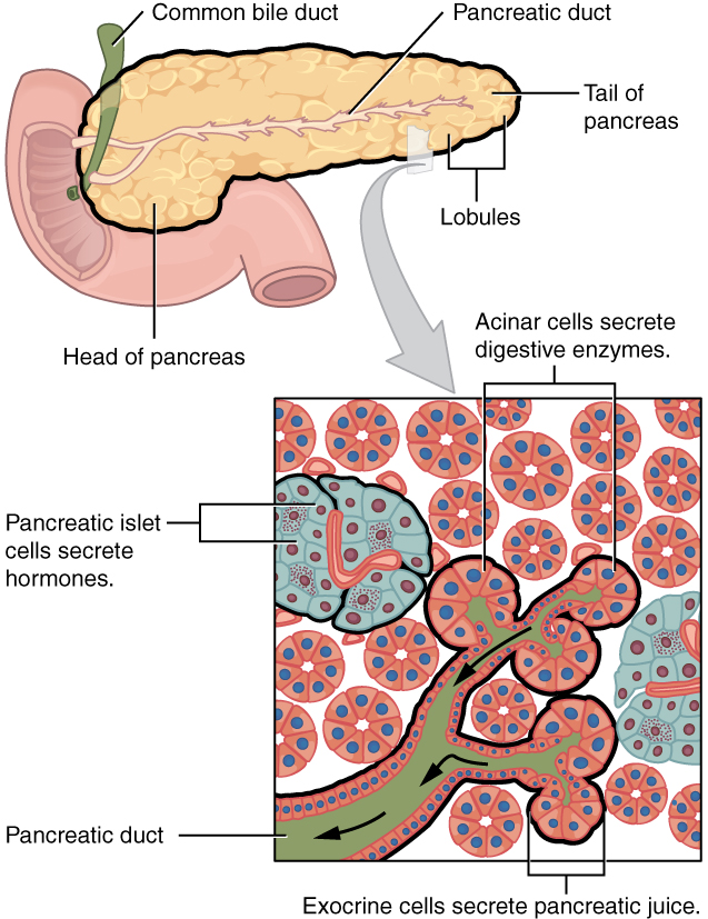
A pancreaticoduodenectomy, also known as a Whipple procedure, is a major surgical operation most often performed to remove cancerous tumours from the head of the pancreas. It is also used for the treatment of pancreatic or duodenal trauma, or chronic pancreatitis. Due to the shared blood supply of organs in the proximal gastrointestinal system, surgical removal of the head of the pancreas also necessitates removal of the duodenum, proximal jejunum, gallbladder, and, occasionally, part of the stomach. Anatomy involved in the procedure The most common technique of a pancreaticoduodenectomy consists of the en bloc removal of the distal segment (antrum) of the stomach, the first and second portions of the duodenum, the head of the w:pancreas#Structure, pancreas, the common bile duct, and the gallbladder. Lymph nodes in the area are often removed during the operation as well (lymphadenectomy). However, not all lymph nodes are removed in the most common type of pancreaticoduodenectom ... [...More Info...] [...Related Items...] OR: [Wikipedia] [Google] [Baidu] |
Pancreatic Cancer
Pancreatic cancer arises when cell (biology), cells in the pancreas, a glandular organ behind the stomach, begin to multiply out of control and form a Neoplasm, mass. These cancerous cells have the malignant, ability to invade other parts of the body. A number of types of pancreatic cancer are known. The most common, pancreatic adenocarcinoma, accounts for about 90% of cases, and the term "pancreatic cancer" is sometimes used to refer only to that type. These adenocarcinomas start within the part of the pancreas that makes digestive enzymes. Several other types of cancer, which collectively represent the majority of the non-adenocarcinomas, can also arise from these cells. About 1–2% of cases of pancreatic cancer are neuroendocrine tumors, which arise from the hormone-producing neuroendocrine cell, cells of the pancreas. These are generally less aggressive than pancreatic adenocarcinoma. Signs and symptoms of the most-common form of pancreatic cancer may include jaundice, ye ... [...More Info...] [...Related Items...] OR: [Wikipedia] [Google] [Baidu] |
Inferior Pancreaticoduodenal Artery
The inferior pancreaticoduodenal artery (the IPDA) is a branch of the superior mesenteric artery. It supplies the head of the pancreas, and the ascending and inferior parts of the duodenum. Rarely, it may have an aneurysm. Structure The inferior pancreaticoduodenal artery is a branch of the superior mesenteric artery. This occurs opposite the upper border of the inferior part of the duodenum. As soon as it branches, it divides into anterior and posterior branches. These run between the head of the pancreas and the lesser curvature of the duodenum. They then join (anastomose) with the anterior and posterior branches of the superior pancreaticoduodenal artery. Variation The inferior pancreaticoduodenal artery may branch from the first intestinal branch of the superior mesenteric artery rather than directly from it. Function The inferior pancreaticoduodenal artery distributes branches to the head of the pancreas and to the ascending and inferior parts of the duodenum. Clinic ... [...More Info...] [...Related Items...] OR: [Wikipedia] [Google] [Baidu] |
Gastrointestinal Stromal Tumor
Gastrointestinal stromal tumors (GISTs) are the most common mesenchymal neoplasms of the gastrointestinal tract. GISTs arise in the smooth muscle pacemaker interstitial cell of Cajal, or similar cells. They are defined as tumors whose behavior is driven by mutations in the KIT gene (85%), PDGFRA gene (10%), or BRAF kinase (rare). 95% of GISTs stain positively for KIT (CD117). Most (66%) occur in the stomach and gastric GISTs have a lower malignant potential than tumors found elsewhere in the GI tract. Classification GIST was introduced as a diagnostic term in 1983. Until the late 1990s, many non-epithelial tumors of the gastrointestinal tract were called "gastrointestinal stromal tumors". Histopathologists were unable to specifically distinguish among types we now know to be dissimilar molecularly. Subsequently, CD34, and later CD117 were identified as markers that could distinguish the various types. Additionally, in the absence of specific therapy, the diagnostic categori ... [...More Info...] [...Related Items...] OR: [Wikipedia] [Google] [Baidu] |
Metastasis
Metastasis is a pathogenic agent's spread from an initial or primary site to a different or secondary site within the host's body; the term is typically used when referring to metastasis by a cancerous tumor. The newly pathological sites, then, are metastases (mets). It is generally distinguished from cancer invasion, which is the direct extension and penetration by cancer cells into neighboring tissues. Cancer occurs after cells are genetically altered to proliferate rapidly and indefinitely. This uncontrolled proliferation by mitosis produces a primary heterogeneic tumour. The cells which constitute the tumor eventually undergo metaplasia, followed by dysplasia then anaplasia, resulting in a malignant phenotype. This malignancy allows for invasion into the circulation, followed by invasion to a second site for tumorigenesis. Some cancer cells known as circulating tumor cells acquire the ability to penetrate the walls of lymphatic or blood vessels, after which they are abl ... [...More Info...] [...Related Items...] OR: [Wikipedia] [Google] [Baidu] |
Pancreatic Tumor
Pancreatic tumors (Pancreatic cancer Pancreatic cancer arises when cell (biology), cells in the pancreas, a glandular organ behind the stomach, begin to multiply out of control and form a Neoplasm, mass. These cancerous cells have the malignant, ability to invade other parts of t ...) are often broken down into exocrine or endocrine tumors. Each different type of pancreatic tumor has a different appearance when examined under a microscope. These unique microscopic features and genetic markers are what allow for proper diagnosis and treatments in patients with pancreatic cancers. Types When talking about pancreatic cancers, the most common type is pancreatic adenocarcinoma, which accounts for greater than 85% of all pancreas cancers. Adenocarcinomas are exocrine tumors of the pancreas, which implies that they begin within the part of the pancreas responsible for creating digestive enzymes. Different subtypes of exocrine adenocarcinomas exist, with the most common being pancrea ... [...More Info...] [...Related Items...] OR: [Wikipedia] [Google] [Baidu] |
Chronic Pancreatitis
Chronic pancreatitis is a long-standing inflammation of the pancreas that alters the organ's normal structure and functions. It can present as episodes of acute inflammation in a previously injured pancreas, or as chronic damage with persistent pain or malabsorption. It is a disease process characterized by irreversible damage to the pancreas as distinct from reversible changes in acute pancreatitis. Signs and symptoms * Upper abdominal pain: Upper abdominal pain which increases after drinking or eating, lessens when fasting or sitting and leaning forward. Some people may not suffer pain. * Nausea and vomiting * Steatorrhea: Frequent, oily, foul-smelling bowel movements. Damage to the pancreas reduces the production of pancreatic enzymes that aid digestion, which can result in malnutrition. Fats and nutrients are not absorbed properly, leading to loose, greasy stool known as steatorrhea. * Weight loss even when eating habits and amounts are normal. * Diabetes type 1: Chronic pan ... [...More Info...] [...Related Items...] OR: [Wikipedia] [Google] [Baidu] |
Periampullary Cancer
Periampullary cancer is a cancer that forms near the ampulla of Vater, an enlargement of the Duct (anatomy), ducts from the liver and pancreas where they join and enter the small intestine. Quoted material is in the Copyright status of work by the U.S. government, public domain. It consists of: # ampullary tumour from ampulla of Vater # cancer of lower common bile duct # duodenal cancer adjacent to ampulla # carcinoma head of pancreas It presents with painless jaundice which may have waxing and waning nature because at times the sloughing of the tumor tissue relieves the obstruction partially. Signs and symptoms of periampullary cancer * Jaundice (yellowing of skin, eyes and urine with pale stools) * Itching * Abdominal pain * Weight loss and loss of appetite * Recurrent vomiting * Black stools * Anaemia Treatment The treatment depends upon the stage of the disease and degree of jaundice. Surgery is the best possible option and can be considered if the cancer is diagnosed a ... [...More Info...] [...Related Items...] OR: [Wikipedia] [Google] [Baidu] |
Graft (surgery)
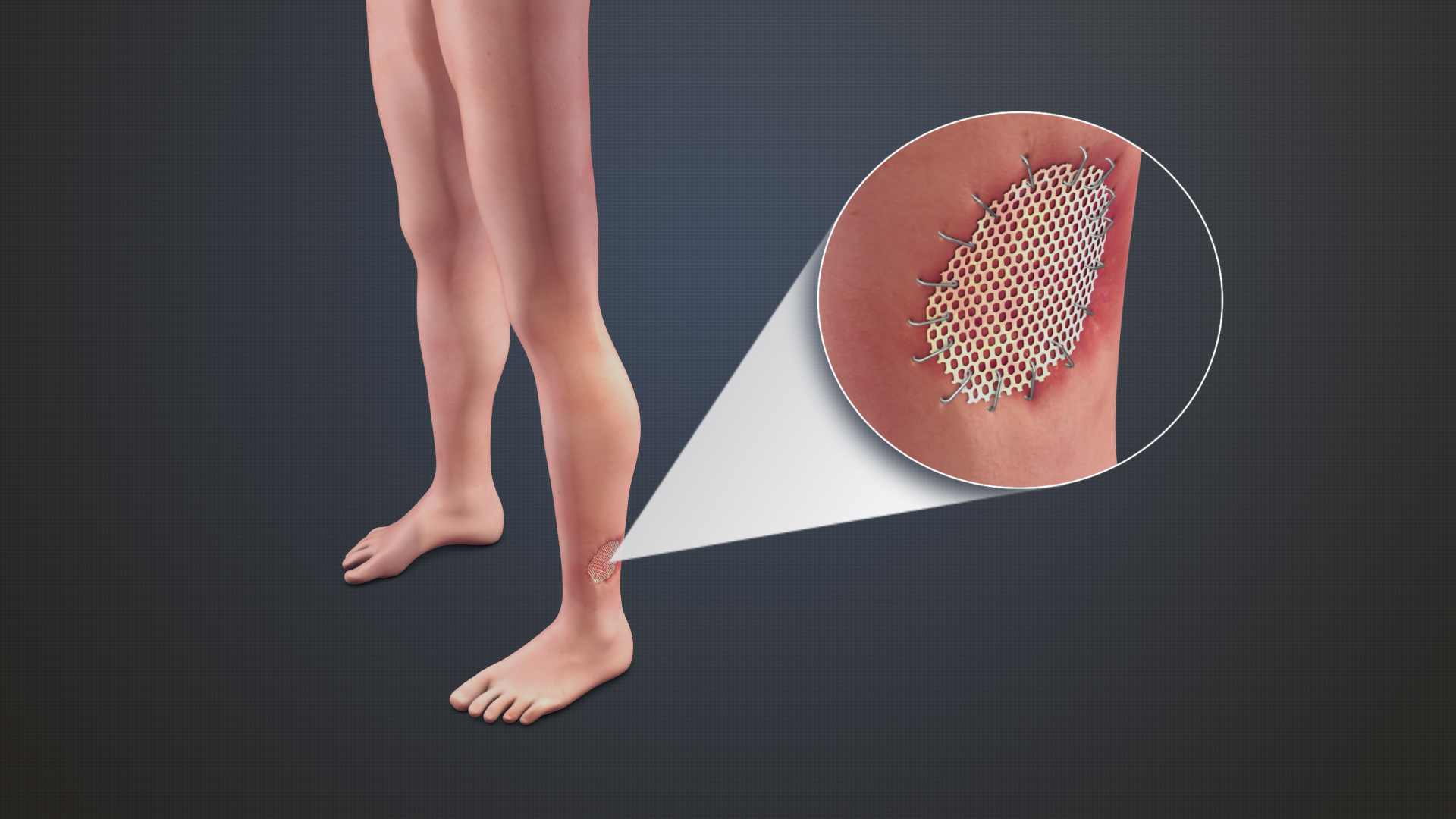
Grafting refers to a surgical procedure to move tissue from one site to another on the body, or from another creature, without bringing its own blood supply with it. Instead, a new blood supply grows in after it is placed. A similar technique where tissue is transferred with the blood supply intact is called a flap. In some instances, a graft can be an artificially manufactured device. Examples of this are a tube to carry blood flow across a defect or from an artery to a vein for use in hemodialysis. Classification Autografts and isografts are usually not considered as foreign and, therefore, do not elicit rejection. Allografts and xenografts may be recognized as foreign by the recipient and rejected. * Autograft: graft taken from one part of the body of an individual and transplanted onto another site in the same individual, e.g., skin graft. * Isograft: graft taken from one individual and placed on another individual of the same genetic constitution, e.g., grafts between ... [...More Info...] [...Related Items...] OR: [Wikipedia] [Google] [Baidu] |
Vascular Surgeons
Vascular surgery is a surgical subspecialty in which diseases of the vascular system, or arteries, veins and lymphatic circulation, are managed by medical therapy, minimally-invasive catheter procedures and surgical reconstruction. The specialty evolved from general and cardiac surgery and includes treatment of the body's other major and essential veins and arteries. Open surgery techniques, as well as endovascular techniques are used to treat vascular diseases. The vascular surgeon is trained in the diagnosis and management of diseases affecting all parts of the vascular system excluding the coronaries and intracranial vasculature. Vascular surgeons often assist other physicians to address traumatic vascular injury, hemorrhage control, and safe exposure of vascular structures. History Early leaders of the field included Russian surgeon Nikolai Korotkov, noted for developing early surgical techniques, American interventional radiologist Charles Theodore Dotter who is credited wi ... [...More Info...] [...Related Items...] OR: [Wikipedia] [Google] [Baidu] |
Inferior Vena Cava
The inferior vena cava is a large vein that carries the deoxygenated blood from the lower and middle body into the right atrium of the heart. It is formed by the joining of the right and the left common iliac veins, usually at the level of the fifth lumbar vertebra. The inferior vena cava is the lower (" inferior") of the two venae cavae, the two large veins that carry deoxygenated blood from the body to the right atrium of the heart: the inferior vena cava carries blood from the lower half of the body whilst the superior vena cava carries blood from the upper half of the body. Together, the venae cavae (in addition to the coronary sinus, which carries blood from the muscle of the heart itself) form the venous counterparts of the aorta. It is a large retroperitoneal vein that lies posterior to the abdominal cavity and runs along the right side of the vertebral column. It enters the right auricle at the lower right, back side of the heart. The name derives from la, vena, "vei ... [...More Info...] [...Related Items...] OR: [Wikipedia] [Google] [Baidu] |
Superior Mesenteric Vein
In human anatomy, the superior mesenteric vein (SMV) is a blood vessel that drains blood from the small intestine (jejunum and ileum). Behind the neck of the pancreas, the superior mesenteric vein combines with the splenic vein to form the hepatic portal vein. The superior mesenteric vein lies to the right of the similarly named artery, the superior mesenteric artery, which originates from the abdominal aorta. Structure Tributaries of the superior mesenteric vein drain the small intestine, large intestine, stomach, pancreas and appendix and include: * Right gastro-omental vein (also known as the right gastro-epiploic vein) * inferior pancreaticoduodenal veins * veins from jejunum * veins from ileum * middle colic vein – drains the transverse colon * right colic vein – drains the ascending colon * ileocolic vein The superior mesenteric vein combines with the splenic vein to form the portal vein. Clinical significance Thrombosis of the superior mesenteric vein is quite rare ... [...More Info...] [...Related Items...] OR: [Wikipedia] [Google] [Baidu] |
Portal Vein
The portal vein or hepatic portal vein (HPV) is a blood vessel that carries blood from the gastrointestinal tract, gallbladder, pancreas and spleen to the liver. This blood contains nutrients and toxins extracted from digested contents. Approximately 75% of total liver blood flow is through the portal vein, with the remainder coming from the hepatic artery proper. The blood leaves the liver to the heart in the hepatic veins. The portal vein is not a true vein, because it conducts blood to capillary beds in the liver and not directly to the heart. It is a major component of the hepatic portal system, one of only two portal venous systems in the body – with the hypophyseal portal system being the other. The portal vein is usually formed by the confluence of the superior mesenteric, splenic veins, inferior mesenteric, left, right gastric veins and the pancreatic vein. Conditions involving the portal vein cause considerable illness and death. An important example of such a condi ... [...More Info...] [...Related Items...] OR: [Wikipedia] [Google] [Baidu] |


