|
Superior Mesenteric Vein
In human anatomy, the superior mesenteric vein (SMV) is a blood vessel that drains blood from the small intestine ( jejunum and ileum). Behind the neck of the pancreas, the superior mesenteric vein combines with the splenic vein to form the hepatic portal vein. The superior mesenteric vein lies to the right of the similarly named artery, the superior mesenteric artery, which originates from the abdominal aorta. Structure Tributaries of the superior mesenteric vein drain the small intestine, large intestine, stomach, pancreas and appendix and include: * Right gastro-omental vein (also known as the right gastro-epiploic vein) * inferior pancreaticoduodenal veins * veins from jejunum * veins from ileum * middle colic vein – drains the transverse colon * right colic vein – drains the ascending colon * ileocolic vein The superior mesenteric vein combines with the splenic vein to form the portal vein. Clinical significance Thrombosis of the superior mesenteric vein is quit ... [...More Info...] [...Related Items...] OR: [Wikipedia] [Google] [Baidu] |
|
 |
Portal Venous System
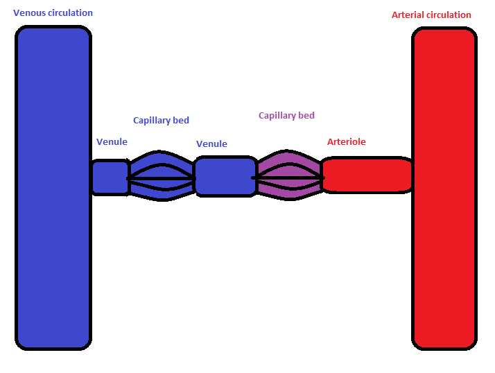
In the circulatory system of animals, a portal venous system occurs when a capillary bed pools into another capillary bed through veins, without first going through the heart. Both capillary beds and the blood vessels that connect them are considered part of the portal venous system. They are relatively uncommon as the majority of capillary beds drain into veins which then drain into the heart, not into another capillary bed. Portal venous systems are considered venous because the blood vessels that join the two capillary beds are either veins or venules. Examples of such systems include the hepatic portal system, the hypophyseal portal system, and (in non-mammals) the renal portal system. Unqualified, ''portal venous system'' often refers to the hepatic portal system. For this reason, ''portal vein'' most commonly refers to the hepatic portal vein. The functional significance of such a system is that it transports products of one region directly to another region in relati ... [...More Info...] [...Related Items...] OR: [Wikipedia] [Google] [Baidu] |
 |
Vermiform Appendix
The appendix (or vermiform appendix; also cecal r caecalappendix; vermix; or vermiform process) is a finger-like, blind-ended tube connected to the cecum, from which it develops in the embryo. The cecum is a pouch-like structure of the large intestine, located at the junction of the small and the large intestines. The term " vermiform" comes from Latin and means "worm-shaped". The appendix was once considered a vestigial organ, but this view has changed since the early 2000s. Research suggests that the appendix may serve an important purpose. In particular, it may serve as a reservoir for beneficial gut bacteria. Structure The human appendix averages in length but can range from . The diameter of the appendix is , and more than is considered a thickened or inflamed appendix. The longest appendix ever removed was long. The appendix is usually located in the lower right quadrant of the abdomen, near the right hip bone. The base of the appendix is located beneath the ... [...More Info...] [...Related Items...] OR: [Wikipedia] [Google] [Baidu] |
 |
Ischemia
Ischemia or ischaemia is a restriction in blood supply to any tissue, muscle group, or organ of the body, causing a shortage of oxygen that is needed for cellular metabolism (to keep tissue alive). Ischemia is generally caused by problems with blood vessels, with resultant damage to or dysfunction of tissue i.e. hypoxia and microvascular dysfunction. It also implies local hypoxia in a part of a body resulting from constriction (such as vasoconstriction, thrombosis, or embolism). Ischemia causes not only insufficiency of oxygen, but also reduced availability of nutrients and inadequate removal of metabolic wastes. Ischemia can be partial (poor perfusion) or total blockage. The inadequate delivery of oxygenated blood to the organs must be resolved either by treating the cause of the inadequate delivery or reducing the oxygen demand of the system that needs it. For example, patients with myocardial ischemia have a decreased blood flow to the heart and are prescribed wit ... [...More Info...] [...Related Items...] OR: [Wikipedia] [Google] [Baidu] |
|
Mesenteric Ischemia
Intestinal ischemia is a medical condition in which injury to the large or small intestine occurs due to not enough blood supply. It can come on suddenly, known as acute intestinal ischemia, or gradually, known as chronic intestinal ischemia. The acute form of the disease often presents with sudden severe abdominal pain and is associated with a high risk of death. The chronic form typically presents more gradually with abdominal pain after eating, unintentional weight loss, vomiting, and fear of eating. Risk factors for acute intestinal ischemia include atrial fibrillation, heart failure, chronic kidney failure, being prone to forming blood clots, and previous myocardial infarction. There are four mechanisms by which poor blood flow occurs: a blood clot from elsewhere getting lodged in an artery, a new blood clot forming in an artery, a blood clot forming in the superior mesenteric vein, and insufficient blood flow due to low blood pressure or spasms of arteries. Chro ... [...More Info...] [...Related Items...] OR: [Wikipedia] [Google] [Baidu] |
|
 |
Thrombosis
Thrombosis (from Ancient Greek "clotting") is the formation of a blood clot inside a blood vessel, obstructing the flow of blood through the circulatory system. When a blood vessel (a vein or an artery) is injured, the body uses platelets (thrombocytes) and fibrin to form a blood clot to prevent blood loss. Even when a blood vessel is not injured, blood clots may form in the body under certain conditions. A clot, or a piece of the clot, that breaks free and begins to travel around the body is known as an embolus. Thrombosis may occur in veins ( venous thrombosis) or in arteries ( arterial thrombosis). Venous thrombosis (sometimes called DVT, deep vein thrombosis) leads to a blood clot in the affected part of the body, while arterial thrombosis (and, rarely, severe venous thrombosis) affects the blood supply and leads to damage of the tissue supplied by that artery (ischemia and necrosis). A piece of either an arterial or a venous thrombus can break off as an embolus, which c ... [...More Info...] [...Related Items...] OR: [Wikipedia] [Google] [Baidu] |
 |
Portal Vein
The portal vein or hepatic portal vein (HPV) is a blood vessel that carries blood from the gastrointestinal tract, gallbladder, pancreas and spleen to the liver. This blood contains nutrients and toxins extracted from digested contents. Approximately 75% of total liver blood flow is through the portal vein, with the remainder coming from the hepatic artery proper. The blood leaves the liver to the heart in the hepatic veins. The portal vein is not a true vein, because it conducts blood to capillary beds in the liver and not directly to the heart. It is a major component of the hepatic portal system, one of only two portal venous systems in the body – with the hypophyseal portal system being the other. The portal vein is usually formed by the confluence of the superior mesenteric, splenic veins, inferior mesenteric, left, right gastric veins and the pancreatic vein. Conditions involving the portal vein cause considerable illness and death. An important example ... [...More Info...] [...Related Items...] OR: [Wikipedia] [Google] [Baidu] |
|
Ileocolic Vein
The ileocolic vein is a vein which drains the ileum, colon, and cecum The cecum or caecum is a pouch within the peritoneum that is considered to be the beginning of the large intestine. It is typically located on the right side of the body (the same side of the body as the appendix, to which it is joined). The w .... Veins of the torso {{circulatory-stub ... [...More Info...] [...Related Items...] OR: [Wikipedia] [Google] [Baidu] |
|
|
Ascending Colon
''Ascending'' is a science fiction novel by the Canadian writer James Alan Gardner, published in 2001 by HarperCollins Publishers under its various imprints.HarperCollins, Avon, HarperCollins Canada, SFBC/Avon; paperback edition 2001, Eos Books. It is the fifth novel in Gardner's " League of Peoples" series. It is a direct sequel to the first novel in the series, ''Expendable'', in that it picks up the dual story of Festina Ramos, Explorer turned admiral, and the transparent glass woman Oar, where the earlier novel left off. Backstory Through the course of ''Ascending'', Gardner adds depth, detail, and perspective to the conceptual background he has established for the "League of Peoples" series. In particular, he explains how human beings and other species in the galaxy are contacted by, and become a part of, the larger galactic community. Sometime in the middle of the 21st century, humanity encounters a mysterious group of beings who call themselves merely "citizens of the Lea ... [...More Info...] [...Related Items...] OR: [Wikipedia] [Google] [Baidu] |
|
|
Right Colic Vein
The right colic vein drains the ascending colon, and is a tributary of the superior mesenteric vein. It travels with its corresponding artery, the right colic artery The right colic artery is an artery of the abdomen, a branch of the superior mesenteric artery supplying the ascending colon. It divides into two terminal branches - an ascending branch and a descending branch - which form anastomoses with the .... Veins of the torso {{Circulatory-stub ... [...More Info...] [...Related Items...] OR: [Wikipedia] [Google] [Baidu] |
|
|
Transverse Colon
In human anatomy, the transverse colon is the longest and most movable part of the colon. Anatomical position It crosses the abdomen from the ascending colon at the right colic flexure (hepatic flexure) with a downward convexity to the descending colon where it curves sharply on itself beneath the lower end of the spleen forming the left colic flexure (splenic flexure). In its course, it describes an arch, the concavity of which is directed backward and a little upward. Toward its splenic end there is often an abrupt U-shaped curve which may descend lower than the main curve. It is almost completely invested by the peritoneum, and is connected to the inferior border of the pancreas by a large and wide duplicature of that membrane, the transverse mesocolon. It is in relation, by its upper surface, with the liver and gall-bladder, the greater curvature of the stomach, and the lower end of the spleen; by its under surface, with the small intestine; by its anterior surface, wi ... [...More Info...] [...Related Items...] OR: [Wikipedia] [Google] [Baidu] |
|
|
Middle Colic Vein
The middle colic vein drains the transverse colon. It is a tributary of the superior mesenteric vein, and follows the path of its corresponding artery, the middle colic artery The middle colic artery is an artery of the abdomen; a branch of the superior mesenteric artery distributed to parts of the ascending and transverse colon. It usually divides into two terminal branches - a left one and a right one - which go on t .... Veins of the torso {{Circulatory-stub ... [...More Info...] [...Related Items...] OR: [Wikipedia] [Google] [Baidu] |
|
|
Veins From Ileum
The ileal veins are tributaries of the superior mesenteric vein In human anatomy, the superior mesenteric vein (SMV) is a blood vessel that drains blood from the small intestine ( jejunum and ileum). Behind the neck of the pancreas, the superior mesenteric vein combines with the splenic vein to form the hepat .... External links Veins of the torso {{circulatory-stub ... [...More Info...] [...Related Items...] OR: [Wikipedia] [Google] [Baidu] |