|
Visual Learning
Visual learning is a learning style in the Fleming VAK/VARK model in which information is presented to a learner in a visual format. Visual learners can utilize graphs, charts, maps, diagrams, and other forms of visual stimulation to effectively interpret information. The Fleming VAK/VARK model also includes kinesthetic learning and auditory learning. There is no evidence that providing visual materials to students identified as having a visual style improves learning. Techniques A review study concluded that using graphic organizers improves student performance in the following areas: ; Retention : Students remember information better and can better recall it when it is represented and learned both visually and verbally. ; Reading comprehension : The use of graphic organizers helps improve the reading comprehension of students. ; Student achievement : Students with and without learning disabilities improve achievement across content areas and grade levels. ; Thinking and lea ... [...More Info...] [...Related Items...] OR: [Wikipedia] [Google] [Baidu] |
Learning Styles
Learning styles refer to a range of theories that aim to account for differences in individuals' learning. Although there is ample evidence that individuals express personal preferences for how they prefer to receive information, few studies have found any validity in using learning styles in education. Many theories share the proposition that humans can be classified according to their "style" of learning, but differ in how the proposed styles should be defined, categorized and assessed. A common concept is that individuals differ in how they learn. The idea of individualized learning styles became popular in the 1970s, and has greatly influenced education despite the criticism that the idea has received from some researchers. Proponents recommend that teachers have to run a needs analysis to assess the learning styles of their students and adapt their classroom methods to best fit each student's learning style. Critics say there is no consistent evidence that identifying an indivi ... [...More Info...] [...Related Items...] OR: [Wikipedia] [Google] [Baidu] |
Visual Field
The visual field is the "spatial array of visual sensations available to observation in introspectionist psychological experiments". Or simply, visual field can be defined as the entire area that can be seen when an eye is fixed straight at a point. The equivalent concept for optical instruments and image sensors is the field of view (FOV). In optometry, ophthalmology, and neurology, a visual field test is used to determine whether the visual field is affected by diseases that cause local scotoma or a more extensive loss of vision or a reduction in sensitivity (increase in threshold). Normal limits The normal (monocular) human visual field extends to approximately 60 degrees nasally (toward the nose, or inward) from the vertical meridian in each eye, to 107 degrees temporally (away from the nose, or outwards) from the vertical meridian, and approximately 70 degrees above and 80 below the horizontal meridian. The binocular visual field is the superimposition of the two monocular ... [...More Info...] [...Related Items...] OR: [Wikipedia] [Google] [Baidu] |
Nerve Impulse
An action potential occurs when the membrane potential of a specific cell location rapidly rises and falls. This depolarization then causes adjacent locations to similarly depolarize. Action potentials occur in several types of animal cells, called excitable cells, which include neurons, muscle cells, and in some plant cells. Certain endocrine cells such as pancreatic beta cells, and certain cells of the anterior pituitary gland are also excitable cells. In neurons, action potentials play a central role in cell-cell communication by providing for—or with regard to saltatory conduction, assisting—the propagation of signals along the neuron's axon toward synaptic boutons situated at the ends of an axon; these signals can then connect with other neurons at synapses, or to motor cells or glands. In other types of cells, their main function is to activate intracellular processes. In muscle cells, for example, an action potential is the first step in the chain of events leadi ... [...More Info...] [...Related Items...] OR: [Wikipedia] [Google] [Baidu] |
Eye Movement
Eye movement includes the voluntary or involuntary movement of the eyes. Eye movements are used by a number of organisms (e.g. primates, rodents, flies, birds, fish, cats, crabs, octopus) to fixate, inspect and track visual objects of interests. A special type of eye movement, rapid eye movement, occurs during REM sleep. The eyes are the visual organs of the human body, and move using a system of six muscles. The retina, a specialised type of tissue containing photoreceptors, senses light. These specialised cells convert light into electrochemical signals. These signals travel along the optic nerve fibers to the brain, where they are interpreted as vision in the visual cortex. Primates and many other vertebrates use three types of voluntary eye movement to track objects of interest: smooth pursuit, vergence shifts and saccades. These types of movements appear to be initiated by a small cortical region in the brain's frontal lobe. This is corroborated by removal of the frontal ... [...More Info...] [...Related Items...] OR: [Wikipedia] [Google] [Baidu] |
Frontal Eye Fields
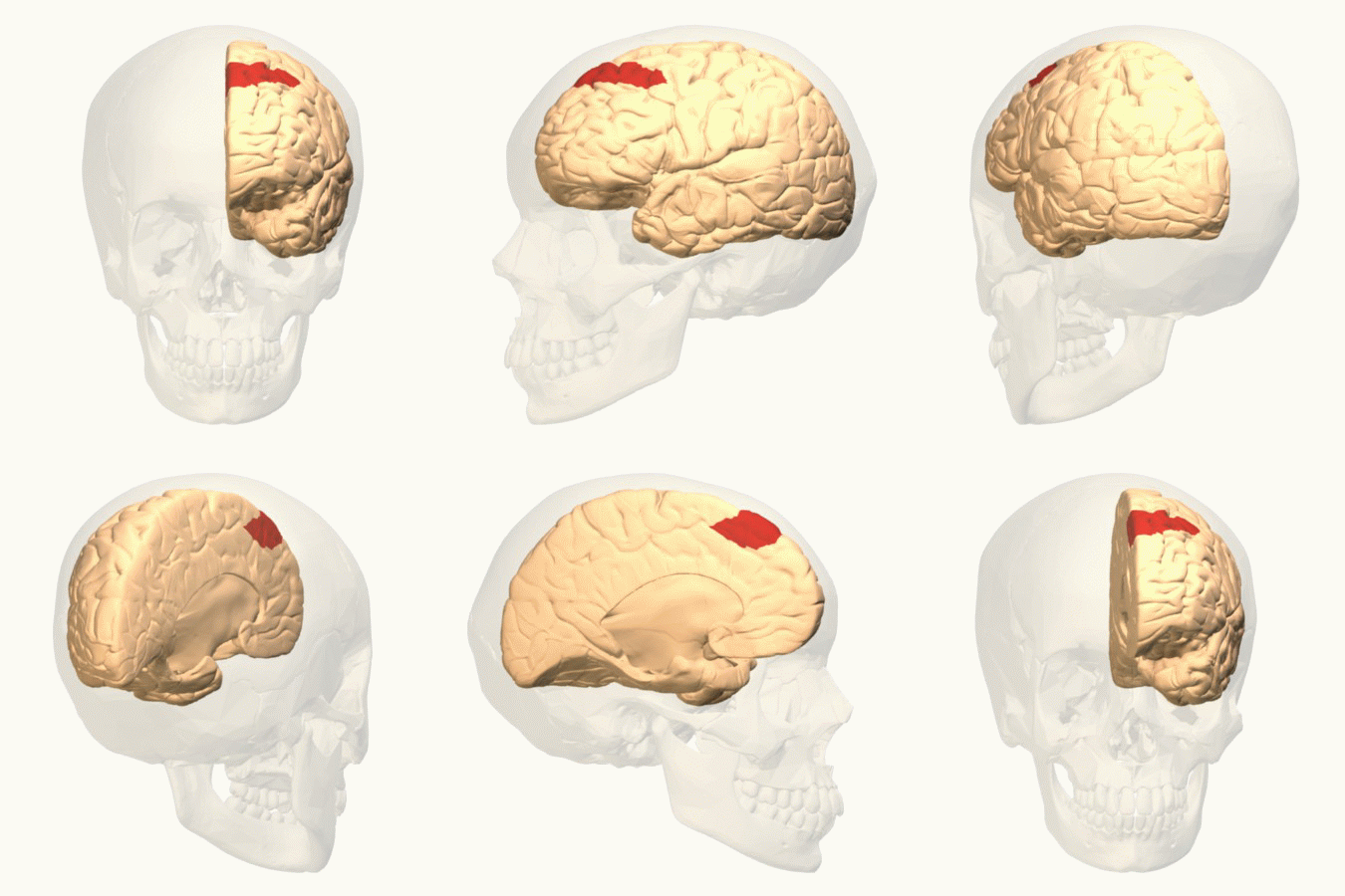
The frontal eye fields (FEF) are a region located in the frontal cortex, more specifically in Brodmann area 8 or BA8, of the primate brain. In humans, it can be more accurately said to lie in a region around the intersection of the middle frontal gyrus with the precentral gyrus, consisting of a frontal and parietal portion. The FEF is responsible for saccadic eye movements for the purpose of visual field perception and awareness, as well as for voluntary eye movement. The FEF communicates with extraocular muscles indirectly via the paramedian pontine reticular formation. Destruction of the FEF causes deviation of the eyes to the ipsilateral side. Function The cortical area called frontal eye field (FEF) plays an important role in the control of visual attention and eye movements. Electrical stimulation in the FEF elicits saccadic eye movements. The FEF have a topographic structure and represents saccade targets in retinotopic coordinates. The frontal eye field is reported to b ... [...More Info...] [...Related Items...] OR: [Wikipedia] [Google] [Baidu] |
Superior Colliculus
In neuroanatomy, the superior colliculus () is a structure lying on the roof of the mammalian midbrain. In non-mammalian vertebrates, the homologous structure is known as the optic tectum, or optic lobe. The adjective form ''tectal'' is commonly used for both structures. In mammals, the superior colliculus forms a major component of the midbrain. It is a paired structure and together with the paired inferior colliculi forms the corpora quadrigemina. The superior colliculus is a layered structure, with a pattern that is similar to all mammals. The layers can be grouped into the superficial layers ( stratum opticum and above) and the deeper remaining layers. Neurons in the superficial layers receive direct input from the retina and respond almost exclusively to visual stimuli. Many neurons in the deeper layers also respond to other modalities, and some respond to stimuli in multiple modalities. The deeper layers also contain a population of motor-related neurons, capable of activat ... [...More Info...] [...Related Items...] OR: [Wikipedia] [Google] [Baidu] |
Gray Matter
Grey matter is a major component of the central nervous system, consisting of neuronal cell bodies, neuropil (dendrites and unmyelinated axons), glial cells (astrocytes and oligodendrocytes), synapses, and capillaries. Grey matter is distinguished from white matter in that it contains numerous cell bodies and relatively few myelinated axons, while white matter contains relatively few cell bodies and is composed chiefly of long-range myelinated axons. The colour difference arises mainly from the whiteness of myelin. In living tissue, grey matter actually has a very light grey colour with yellowish or pinkish hues, which come from capillary blood vessels and neuronal cell bodies. Structure Grey matter refers to unmyelinated neurons and other cells of the central nervous system. It is present in the brain, brainstem and cerebellum, and present throughout the spinal cord. Grey matter is distributed at the surface of the cerebral hemispheres (cerebral cortex) and of the cerebellum ... [...More Info...] [...Related Items...] OR: [Wikipedia] [Google] [Baidu] |
Visual Memory
Visual memory describes the relationship between perceptual processing and the encoding, storage and retrieval of the resulting neural representations. Visual memory occurs over a broad time range spanning from eye movements to years in order to visually navigate to a previously visited location.Berryhill, M. (2008, May 09). Visual memory and the brain. Retrieved from http://www.visionsciences.org/symposia2008_4.html Visual memory is a form of memory which preserves some characteristics of our senses pertaining to visual experience. We are able to place in memory visual information which resembles objects, places, animals or people in a mental image. The experience of visual memory is also referred to as the mind's eye through which we can retrieve from our memory a mental image of original objects, places, animals or people. Visual memory is one of several cognitive systems, which are all interconnected parts that combine to form the human memory. Types of palinopsia, the pers ... [...More Info...] [...Related Items...] OR: [Wikipedia] [Google] [Baidu] |
Schema (psychology)
In psychology and cognitive science, a schema (plural ''schemata'' or ''schemas'') describes a pattern of thought or behavior that organizes categories of information and the relationships among them. It can also be described as a mental structure of preconceived ideas, a framework representing some aspect of the world, or a system of organizing and perceiving new information, such as a mental schema or conceptual model. Schemata influence attention and the absorption of new knowledge: people are more likely to notice things that fit into their schema, while re-interpreting contradictions to the schema as exceptions or distorting them to fit. Schemata have a tendency to remain unchanged, even in the face of contradictory information. Schemata can help in understanding the world and the rapidly changing environment. People can organize new perceptions into schemata quickly as most situations do not require complex thought when using schema, since automatic thought is all that is r ... [...More Info...] [...Related Items...] OR: [Wikipedia] [Google] [Baidu] |
Limbic
The limbic system, also known as the paleomammalian cortex, is a set of brain structures located on both sides of the thalamus, immediately beneath the medial temporal lobe of the cerebrum primarily in the forebrain.Schacter, Daniel L. 2012. ''Psychology''.sec. 3.20 It supports a variety of functions including emotion, behavior, long-term memory, and olfaction. Emotional life is largely housed in the limbic system, and it critically aids the formation of memories. With a primordial structure, the limbic system is involved in lower order emotional processing of input from sensory systems and consists of the amygdaloid nuclear complex (amygdala), mammillary bodies, stria medullaris, central gray and dorsal and ventral nuclei of Gudden. This processed information is often relayed to a collection of structures from the telencephalon, diencephalon, and mesencephalon, including the prefrontal cortex, cingulate gyrus, limbic thalamus, hippocampus including the parahippocampal gyrus an ... [...More Info...] [...Related Items...] OR: [Wikipedia] [Google] [Baidu] |
Neostriatum
The striatum, or corpus striatum (also called the striate nucleus), is a nucleus (a cluster of neurons) in the subcortical basal ganglia of the forebrain. The striatum is a critical component of the motor and reward systems; receives glutamatergic and dopaminergic inputs from different sources; and serves as the primary input to the rest of the basal ganglia. Functionally, the striatum coordinates multiple aspects of cognition, including both motor and action planning, decision-making, motivation, reinforcement, and reward perception. The striatum is made up of the caudate nucleus and the lentiform nucleus. The lentiform nucleus is made up of the larger putamen, and the smaller globus pallidus. Strictly speaking the globus pallidus is part of the striatum. It is common practice, however, to implicitly exclude the globus pallidus when referring to striatal structures. In primates, the striatum is divided into a ventral striatum, and a dorsal striatum, subdivisions that ... [...More Info...] [...Related Items...] OR: [Wikipedia] [Google] [Baidu] |
Neocortex
The neocortex, also called the neopallium, isocortex, or the six-layered cortex, is a set of layers of the mammalian cerebral cortex involved in higher-order brain functions such as sensory perception, cognition, generation of motor commands, spatial reasoning and language. The neocortex is further subdivided into the true isocortex and the proisocortex. In the human brain, the neocortex is the largest part of the cerebral cortex (the outer layer of the cerebrum). The neocortex makes up the largest part of the cerebral cortex, with the allocortex making up the rest. The neocortex is made up of six layers, labelled from the outermost inwards, I to VI. Etymology The term is from ''cortex'', Latin, " bark" or "rind", combined with ''neo-'', Greek, "new". ''Neopallium'' is a similar hybrid, from Latin ''pallium'', "cloak". ''Isocortex'' and ''allocortex'' are hybrids with Greek ''isos'', "same", and ''allos'', "other". Anatomy The neocortex is the most developed in its organisat ... [...More Info...] [...Related Items...] OR: [Wikipedia] [Google] [Baidu] |


