|
Frontal Eye Fields
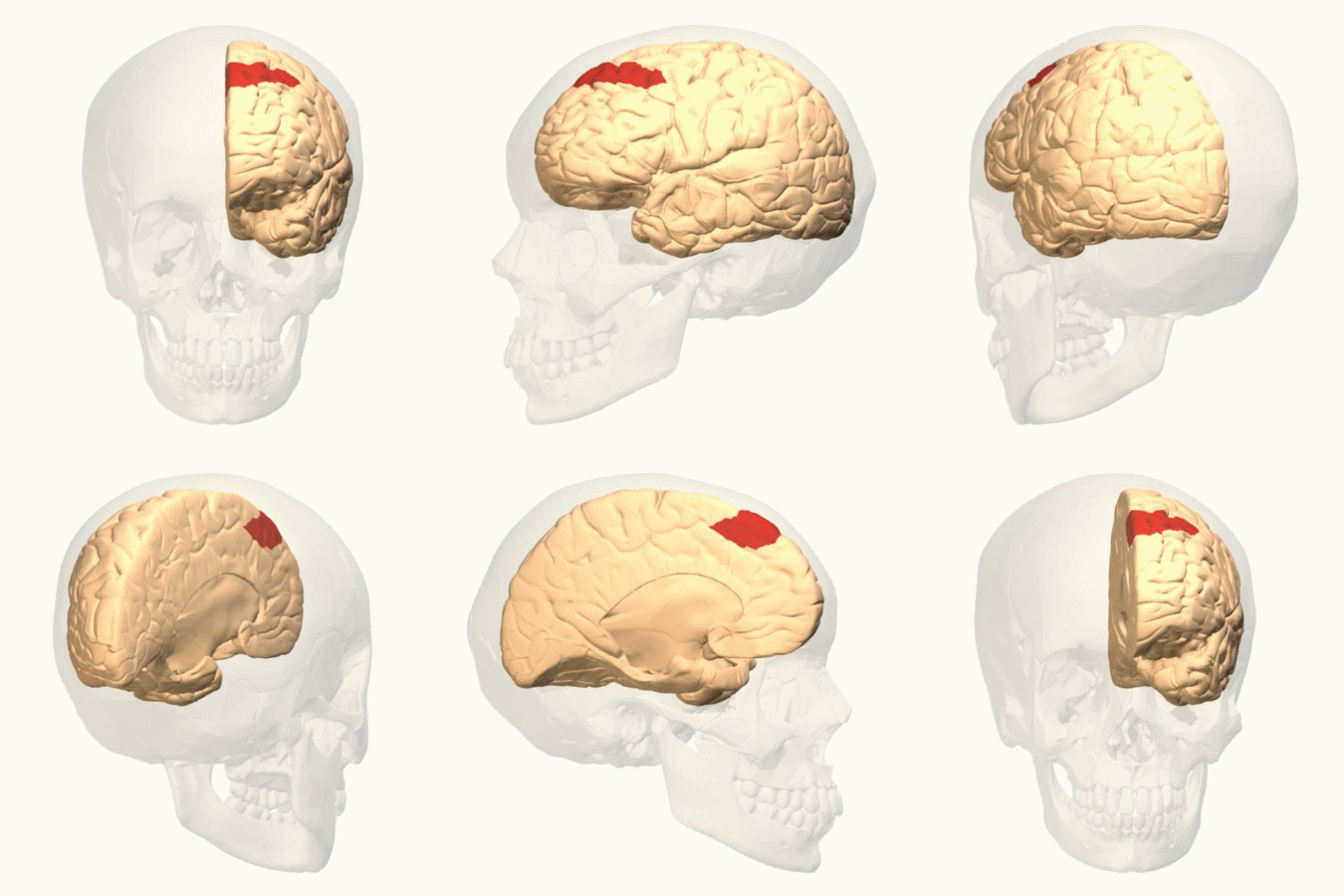
The frontal eye fields (FEF) are a region located in the frontal cortex, more specifically in Brodmann area 8 or BA8, of the primate brain. In humans, it can be more accurately said to lie in a region around the intersection of the middle frontal gyrus with the precentral gyrus, consisting of a frontal and parietal portion. The FEF is responsible for saccadic eye movements for the purpose of visual field perception and awareness, as well as for voluntary eye movement. The FEF communicates with extraocular muscles indirectly via the paramedian pontine reticular formation. Destruction of the FEF causes deviation of the eyes to the ipsilateral side. Function The cortical area called frontal eye field (FEF) plays an important role in the control of visual attention and eye movements. Electrical stimulation in the FEF elicits saccadic eye movements. The FEF have a topographic structure and represents saccade targets in retinotopic coordinates. The frontal eye field is reported to b ... [...More Info...] [...Related Items...] OR: [Wikipedia] [Google] [Baidu] |
Frontal Cortex
The frontal lobe is the largest of the four major lobes of the brain in mammals, and is located at the front of each cerebral hemisphere (in front of the parietal lobe and the temporal lobe). It is parted from the parietal lobe by a groove between tissues called the central sulcus and from the temporal lobe by a deeper groove called the lateral sulcus (Sylvian fissure). The most anterior rounded part of the frontal lobe (though not well-defined) is known as the frontal pole, one of the three poles of the cerebrum. The frontal lobe is covered by the frontal cortex. The frontal cortex includes the premotor cortex, and the primary motor cortex – parts of the motor cortex. The front part of the frontal cortex is covered by the prefrontal cortex. There are four principal gyri in the frontal lobe. The precentral gyrus is directly anterior to the central sulcus, running parallel to it and contains the primary motor cortex, which controls voluntary movements of specific body ... [...More Info...] [...Related Items...] OR: [Wikipedia] [Google] [Baidu] |
Superior Colliculus
In neuroanatomy, the superior colliculus () is a structure lying on the roof of the mammalian midbrain. In non-mammalian vertebrates, the homologous structure is known as the optic tectum, or optic lobe. The adjective form '' tectal'' is commonly used for both structures. In mammals, the superior colliculus forms a major component of the midbrain. It is a paired structure and together with the paired inferior colliculi forms the corpora quadrigemina. The superior colliculus is a layered structure, with a pattern that is similar to all mammals. The layers can be grouped into the superficial layers ( stratum opticum and above) and the deeper remaining layers. Neurons in the superficial layers receive direct input from the retina and respond almost exclusively to visual stimuli. Many neurons in the deeper layers also respond to other modalities, and some respond to stimuli in multiple modalities. The deeper layers also contain a population of motor-related neurons, capable of acti ... [...More Info...] [...Related Items...] OR: [Wikipedia] [Google] [Baidu] |
Brodmann Areas
A Brodmann area is a region of the cerebral cortex, in the human or other primate brain, defined by its cytoarchitecture, or histological structure and organization of cells. History Brodmann areas were originally defined and numbered by the German anatomist Korbinian Brodmann based on the cytoarchitectural organization of neurons he observed in the cerebral cortex using the Nissl method of cell staining. Brodmann published his maps of cortical areas in humans, monkeys, and other species in 1909, along with many other findings and observations regarding the general cell types and laminar organization of the mammalian cortex. The same Brodmann area number in different species does not necessarily indicate homologous areas. A similar, but more detailed cortical map was published by Constantin von Economo and Georg N. Koskinas in 1925. Present importance Brodmann areas have been discussed, debated, refined, and renamed exhaustively for nearly a century and remain the most ... [...More Info...] [...Related Items...] OR: [Wikipedia] [Google] [Baidu] |
Lateral Intraparietal Cortex
The lateral intraparietal cortex (area LIP) is found in the intraparietal sulcus of the brain. This area is most likely involved in eye movement, as electrical stimulation evokes saccades (quick movements) of the eyes. It is also thought to contribute to working memory associated with guiding eye movement, examined using a delayed saccade task described below:Pesaran, B., Pezaris, J. S., Sahani, M., Mitra, P. P., & Andersen, R. A. (2002). Temporal structure in neuronal activity during working memory in macaque parietal cortex. Nature neuroscience, 5(8), 805-811. #A subject focuses on a fixation point at the center of a computer screen. #A target (for instance a shape) is presented at a peripheral location on the screen. #The target is removed and followed by a variable-length delay period. #The initial focus point in the middle of the screen is removed. #The subject's task is to make a saccade to the location of the target. Neurons in area LIP have been shown to start responding ... [...More Info...] [...Related Items...] OR: [Wikipedia] [Google] [Baidu] |
Intraparietal Sulcus
The intraparietal sulcus (IPS) is located on the lateral surface of the parietal lobe, and consists of an oblique and a horizontal portion. The IPS contains a series of functionally distinct subregions that have been intensively investigated using both single cell neurophysiology in primates and human functional neuroimaging. Its principal functions are related to perceptual-motor coordination (e.g., directing eye movements and reaching) and visual attention, which allows for visually-guided pointing, grasping, and object manipulation that can produce a desired effect. The IPS is also thought to play a role in other functions, including processing symbolic numerical information, visuospatial working memory and interpreting the intent of others. Function Five regions of the intraparietal sulcus (IPS): anterior, lateral, ventral, caudal, and medial * LIP & VIP: involved in visual attention and saccadic eye movements * VIP & MIP: visual control of reaching and pointing * AIP: visu ... [...More Info...] [...Related Items...] OR: [Wikipedia] [Google] [Baidu] |
Supplementary Eye Fields
Supplementary eye field (SEF) is the name for the anatomical area of the dorsal medial frontal lobe of the primate cerebral cortex that is indirectly involved in the control of saccadic eye movements. Evidence for a supplementary eye field was first shown by Schlag, and Schlag-Rey.Schlag J, Schlag-Rey M.(1987) Evidence for a supplementary eye field. J Neurophysiol. 57(1):179-200. Current research strives to explore the SEF's contribution to visual search and its role in visual salience.Purcell, B. A., Weigand, P. K., & Schall, J. D. (2012). Supplementary Eye Field during Visual Search: Salience, Cognitive Control, and Performance Monitoring. Journal of Neuroscience, 32(30), 10273-10285. doi: Doi 10.1523/Jneurosci.6386-11.2012Stuphorn V, Brown JW, Schall JD. Role of Supplementary Eye Field in Saccade Initiation: Executive, Not Direct, Control. J Neurophysiol. Feb 2010;103(2):801-816 The SEF constitutes together with the frontal eye fields (FEF), the intraparietal sulcus (IPS), ... [...More Info...] [...Related Items...] OR: [Wikipedia] [Google] [Baidu] |
Smooth Pursuit
In the scientific study of vision, smooth pursuit describes a type of eye movement in which the eyes remain fixated on a moving object. It is one of two ways that visual animals can voluntarily shift gaze, the other being saccadic eye movements. Pursuit differs from the vestibulo-ocular reflex, which only occurs during movements of the head and serves to stabilize gaze on a stationary object. Most people are unable to initiate pursuit without a moving visual signal. The pursuit of targets moving with velocities of greater than 30°/s tends to require catch-up saccades. Smooth pursuit is asymmetric: most humans and primates tend to be better at horizontal than vertical smooth pursuit, as defined by their ability to pursue smoothly without making ''catch-up saccades''. Most humans are also better at downward than upward pursuit. Pursuit is modified by ongoing visual feedback. Measurement There are two basic methods for recording smooth pursuit eye movements, and eye movement in ... [...More Info...] [...Related Items...] OR: [Wikipedia] [Google] [Baidu] |
Gaze (physiology)
The term gaze is frequently used in physiology to describe coordinated motion of the eyes and neck. The lateral gaze is controlled by the paramedian pontine reticular formation (PPRF). The vertical gaze is controlled by the rostral interstitial nucleus of medial longitudinal fasciculus and the interstitial nucleus of Cajal. Conjugate gaze The ''conjugate gaze'' is the motion of both eyes in the same direction at the same time, and conjugate gaze palsy refers to an impairment of this function. The conjugate gaze is controlled by four different mechanisms: * the saccadic system that allows for voluntary direction of the gaze * the pursuit system that allows the subject to follow a moving object * nystagmus Nystagmus is a condition of involuntary (or voluntary, in some cases) eye movement. Infants can be born with it but more commonly acquire it in infancy or later in life. In many cases it may result in reduced or limited vision. Due to the invol ... which includes both vestib ... [...More Info...] [...Related Items...] OR: [Wikipedia] [Google] [Baidu] |
Intraparietal Sulcus
The intraparietal sulcus (IPS) is located on the lateral surface of the parietal lobe, and consists of an oblique and a horizontal portion. The IPS contains a series of functionally distinct subregions that have been intensively investigated using both single cell neurophysiology in primates and human functional neuroimaging. Its principal functions are related to perceptual-motor coordination (e.g., directing eye movements and reaching) and visual attention, which allows for visually-guided pointing, grasping, and object manipulation that can produce a desired effect. The IPS is also thought to play a role in other functions, including processing symbolic numerical information, visuospatial working memory and interpreting the intent of others. Function Five regions of the intraparietal sulcus (IPS): anterior, lateral, ventral, caudal, and medial * LIP & VIP: involved in visual attention and saccadic eye movements * VIP & MIP: visual control of reaching and pointing * AIP: visu ... [...More Info...] [...Related Items...] OR: [Wikipedia] [Google] [Baidu] |
Supplementary Eye Fields
Supplementary eye field (SEF) is the name for the anatomical area of the dorsal medial frontal lobe of the primate cerebral cortex that is indirectly involved in the control of saccadic eye movements. Evidence for a supplementary eye field was first shown by Schlag, and Schlag-Rey.Schlag J, Schlag-Rey M.(1987) Evidence for a supplementary eye field. J Neurophysiol. 57(1):179-200. Current research strives to explore the SEF's contribution to visual search and its role in visual salience.Purcell, B. A., Weigand, P. K., & Schall, J. D. (2012). Supplementary Eye Field during Visual Search: Salience, Cognitive Control, and Performance Monitoring. Journal of Neuroscience, 32(30), 10273-10285. doi: Doi 10.1523/Jneurosci.6386-11.2012Stuphorn V, Brown JW, Schall JD. Role of Supplementary Eye Field in Saccade Initiation: Executive, Not Direct, Control. J Neurophysiol. Feb 2010;103(2):801-816 The SEF constitutes together with the frontal eye fields (FEF), the intraparietal sulcus (IPS), ... [...More Info...] [...Related Items...] OR: [Wikipedia] [Google] [Baidu] |
Primary Visual Cortex
The visual cortex of the brain is the area of the cerebral cortex that processes visual information. It is located in the occipital lobe. Sensory input originating from the eyes travels through the lateral geniculate nucleus in the thalamus and then reaches the visual cortex. The area of the visual cortex that receives the sensory input from the lateral geniculate nucleus is the primary visual cortex, also known as visual area 1 ( V1), Brodmann area 17, or the striate cortex. The extrastriate areas consist of visual areas 2, 3, 4, and 5 (also known as V2, V3, V4, and V5, or Brodmann area 18 and all Brodmann area 19). Both hemispheres of the brain include a visual cortex; the visual cortex in the left hemisphere receives signals from the right visual field, and the visual cortex in the right hemisphere receives signals from the left visual field. Introduction The primary visual cortex (V1) is located in and around the calcarine fissure in the occipital lobe. Each hemisphere' ... [...More Info...] [...Related Items...] OR: [Wikipedia] [Google] [Baidu] |
Medial Dorsal Nucleus
The medial dorsal nucleus (or dorsomedial nucleus of thalamus) is a large nucleus in the thalamus. It is believed to play a role in memory. Structure It relays inputs from the amygdala and olfactory cortex and projects to the prefrontal cortex and the limbic system and in turn relays them to the prefrontal association cortex. As a result, it plays a crucial role in attention, planning, organization, abstract thinking, multi-tasking, and active memory. The connections of the medial dorsal nucleus have even been used to delineate the prefrontal cortex of the Göttingen minipig brain. By stereology the number of brain cells in the region has been estimated to around 6.43 million neurons in the adult human brain and 36.3 million glial cell Glia, also called glial cells (gliocytes) or neuroglia, are non-neuronal cells in the central nervous system (brain and spinal cord) and the peripheral nervous system that do not produce electrical impulses. They maintain homeostasis, for ... [...More Info...] [...Related Items...] OR: [Wikipedia] [Google] [Baidu] |


