|
Versican
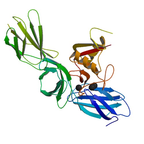
Versican is a large extracellular matrix proteoglycan that is present in a variety of human tissues. It is encoded by the ''VCAN'' gene. Versican is a large chondroitin sulfate proteoglycan with an apparent molecular mass of more than 1000kDa. In 1989, Zimmermann and Ruoslahti cloned and sequenced the core protein of fibroblast chondroitin sulfate proteoglycan. They designated it versican in recognition of its versatile modular structure. Versican belongs to the lectican protein family, with aggrecan (abundant in cartilage), brevican and neurocan (nervous system proteoglycans) as other members. Versican is also known as chondroitin sulfate proteoglycan core protein 2 or chondroitin sulfate proteoglycan 2 (CSPG2), and PG-M. Structure These proteoglycans share a homologous globular N-terminal, C-terminal, and glycosaminoglycan (GAG) binding regions. The N-terminal (G1) globular domain consists of Ig-like loop and two link modules, and has Hyaluronan (HA) binding properti ... [...More Info...] [...Related Items...] OR: [Wikipedia] [Google] [Baidu] |
Chondroitin Sulfate Proteoglycan
Chondroitin sulfate proteoglycans (CSPGs) are proteoglycans consisting of a protein core and a chondroitin sulfate side chain. They are known to be structural components of a variety of human tissues, including cartilage, and also play key roles in neural development and glial scar formation. They are known to be involved in certain cell processes, such as cell adhesion, cell growth, receptor binding, cell migration, and interaction with other extracellular matrix constituents. They are also known to interact with laminin, fibronectin, tenascin, and collagen. CSPGs are generally secreted from cells. Importantly, CSPGs are known to inhibit axon regeneration after spinal cord injury. CSPGs contribute to glial scar formation post injury, acting as a barrier against new axons growing into the injury site. CSPGs play a crucial role in explaining why the spinal cord doesn't self-regenerate after an injury. General structure Chondroitin sulfate proteoglycans are composed of a core protei ... [...More Info...] [...Related Items...] OR: [Wikipedia] [Google] [Baidu] |
Perineuronal Net
Perineuronal nets (PNNs) are specialized extracellular matrix structures responsible for synaptic stabilization in the adult brain. PNNs are found around certain neuron cell bodies and proximal neurites in the central nervous system. PNNs play a critical role in the closure of the childhood critical period, and their digestion can cause restored critical period-like synaptic plasticity in the adult brain. They are largely negatively charged and composed of chondroitin sulfate proteoglycans, molecules that play a key role in development and plasticity during postnatal development and in the adult. PNNs appear to be mainly present in the cortex, hippocampus, thalamus, brainstem, and the spinal cord. Studies of the rat brain have shown that the cortex contains high numbers of PNNs in the motor and primary sensory areas and relatively fewer in the association and limbic cortices. In the cortex, PNNs are associated mostly with inhibitory interneurons and are thought to be respon ... [...More Info...] [...Related Items...] OR: [Wikipedia] [Google] [Baidu] |
Lectican
Lecticans, also known as hyalectans, are a family of proteoglycans (a type protein that is attached to chains of negatively charged polysaccharides) that are components of the extracellular matrix. There are four members of the lectican family: aggrecan, brevican, neurocan, and versican. Lecticans interact with hyaluronic acid and tenascin-R to form a ternary complex. Tissue distribution Aggrecan is a major component of extracellular matrix in cartilage whereas versican is widely expressed in a number of connective tissues including those in vascular smooth muscle, skin epithelial cells, and the cells of central and peripheral nervous system. The expression of neurocan and brevican is largely restricted to neural tissues. Structure All four lecticans contain an N-terminal globular domain (G1 domain) that in turn contains an immunoglobulin V-set domain and a Link domain that binds hyaluronic acid; a long extended central domain (CS) that is modified with covalently atta ... [...More Info...] [...Related Items...] OR: [Wikipedia] [Google] [Baidu] |
Proteoglycan
Proteoglycans are proteins that are heavily glycosylated. The basic proteoglycan unit consists of a "core protein" with one or more covalently attached glycosaminoglycan (GAG) chain(s). The point of attachment is a serine (Ser) residue to which the glycosaminoglycan is joined through a tetrasaccharide bridge (e.g. chondroitin sulfate- GlcA- Gal-Gal- Xyl-PROTEIN). The Ser residue is generally in the sequence -Ser-Gly-X-Gly- (where X can be any amino acid residue but proline), although not every protein with this sequence has an attached glycosaminoglycan. The chains are long, linear carbohydrate polymers that are negatively charged under physiological conditions due to the occurrence of sulfate and uronic acid groups. Proteoglycans occur in connective tissue. Types Proteoglycans are categorized by their relative size (large and small) and the nature of their glycosaminoglycan chains. Types include: Certain members are considered members of the "small leucine-rich proteoglyc ... [...More Info...] [...Related Items...] OR: [Wikipedia] [Google] [Baidu] |
Aggrecan
Aggrecan (ACAN), also known as cartilage-specific proteoglycan core protein (CSPCP) or chondroitin sulfate proteoglycan 1, is a protein that in humans is encoded by the ''ACAN'' gene. This gene is a member of the lectican (chondroitin sulfate proteoglycan) family. The encoded protein is an integral part of the extracellular matrix in cartilagenous tissue and it withstands compression in cartilage. Aggrecan is a proteoglycan, or a protein modified with large carbohydrates; the human form of the protein is 2316 amino acids long and can be expressed in multiple isoforms due to alternative splicing. Aggrecan was named for its ability to form large aggregates in the cartilage tissue (a large aggregating proteoglycan). Structure Aggrecan is a high molecular weight (1x10⁶ < M < 3x10⁶) proteoglycan. It exhibits a bottlebrush structure, in which |
Myeloid Cell
A myelocyte is a young cell of the granulocytic series, occurring normally in bone marrow (can be found in circulating blood when caused by certain diseases). Structure When stained with the usual dyes, the cytoplasm is distinctly basophilic and relatively more abundant than in myeloblasts or promyelocytes, even though myelocytes are smaller cells. Numerous cytoplasmic granules are present in the more mature forms of myelocytes. Neutrophilic and eosinophilic granules are peroxidase-positive, while basophilic granules are not. The nuclear chromatin is coarser than that observed in a promyelocyte, but it is relatively faintly stained and lacks a well-defined membrane. The nucleus is fairly regular in contour (not indented), and seems to be 'buried' beneath the numerous cytoplasmic granules. (If the nucleus were indented, it would likely be a metamyelocyte.) Measurement There is an internationally agreed method of counting blasts, with results from M1 upwards. Development ... [...More Info...] [...Related Items...] OR: [Wikipedia] [Google] [Baidu] |
TLR2
Toll-like receptor 2 also known as TLR2 is a protein that in humans is encoded by the ''TLR2'' gene. TLR2 has also been designated as CD282 (cluster of differentiation 282). TLR2 is one of the toll-like receptors and plays a role in the immune system. TLR2 is a membrane protein, a receptor, which is expressed on the surface of certain cells and recognizes foreign substances and passes on appropriate signals to the cells of the immune system. Function The protein encoded by this gene is a member of the Toll-like receptor (TLR) family, which plays a fundamental role in pathogen recognition and activation of innate immunity. TLRs are highly conserved from ''Drosophila'' to humans and share structural and functional similarities. They recognize pathogen-associated molecular patterns (PAMPs) that are expressed on infectious agents, and mediate the production of cytokines necessary for the development of effective immunity. The various TLRs exhibit different patterns of expression. Th ... [...More Info...] [...Related Items...] OR: [Wikipedia] [Google] [Baidu] |
Lewis Lung Carcinoma
Lewis lung carcinoma is a tumor that spontaneously developed as an epidermoid carcinoma in the lung of a C57BL mouse. It was discovered in 1951 by Dr. Margaret Lewis of the Wistar Institute and became one of the first transplantable tumors. Models Syngeneic According to a 2015 review article, Lewis lung carcinoma is the only reproducible syngeneic lung cancer model, meaning that it is the only reproducible lung cancer model that utilizes a transplant that is immunologically compatible. Syngeneic models have proven to be useful in predicting clinical benefit of therapy in preclinical experiments. However, there has been criticism directed towards syngeneic model usage when attempting to translate therapies from another species to humans. For example, cancer therapies that exhibited promising results in mouse models can and have failed in clinical trials due to physiological differences in the activity of the targeted gene product. The activity of the mouse product did not transla ... [...More Info...] [...Related Items...] OR: [Wikipedia] [Google] [Baidu] |
Carbohydrate-binding Module
In molecular biology, a carbohydrate-binding module (CBM) is a protein domain found in carbohydrate-active enzymes (for example glycoside hydrolases). The majority of these domains have carbohydrate-binding activity. Some of these domains are found on cellulosome, cellulosomal scaffoldin proteins. CBMs were previously known as cellulose-binding domains. CBMs are classified into numerous families, based on amino acid sequence similarity. There are currently (June 2011) 64 families of CBM in the CAZy database. CBMs of microbe, microbial glycoside hydrolases play a central role in the recycling of Photosynthesis, photosynthetically fixed carbon through their Binding (molecular), binding to specific plant structural polysaccharides. CBMs can recognise both crystalline and amorphous cellulose forms. CBMs are the most common non-catalytic modules associated with enzymes active in plant cell-wall hydrolysis. Many putative CBMs have been identified by amino acid sequence ... [...More Info...] [...Related Items...] OR: [Wikipedia] [Google] [Baidu] |
Breast
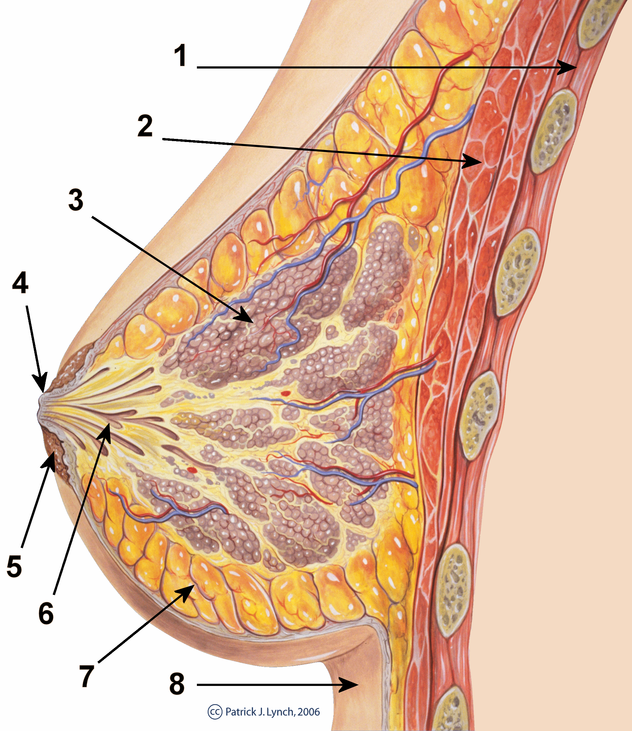
The breast is one of two prominences located on the upper ventral region of a primate's torso. Both females and males develop breasts from the same embryological tissues. In females, it serves as the mammary gland, which produces and secretes milk to feed infants. Subcutaneous fat covers and envelops a network of ducts that converge on the nipple, and these tissues give the breast its size and shape. At the ends of the ducts are lobules, or clusters of alveoli, where milk is produced and stored in response to hormonal signals. During pregnancy, the breast responds to a complex interaction of hormones, including estrogens, progesterone, and prolactin, that mediate the completion of its development, namely lobuloalveolar maturation, in preparation of lactation and breastfeeding. Humans are the only animals with permanent breasts. At puberty, estrogens, in conjunction with growth hormone, cause permanent breast growth in female humans. This happens only to a much lesser ... [...More Info...] [...Related Items...] OR: [Wikipedia] [Google] [Baidu] |

.png)


