|
Villoglandular Adenocarcinoma Of The Cervix
Villoglandular adenocarcinoma of the cervix is a rare type of cervical cancer that, in relation to other cervical cancers, is typically found in younger women and has a better prognosis. A similar lesion, ''villoglandular adenocarcinoma of the endometrium'', may arise from the inner lining of the uterus, the endometrium. Signs and symptoms The signs and symptoms are similar to other cervical cancers and may include post- coital bleeding and/or pain during intercourse ( dyspareunia). Early lesions may be completely asymptomatic. Cause Diagnosis The diagnosis is based on tissue examination, e.g. biopsy. The name of the lesion describes it microscopic appearance. It has nipple-like structures with fibrovascular cores ( papillae) that are long in relation to their width ( villus-like), which are covered with a glandular pseudostratified columnar epithelium. File:Villoglandular adenocarcinoma - very low mag.jpg, Very low magnification File:Villoglandular adenocarcinoma - interm ... [...More Info...] [...Related Items...] OR: [Wikipedia] [Google] [Baidu] |
Micrograph
A micrograph or photomicrograph is a photograph or digital image taken through a microscope or similar device to show a magnified image of an object. This is opposed to a macrograph or photomacrograph, an image which is also taken on a microscope but is only slightly magnified, usually less than 10 times. Micrography is the practice or art of using microscopes to make photographs. A micrograph contains extensive details of microstructure. A wealth of information can be obtained from a simple micrograph like behavior of the material under different conditions, the phases found in the system, failure analysis, grain size estimation, elemental analysis and so on. Micrographs are widely used in all fields of microscopy. Types Photomicrograph A light micrograph or photomicrograph is a micrograph prepared using an optical microscope, a process referred to as ''photomicroscopy''. At a basic level, photomicroscopy may be performed simply by connecting a camera to a microscope, th ... [...More Info...] [...Related Items...] OR: [Wikipedia] [Google] [Baidu] |
Chorionic Villi
Chorionic villi are villi that sprout from the chorion to provide maximal contact area with maternal blood. They are an essential element in pregnancy from a histomorphologic perspective, and are, by definition, a product of conception. Branches of the umbilical arteries carry embryonic blood to the villi. After circulating through the capillaries of the villi, blood returns to the embryo through the umbilical vein. Thus, villi are part of the border between maternal and fetal blood during pregnancy. Structure Villi can also be classified by their relations: * Floating villi float freely in the intervillous space. They exhibit a bi-layered epithelium consisting of cytotrophoblasts with overlaying syncytium ( syncytiotrophoblast). * Anchoring (stem) villi stabilize mechanical integrity of the placental-maternal interface. Development The chorion undergoes rapid proliferation and forms numerous processes, the chorionic villi, which invade and destroy the uterine decidua and a ... [...More Info...] [...Related Items...] OR: [Wikipedia] [Google] [Baidu] |
Villous Adenoma
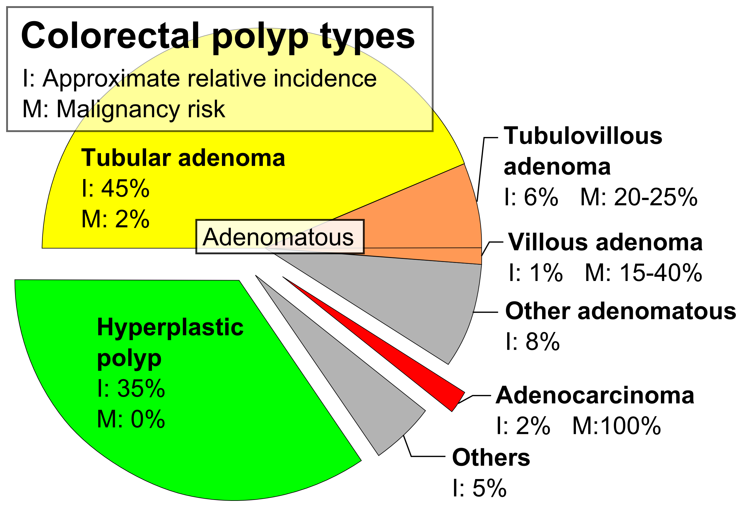
The colorectal adenoma is a benign glandular tumor of the colon and the rectum. It is a precursor lesion of the colorectal adenocarcinoma (colon cancer). They often manifest as colorectal polyps. Comparison table Tubular adenoma In contrast to hyperplastic polyps, these display dysplasia. Tubulovillous adenoma ''Tubulovillous adenoma'', ''TVA'' are considered to have a higher risk of becoming malignant (cancerous) than tubular adenomas. Villous adenoma These adenomas may become malignant (cancerous). Villous adenomas have been demonstrated to contain malignant portions in about 15–25% of cases, approaching 40% in those over 4 cm in diameter. Colonic resection may be required for large lesions. These can also lead to secretory diarrhea with large volume liquid stools with few formed elements. They are commonly described as secreting large amounts of mucus, resulting in hypokalaemia in patients. On endoscopy, a "cauliflower' like mass is described due to villi stretching ... [...More Info...] [...Related Items...] OR: [Wikipedia] [Google] [Baidu] |
Glassy Cell Carcinoma Of The Cervix
Glassy cell carcinoma of the cervix, also glassy cell carcinoma, is a rare aggressive malignant tumour of the uterine cervix. The tumour gets its name from its microscopic appearance; its cytoplasm has a glass-like appearance. Signs and symptoms The signs and symptoms are similar to other cervical cancers and may include post- coital bleeding and/or pain during intercourse ( dyspareunia). Early lesions may be completely asymptomatic. Cause Diagnosis The diagnosis is based on tissue examination, e.g. biopsy. Under the microscope, glassy cell carcinoma tumours are composed of cells with a glass-like cytoplasm, typically associated with an inflammatory infiltrate abundant in eosinophils and very mitotically active. PAS staining highlights the plasma membrane. Treatment The treatment is dependent on the stage. Advanced tumours are treated with surgery (radical hysterectomy and bilateral salpingo-opherectomy), radiation therapy and chemotherapy. See also * Cervix * Cervical ca ... [...More Info...] [...Related Items...] OR: [Wikipedia] [Google] [Baidu] |
Endometrial Cancer
Endometrial cancer is a cancer that arises from the endometrium (the lining of the uterus or womb). It is the result of the abnormal growth of cells that have the ability to invade or spread to other parts of the body. The first sign is most often vaginal bleeding not associated with a menstrual period. Other symptoms include pain with urination, pain during sexual intercourse, or pelvic pain. Endometrial cancer occurs most commonly after menopause. Approximately 40% of cases are related to obesity. Endometrial cancer is also associated with excessive estrogen exposure, high blood pressure and diabetes. Whereas taking estrogen alone increases the risk of endometrial cancer, taking both estrogen and a progestogen in combination, as in most birth control pills, decreases the risk. Between two and five percent of cases are related to genes inherited from the parents. Endometrial cancer is sometimes loosely referred to as "uterine cancer", although it is distinct from other fo ... [...More Info...] [...Related Items...] OR: [Wikipedia] [Google] [Baidu] |
Cervicectomy
In gynecologic oncology, trachelectomy, also called cervicectomy, is a surgical removal of the uterine cervix. As the uterine body is preserved, this type of surgery is a fertility preserving surgical alternative to a radical hysterectomy and applicable in selected younger women with early cervical cancer. Types Trachelectomies, broadly, can be divided into the ''simple'' and ''radical'' variants. Radical The formal name of this operation is ''radical vaginal trachelectomy (RVT)'' and also known as the ''Dargent operation'' and ''radical trachelectomy''. The word ''radical'' is used as, in addition to the cervix (like in radical hysterectomies), the parametria (tissue adjacent to the cervix) and vaginal cuff (the end of the vagina close to the cervix) are also excised as a part of the operation. It is usually done with a lymphadenectomy, to assess for tumour spread to the lymph nodes. This operation was pioneered by the renowned French Obstetrician-Gynecologist Surgeon, Daniel ... [...More Info...] [...Related Items...] OR: [Wikipedia] [Google] [Baidu] |
Cervical Conization
Cervical conization ( CPT codes 57520 (Cold Knife) and 57522 (Loop Excision)) refers to an excision of a cone-shaped sample of tissue from the mucous membrane of the cervix. Conization may be used for either diagnostic purposes as part of a biopsy or therapeutic purposes to remove pre-cancerous cells. Types include: * Cold knife conization (CKC): usually outpatient, occasionally inpatient * Loop electrical excision procedure (LEEP): usually outpatient. Conization of the cervix is a common treatment for dysplasia following abnormal results from a pap smear. Side effects Cervical conization causes a risk for subsequent pregnancies ending up in preterm birth of approximately 30% on average, due to cervical incompetence Cervical weakness, also called cervical incompetence or cervical insufficiency, is a medical condition of pregnancy in which the cervix begins to dilate (widen) and efface (thin) before the pregnancy has reached term. Definitions of cervical wea .... See also * ... [...More Info...] [...Related Items...] OR: [Wikipedia] [Google] [Baidu] |
Fertility
Fertility is the capability to produce offspring through reproduction following the onset of sexual maturity. The fertility rate is the average number of children born by a female during her lifetime and is quantified demographically. Fertility is addressed when there is a difficulty or an inability to reproduce naturally, which is referred to as infertility. Infertility is widespread, with fertility specialists available all over the world to assist mothers and couples who experience difficulties having a baby. Human fertility depends on factors of nutrition, sexual behaviour, consanguinity, culture, instinct, endocrinology, timing, economics, personality, way of life, and emotions. Fertility differs from fecundity, which is defined as the ''potential'' for reproduction (influenced by gamete production, fertilization and carrying a pregnancy to term). Where a woman or the lack of fertility is infertility while a lack of fecundity would be called sterility. Demography I ... [...More Info...] [...Related Items...] OR: [Wikipedia] [Google] [Baidu] |
Cancer Stage
Cancer is a group of diseases involving abnormal cell growth with the potential to invade or spread to other parts of the body. These contrast with benign tumors, which do not spread. Possible signs and symptoms include a lump, abnormal bleeding, prolonged cough, unexplained weight loss, and a change in bowel movements. While these symptoms may indicate cancer, they can also have other causes. Over 100 types of cancers affect humans. Tobacco use is the cause of about 22% of cancer deaths. Another 10% are due to obesity, poor diet, lack of physical activity or excessive drinking of alcohol. Other factors include certain infections, exposure to ionizing radiation, and environmental pollutants. In the developing world, 15% of cancers are due to infections such as ''Helicobacter pylori'', hepatitis B, hepatitis C, human papillomavirus infection, Epstein–Barr virus and human immunodeficiency virus (HIV). These factors act, at least partly, by changing the genes of a cell. ... [...More Info...] [...Related Items...] OR: [Wikipedia] [Google] [Baidu] |
Pseudostratified Columnar Epithelium
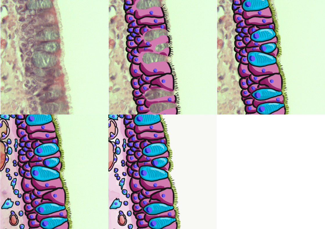
A pseudostratified epithelium is a type of epithelium that, though comprising only a single layer of cells, has its cell nuclei positioned in a manner suggestive of stratified epithelia. As it rarely occurs as squamous or cuboidal epithelia, it is usually considered synonymous with the term pseudostratified columnar epithelium. The term ''pseudostratified'' is derived from the appearance of this epithelium in the section which conveys the erroneous (''pseudo'' means almost or approaching) impression that there is more than one layer of cells, when in fact this is a true simple epithelium since all the cells rest on the basement membrane. The nuclei of these cells, however, are disposed at different levels, thus creating the illusion of cellular stratification. All cells are not of equal size and not all cells extend to the luminal/apical surface; such cells are capable of cell division providing replacements for cells lost or damaged. Pseudostratified epithelia function in s ... [...More Info...] [...Related Items...] OR: [Wikipedia] [Google] [Baidu] |
H&E Stain
Hematoxylin and eosin stain ( or haematoxylin and eosin stain or hematoxylin-eosin stain; often abbreviated as H&E stain or HE stain) is one of the principal tissue stains used in histology. It is the most widely used stain in medical diagnosis and is often the gold standard. For example, when a pathologist looks at a biopsy of a suspected cancer, the histological section is likely to be stained with H&E. H&E is the combination of two histological stains: hematoxylin and eosin. The hematoxylin stains cell nuclei a purplish blue, and eosin stains the extracellular matrix and cytoplasm pink, with other structures taking on different shades, hues, and combinations of these colors. Hence a pathologist can easily differentiate between the nuclear and cytoplasmic parts of a cell, and additionally, the overall patterns of coloration from the stain show the general layout and distribution of cells and provides a general overview of a tissue sample's structure. Thus, pattern recogniti ... [...More Info...] [...Related Items...] OR: [Wikipedia] [Google] [Baidu] |


