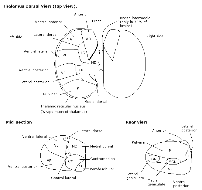
|
Ventral Posteromedial
The ventral posteromedial nucleus (VPM) is a Nucleus (neuroanatomy), nucleus of the thalamus. Inputs and outputs The VPM contains synapses between second and third order neurons from the Anterior trigeminothalamic tract, anterior (ventral) trigeminothalamic tract and Dorsal Trigeminothalamic Tract, posterior (dorsal) trigeminothalamic tract. These neurons convey sensory information from the face and oral cavity. Third order neurons in the trigeminothalamic systems project to the postcentral gyrus. The VPM also receives taste afferent information from the solitary tract and projects to the cortical gustatory area. Subareas VPMpc The parvicellular part of the ventroposterior medial nucleus (VPMpc) is argued by some as not an actually part of the VPM, because it does not project to the somatosensory cortex as the remainder of the VPM does, and therefore should be called the ventromedial nucleus (VMb) instead. Sources * Kiernan, J., & Rajakumar, R. (2013). ''Barr's the human ... [...More Info...] [...Related Items...] OR: [Wikipedia] [Google] [Baidu] |
Midline Nuclear Group
The midline nuclear group (or midline thalamic nuclei) is a region of the thalamus consisting of the following nuclei: * paraventricular nucleus of thalamus (''nucleus paraventricularis thalami'') - not to be confused with paraventricular nucleus of hypothalamus * paratenial nucleus The paratenial nucleus, or parataenial nucleus ( la, nucleus parataenialis), is a component of the midline nuclear group in the thalamus. It is sometimes subdivided into the nucleus parataenialis interstitialis and nucleus parataenialis parvocellu ... (''nucleus parataenialis'') * nucleus reuniens * rhomboid nucleus (''nucleus commissuralis rhomboidalis'') * subfascicular nucleus (''nucleus subfascicularis'') The midline nuclei are often called "nonspecific" in that they project widely to the cortex and elsewhere. This has led to the assumption that they may be involved in general functions such as alerting. However, anatomical connections might suggest more specific functions, with the paraventr ... [...More Info...] [...Related Items...] OR: [Wikipedia] [Google] [Baidu] |
Lateral Geniculate Nucleus
In neuroanatomy, the lateral geniculate nucleus (LGN; also called the lateral geniculate body or lateral geniculate complex) is a structure in the thalamus and a key component of the mammalian visual pathway. It is a small, ovoid, ventral projection of the thalamus where the thalamus connects with the optic nerve. There are two LGNs, one on the left and another on the right side of the thalamus. In humans, both LGNs have six layers of neurons (grey matter) alternating with optic fibers (white matter). The LGN receives information directly from the ascending retinal ganglion cells via the optic tract and from the reticular activating system. Neurons of the LGN send their axons through the optic radiation, a direct pathway to the primary visual cortex. In addition, the LGN receives many strong feedback connections from the primary visual cortex. In humans as well as other mammals, the two strongest pathways linking the eye to the brain are those projecting to the dorsal part of th ... [...More Info...] [...Related Items...] OR: [Wikipedia] [Google] [Baidu] |
Solitary Tract
The solitary tract (tractus solitarius, or fasciculus solitarius), is a compact fiber bundle that extends longitudinally through the posterolateral region of the medulla oblongata. The solitary tract is surrounded by the solitary nucleus, and descends to the upper cervical segments of the spinal cord. It was first named by Theodor Meynert in 1872. Composition The solitary tract is made up of primary sensory fibers and descending fibers of the vagus, glossopharyngeal, and facial nerves. Function The solitary tract conveys afferent information from stretch receptors and chemoreceptors in the walls of the cardiovascular, respiratory, and intestinal tracts. Afferent fibers from cranial nerves 7, 9 and 10 convey taste ( SVA) in its rostral portion, and general visceral sense ( general visceral afferent fibers, GVA) in its caudal part. Taste buds in the mucosa of the tongue can also generate impulses in the rostral regions of the solitary tract. The efferent fibers are distrib ... [...More Info...] [...Related Items...] OR: [Wikipedia] [Google] [Baidu] |
Dorsal Trigeminothalamic Tract
The dorsal trigeminal tract, dorsal trigeminothalamic tract, or posterior trigeminothalamic tract, is composed of second-order neuronal axons. These fibers carry sensory information about discriminative touch and conscious proprioception in the oral cavity from the principal (chief sensory) nucleus of the trigeminal nerve to the ventral posteromedial (VPM) nucleus of the thalamus. The dorsal trigeminothalamic tract is also called the posterior trigeminal lemniscus. Pathway The first-order neurons (from the trigeminal ganglion) enter the pons and synapse on second-order neurons in the principal (chief sensory) nucleus. Axons of the second-order neurons then decussate to enter the trigeminal lemniscus in the midbrain and then ascend to the ventral posteromedial nucleus of the contralateral thalamus, forming the ventral trigeminothalamic tract. A subset of these fibers do not decussate and travel to the ipsilateral ventral posteromedial nucleus of the thalamus. These non-decu ... [...More Info...] [...Related Items...] OR: [Wikipedia] [Google] [Baidu] |
Anterior Trigeminothalamic Tract
The ventral trigeminal tract, ventral trigeminothalamic tract, anterior trigeminal tract, or anterior trigeminothalamic tract, is a tract composed of second order neuronal axons. These fibers carry sensory information about discriminative and crude touch, conscious proprioception, pain, and temperature from the head, face, and oral cavity. The ventral trigeminal tract connects the two major components of the brainstem trigeminal complex – the principal, or main sensory nucleus and the spinal trigeminal nucleus, to the ventral posteromedial nucleus of the thalamus. The ventral trigeminal tract is also called the anterior trigeminal lemniscus. Structure The first order neurons (from the trigeminal ganglion) enter the pons and synapse in the principal (chief sensory) nucleus or spinal trigeminal nucleus. Axons of the second order neurons cross the midline and terminate in the ventral posteromedial nucleus of the contralateral thalamus (as opposed to the ventral posterolateral nucl ... [...More Info...] [...Related Items...] OR: [Wikipedia] [Google] [Baidu] |
Thalamus
The thalamus (from Greek θάλαμος, "chamber") is a large mass of gray matter located in the dorsal part of the diencephalon (a division of the forebrain). Nerve fibers project out of the thalamus to the cerebral cortex in all directions, allowing hub-like exchanges of information. It has several functions, such as the relaying of sensory signals, including motor signals to the cerebral cortex and the regulation of consciousness, sleep, and alertness. Anatomically, it is a paramedian symmetrical structure of two halves (left and right), within the vertebrate brain, situated between the cerebral cortex and the midbrain. It forms during embryonic development as the main product of the diencephalon, as first recognized by the Swiss embryologist and anatomist Wilhelm His Sr. in 1893. Anatomy The thalamus is a paired structure of gray matter located in the forebrain which is superior to the midbrain, near the center of the brain, with nerve fibers projecting out to the ... [...More Info...] [...Related Items...] OR: [Wikipedia] [Google] [Baidu] |
Nucleus (neuroanatomy)
In neuroanatomy, a nucleus (plural form: nuclei) is a cluster of neurons in the central nervous system, located deep within the cerebral hemispheres and brainstem. The neurons in one nucleus usually have roughly similar connections and functions. Nuclei are connected to other nuclei by tracts, the bundles (fascicles) of axons (nerve fibers) extending from the cell bodies. A nucleus is one of the two most common forms of nerve cell organization, the other being layered structures such as the cerebral cortex or cerebellar cortex. In anatomical sections, a nucleus shows up as a region of gray matter, often bordered by white matter. The vertebrate brain contains hundreds of distinguishable nuclei, varying widely in shape and size. A nucleus may itself have a complex internal structure, with multiple types of neurons arranged in clumps (subnuclei) or layers. The term "nucleus" is in some cases used rather loosely, to mean simply an identifiably distinct group of neurons, even if they ... [...More Info...] [...Related Items...] OR: [Wikipedia] [Google] [Baidu] |
Medial Geniculate Nucleus
The medial geniculate nucleus (MGN) or medial geniculate body (MGB) is part of the auditory thalamus and represents the thalamic relay between the inferior colliculus (IC) and the auditory cortex (AC). It is made up of a number of sub-nuclei that are distinguished by their neuronal morphology and density, by their afferent and efferent connections, and by the coding properties of their neurons. It is thought that the MGN influences the direction and maintenance of attention. Divisions The MGN has three major divisions; ventral (VMGN), dorsal (DMGN) and medial (MMGN). Whilst the VMGN is specific to auditory information processing, the DMGN and MMGN also receive information from non-auditory pathways. Ventral subnucleus Cell types There are two main cell types in the ventral subnucleus of the medial geniculate body (VMGN): * Thalamocortical relay cells (or principal neurons): The dendritic input to these cells comes from two sets of dendritic trees oriented on opposite poles of th ... [...More Info...] [...Related Items...] OR: [Wikipedia] [Google] [Baidu] |
Metathalamus
The thalamus (from Greek θάλαμος, "chamber") is a large mass of gray matter located in the dorsal part of the diencephalon (a division of the forebrain). Nerve fibers project out of the thalamus to the cerebral cortex in all directions, allowing hub-like exchanges of information. It has several functions, such as the relaying of sensory signals, including motor signals to the cerebral cortex and the regulation of consciousness, sleep, and alertness. Anatomically, it is a paramedian symmetrical structure of two halves (left and right), within the vertebrate brain, situated between the cerebral cortex and the midbrain. It forms during embryonic development as the main product of the diencephalon, as first recognized by the Swiss embryologist and anatomist Wilhelm His Sr. in 1893. Anatomy The thalamus is a paired structure of gray matter located in the forebrain which is superior to the midbrain, near the center of the brain, with nerve fibers projecting out to the ce ... [...More Info...] [...Related Items...] OR: [Wikipedia] [Google] [Baidu] |
Anterior Nuclei Of Thalamus
The anterior nuclei of thalamus (or anterior nuclear group) are a collection of nuclei at the rostral end of the dorsal thalamus. They comprise the anteromedial, anterodorsal, and anteroventral nuclei. Inputs and outputs The anterior nuclei receive afferents from the mammillary bodies via the mammillothalamic tract and from the subiculum via the fornix. In turn, they project to the cingulate gyrus. The anterior nuclei of the thalamus display functions pertaining to memory. Persons displaying lesions in the anterior thalamus, preventing input from the pathway involving the hippocampus, mammillary bodies and the MTT, display forms of amnesia, supporting the anterior thalamus's involvement in episodic memory. However, although the hypothalamus projects to both the mammillary bodies and the anterior nuclei of the thalamus, the anterior nuclei receive input from hippocampal cells deep to the pyramidal cells projecting to the mammillary bodies. These nuclei are considered to be ass ... [...More Info...] [...Related Items...] OR: [Wikipedia] [Google] [Baidu] |
Pulvinar Nuclei
The pulvinar nuclei or nuclei of the pulvinar (nuclei pulvinares) are the nuclei (cell bodies of neurons) located in the thalamus (a part of the vertebrate brain). As a group they make up the collection called the pulvinar of the thalamus (pulvinar thalami), usually just called the pulvinar. The pulvinar is usually grouped as one of the ''lateral thalamic nuclei'' in rodents and carnivores, and stands as an independent complex in primates. Structure By convention, the pulvinar is divided into four nuclei: Their connectomic details are as follows: * The ''lateral'' and ''inferior'' pulvinar nuclei have widespread connections with early visual cortical areas. * The dorsal part of the ''lateral'' pulvinar nucleus predominantly has connections with posterior parietal cortex and the dorsal stream cortical areas. * The ''medial'' pulvinar nucleus has widespread connections with cingulate, posterior parietal, premotor and prefrontal cortical areas. * The pulvinar also has input fr ... [...More Info...] [...Related Items...] OR: [Wikipedia] [Google] [Baidu] |
Lateral Nuclear Group
The lateral nuclear group is a collection of nuclei on the lateral side of the thalamus. According to MeSH A mesh is a barrier made of connected strands of metal, fiber, or other flexible or ductile materials. A mesh is similar to a web or a net in that it has many attached or woven strands. Types * A plastic mesh may be extruded, oriented, exp ..., it consists of the following: * lateral dorsal nucleus * lateral posterior nucleus * pulvinar Thalamic nuclei {{Neuroanatomy-stub ... [...More Info...] [...Related Items...] OR: [Wikipedia] [Google] [Baidu] |
