|
VATER Association
The VACTERL association (also VATER association, and less accurately VACTERL syndrome) refers to a recognized group of birth defects which tend to co-occur (see below). This pattern is a recognized association, as opposed to a syndrome, because there is no known pathogenetic cause to explain the grouped incidence. Each child with this condition can be unique. At present this condition is treated after birth with issues being approached one at a time. Some infants are born with symptoms that cannot be treated and they do not survive. Also, VACTERL association can be linked to other similar conditions such as Klippel Feil and Goldenhar syndrome including crossovers of conditions. No specific genetic or chromosome problem has been identified with VACTERL association. VACTERL can be seen with some chromosomal defects such as Trisomy 18 and is more frequently seen in babies of diabetic mothers. VACTERL association, however, is most likely caused by multiple factors. VACTERL associat ... [...More Info...] [...Related Items...] OR: [Wikipedia] [Google] [Baidu] |
Radial Aplasia
Radial aplasia is a congenital defect which affects the formation of the radius bone in the arm. The radius is the lateral bone (thumb side) which connects the humerus of the upper arm to the wrist via articulation with the carpal bones. A child born with this condition has either a short or absent radius bone in one or both of his or her arms. Radial aplasia also results in the thumb being either partly formed or completely absent from the hand, which can result in difficulties performing activities of daily living. Radial aplasia is connected with the condition VACTERL association The VACTERL association (also VATER association, and less accurately VACTERL syndrome) refers to a recognized group of birth defects which tend to co-occur (see below). This pattern is a recognized association, as opposed to a syndrome, because th ..., under the 'L' for limb malformations. Radial aplasia is not inherited. The cause for radial aplasia is unknown, but it widely believed to occur within ... [...More Info...] [...Related Items...] OR: [Wikipedia] [Google] [Baidu] |
Scoliosis
Scoliosis is a condition in which a person's spine has a sideways curve. The curve is usually "S"- or "C"-shaped over three dimensions. In some, the degree of curve is stable, while in others, it increases over time. Mild scoliosis does not typically cause problems, but more severe cases can affect breathing and movement. Pain is usually present in adults, and can worsen with age. The cause of most cases is unknown, but it is believed to involve a combination of genetic and environmental factors. Risk factors include other affected family members. It can also occur due to another condition such as muscle spasms, cerebral palsy, Marfan syndrome, and tumors such as neurofibromatosis. Diagnosis is confirmed with X-rays. Scoliosis is typically classified as either structural in which the curve is fixed, or functional in which the underlying spine is normal. Treatment depends on the degree of curve, location, and cause. Minor curves may simply be watched periodically. Treatme ... [...More Info...] [...Related Items...] OR: [Wikipedia] [Google] [Baidu] |
Kidney Transplant
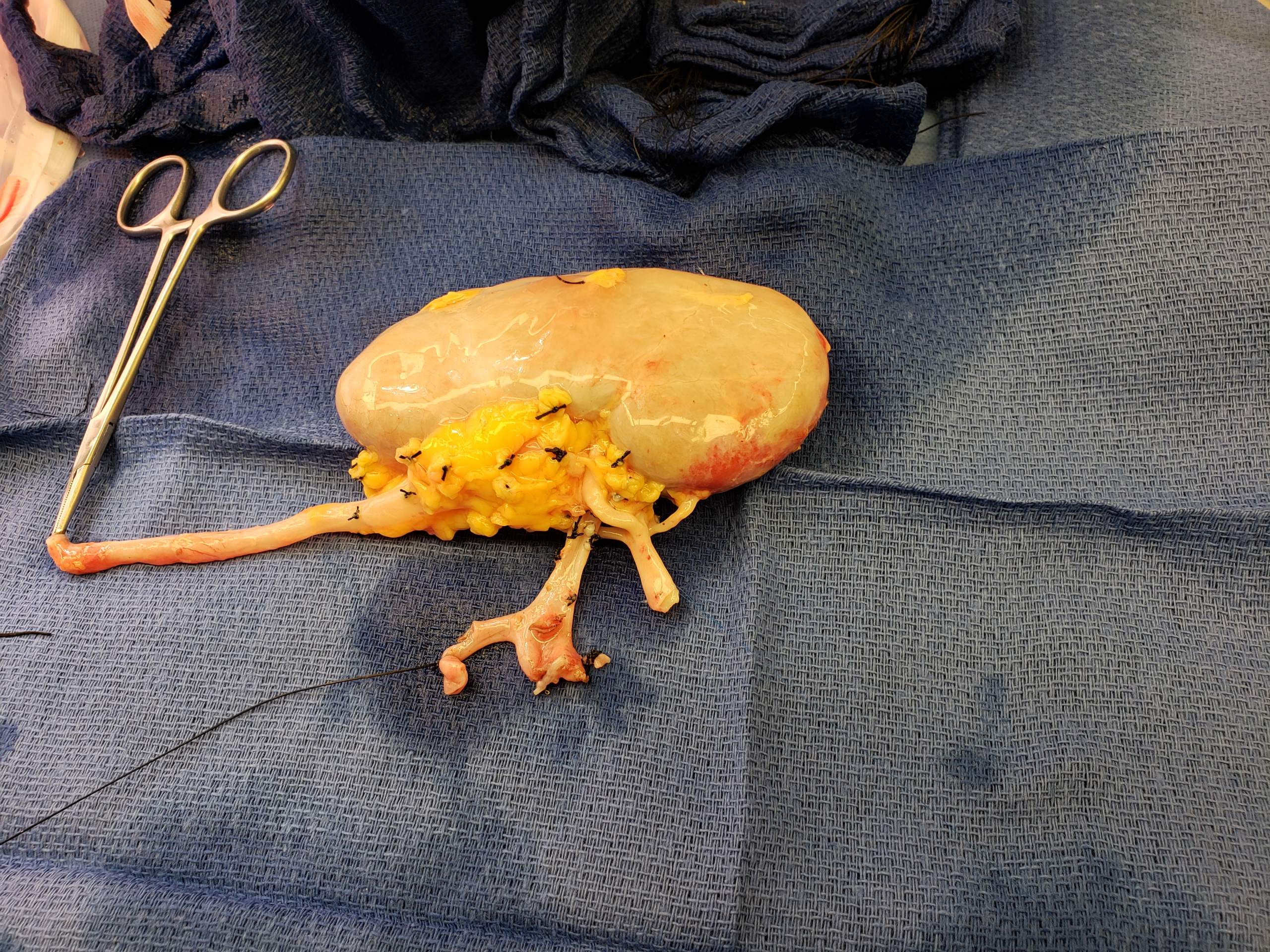
Kidney transplant or renal transplant is the organ transplant of a kidney into a patient with end-stage kidney disease (ESRD). Kidney transplant is typically classified as deceased-donor (formerly known as cadaveric) or living-donor transplantation depending on the source of the donor organ. Living-donor kidney transplants are further characterized as genetically related (living-related) or non-related (living-unrelated) transplants, depending on whether a biological relationship exists between the donor and recipient. Before receiving a kidney transplant, a person with ESRD must undergo a thorough medical evaluation to make sure that they are healthy enough to undergo transplant surgery. If they are deemed a good candidate, they can be placed on a waiting list to receive a kidney from a deceased donor. Once they are placed on the waiting list, they can receive a new kidney very quickly, or they may have to wait many years; in the United States, the average waiting time is three t ... [...More Info...] [...Related Items...] OR: [Wikipedia] [Google] [Baidu] |
Kidney Failure
Kidney failure, also known as end-stage kidney disease, is a medical condition in which the kidneys can no longer adequately filter waste products from the blood, functioning at less than 15% of normal levels. Kidney failure is classified as either acute kidney failure, which develops rapidly and may resolve; and chronic kidney failure, which develops slowly and can often be irreversible. Symptoms may include leg swelling, feeling tired, vomiting, loss of appetite, and confusion. Complications of acute and chronic failure include uremia, high blood potassium, and volume overload. Complications of chronic failure also include heart disease, high blood pressure, and anemia. Causes of acute kidney failure include low blood pressure, blockage of the urinary tract, certain medications, muscle breakdown, and hemolytic uremic syndrome. Causes of chronic kidney failure include diabetes, high blood pressure, nephrotic syndrome, and polycystic kidney disease. Diagnosis of acute failure ... [...More Info...] [...Related Items...] OR: [Wikipedia] [Google] [Baidu] |
Renal Agenesis
Renal agenesis is a medical condition in which one (unilateral) or both (bilateral) fetal kidneys fail to develop. Unilateral and bilateral renal agenesis in humans, mice and zebra fish has been linked to mutations in the gene GREB1L. It has also been associated with mutations in the genes ''RET proto-oncogene, RET'' or ''UPK3A'' in humans and mice respectively. Type Bilateral Bilateral renal agenesis is a condition in which both kidneys of a fetus fail to develop during gestation. It is incompatible with life. It is one causative agent of Potter sequence. This absence of kidneys causes oligohydramnios, a deficiency of amniotic fluid in a pregnant woman, which can place extra pressure on the developing baby and cause further malformations. The condition is frequently, but not always the result of a genetic disorder, and is more common in infants born to one or more parents with a malformed or absent kidney. Unilateral This is much more common, but is not usually of any major hea ... [...More Info...] [...Related Items...] OR: [Wikipedia] [Google] [Baidu] |
Single Umbilical Artery
Occasionally, there is only the one single umbilical artery (SUA) present in the umbilical cord. This is sometimes also called a two-vessel umbilical cord, or two-vessel cord. Approximately, this affects between 1 in 100 and 1 in 500 pregnancies, making it the most common umbilical abnormality. Its cause is not known. Most cords have one vein and two arteries. The vein carries oxygenated blood from the placenta to the baby and the arteries carry deoxygenated blood from the baby to the placenta. In approximately 1% of pregnancies there are only two vessels —usually a single vein and single artery. In about 75% of those cases, the baby is entirely normal and healthy. One artery can support a pregnancy and does not necessarily indicate problems. For the other 25%, a 2-vessel cord is a sign that the baby has other abnormalities—sometimes life-threatening and sometimes not. Doctors and midwives often suggest parents take the added precaution of having regular growth scans near term ... [...More Info...] [...Related Items...] OR: [Wikipedia] [Google] [Baidu] |
Tracheoesophageal Fistula
A tracheoesophageal fistula (TEF, or TOF; see spelling differences) is an abnormal connection (fistula) between the esophagus and the trachea. TEF is a common congenital abnormality, but when occurring late in life is usually the sequela of surgical procedures such as a laryngectomy. Presentation Tracheoesophageal fistula is suggested in a newborn by copious salivation associated with choking, coughing, vomiting, and cyanosis coincident with the onset of feeding. Esophageal atresia and the subsequent inability to swallow typically cause polyhydramnios in utero. Rarely it may present in an adult. Complications Surgical repair can sometimes result in complications, including: * Stricture, due to gastric acid erosion of the shortened esophagus * Leak of contents at the point of anastomosis * Recurrence of fistula * Gastro-esophageal reflux disease * Dysphagia * Asthma-like symptoms, such as persistent coughing/wheezing * Recurrent chest infections * Tracheomalacia Associations ... [...More Info...] [...Related Items...] OR: [Wikipedia] [Google] [Baidu] |
Oesophageal Atresia
The esophagus (American English) or oesophagus (British English; both ), non-technically known also as the food pipe or gullet, is an organ in vertebrates through which food passes, aided by peristaltic contractions, from the pharynx to the stomach. The esophagus is a fibromuscular tube, about long in adults, that travels behind the trachea and heart, passes through the diaphragm, and empties into the uppermost region of the stomach. During swallowing, the epiglottis tilts backwards to prevent food from going down the larynx and lungs. The word ''oesophagus'' is from Ancient Greek οἰσοφάγος (oisophágos), from οἴσω (oísō), future form of φέρω (phérō, “I carry”) + ἔφαγον (éphagon, “I ate”). The wall of the esophagus from the lumen outwards consists of mucosa, submucosa (connective tissue), layers of muscle fibers between layers of fibrous tissue, and an outer layer of connective tissue. The mucosa is a stratified squamous epithelium ... [...More Info...] [...Related Items...] OR: [Wikipedia] [Google] [Baidu] |
Transposition Of The Great Arteries
Transposition of the great vessels (TGV) is a group of congenital heart defects involving an abnormal spatial arrangement of any of the great vessels: superior and/or inferior venae cavae, pulmonary artery, pulmonary veins, and aorta. Congenital heart diseases involving only the primary arteries (pulmonary artery and aorta) belong to a sub-group called transposition of the great arteries (TGA), which is considered the most common congenital heart lesion that presents in neonates. Types Transposed vessels can present with atriovenous, ventriculoarterial and/or arteriovenous discordance. The effects may range from a slight change in blood pressure to an interruption in circulation depending on the nature and degree of the misplacement, and on which specific vessels are involved. Although "transposed" literally means "swapped", many types of TGV involve vessels that are in abnormal positions, while not actually being swapped with each other. The terms TGV and TGA are most c ... [...More Info...] [...Related Items...] OR: [Wikipedia] [Google] [Baidu] |
Persistent Truncus Arteriosus
Persistent truncus arteriosus (PTA), often referred to simply as truncus arteriosus, is a rare form of congenital heart disease that presents at birth. In this condition, the embryological structure known as the truncus arteriosus (embryology), truncus arteriosus fails to properly divide into the pulmonary trunk and aorta. This results in one arterial trunk arising from the heart and providing mixed blood to the coronary arteries, pulmonary arteries, and systemic circulation. For the ICD-11, International Classification of Diseases (ICD-11), the International Paediatric and Congenital Cardiac Code (IPCCC) was developed to standardize the nomenclature of congenital heart disease. Under this system, English is now the official language, and persistent truncus arteriosus should properly be termed common arterial trunk. Causes Most of the time, this defect occurs spontaneously. Genetic disorders and teratogens (viruses, metabolic imbalance, and industrial or pharmacological agents) ha ... [...More Info...] [...Related Items...] OR: [Wikipedia] [Google] [Baidu] |
Tetralogy Of Fallot
Tetralogy of Fallot (TOF), formerly known as Steno-Fallot tetralogy, is a congenital heart defect characterized by four specific cardiac defects. Classically, the four defects are: *pulmonary stenosis, which is narrowing of the exit from the right ventricle; * a ventricular septal defect, which is a hole allowing blood to flow between the two ventricles; * right ventricular hypertrophy, which is thickening of the right ventricular muscle; and * an overriding aorta, which is where the aorta expands to allow blood from both ventricles to enter. At birth, children may be asymptomatic or present with many severe symptoms. Later in infancy, there are typically episodes of bluish colour to the skin due to a lack of sufficient oxygenation, known as cyanosis. When affected babies cry or have a bowel movement, they may undergo a "tet spell" where they turn cyanotic, have difficulty breathing, become limp, and occasionally lose consciousness. Other symptoms may include a heart murmur, ... [...More Info...] [...Related Items...] OR: [Wikipedia] [Google] [Baidu] |
Atrial Septal Defect
Atrial septal defect (ASD) is a congenital heart defect in which blood flows between the atria (upper chambers) of the heart. Some flow is a normal condition both pre-birth and immediately post-birth via the foramen ovale; however, when this does not naturally close after birth it is referred to as a patent (open) foramen ovale (PFO). It is common in patients with a congenital atrial septal aneurysm (ASA). After PFO closure the atria normally are separated by a dividing wall, the interatrial septum. If this septum is defective or absent, then oxygen-rich blood can flow directly from the left side of the heart to mix with the oxygen-poor blood in the right side of the heart; or the opposite, depending on whether the left or right atrium has the higher blood pressure. In the absence of other heart defects, the left atrium has the higher pressure. This can lead to lower-than-normal oxygen levels in the arterial blood that supplies the brain, organs, and tissues. However, an ASD m ... [...More Info...] [...Related Items...] OR: [Wikipedia] [Google] [Baidu] |


