|
Uterine Artery
The uterine artery is an artery that supplies blood to the uterus in females. Structure The uterine artery usually arises from the anterior division of the internal iliac artery. It travels to the uterus, crossing the ureter anteriorly, to the uterus by traveling in the cardinal ligament. It travels through the parametrium of the inferior broad ligament of the uterus. It commonly anastomoses (connects with) the ovarian artery. The uterine artery is the major blood supply to the uterus and enlarges significantly during pregnancy. Branches and organs supplied * round ligament of the uterus * ovary ("ovarian branches") * uterus ( arcuate vessels) * vagina (Vaginal branches of uterine artery) * uterine tube ("tubal branch") Anatomical variants Uterine artery can arise from the first branch of inferior gluteal artery. It can can also arise as the 2nd or 3rd branch from the inferior gluteal artery. On the other hand, uterine artery can be first branch from internal iliac artery befor ... [...More Info...] [...Related Items...] OR: [Wikipedia] [Google] [Baidu] |
Ovarian Artery
The ovarian artery is an artery that supplies oxygenated blood to the ovary in females. It arises from the abdominal aorta below the renal artery. It can be found within the suspensory ligament of the ovary, anterior to the ovarian vein and ureter. Structure The ovarian arteries are paired structures that arise from the abdominal aorta, usually at the level of L2. After emerging from the aorta, the artery travels within the suspensory ligament of the ovary and enters the mesovarium. The ovarian arteries are the corresponding arteries in the female to the testicular artery in the male. They are shorter than the testicular arteries, as the testicular arteries courses through the abdominal wall to the external scrotum. The origin and course of the first part of each artery are the same as those of the testicular artery, but on arriving at the upper opening of the lesser pelvis the ovarian artery passes inward, between the two layers of the ovariopelvic ligament and of the broad ... [...More Info...] [...Related Items...] OR: [Wikipedia] [Google] [Baidu] |
Uterine Artery
The uterine artery is an artery that supplies blood to the uterus in females. Structure The uterine artery usually arises from the anterior division of the internal iliac artery. It travels to the uterus, crossing the ureter anteriorly, to the uterus by traveling in the cardinal ligament. It travels through the parametrium of the inferior broad ligament of the uterus. It commonly anastomoses (connects with) the ovarian artery. The uterine artery is the major blood supply to the uterus and enlarges significantly during pregnancy. Branches and organs supplied * round ligament of the uterus * ovary ("ovarian branches") * uterus ( arcuate vessels) * vagina (Vaginal branches of uterine artery) * uterine tube ("tubal branch") Anatomical variants Uterine artery can arise from the first branch of inferior gluteal artery. It can can also arise as the 2nd or 3rd branch from the inferior gluteal artery. On the other hand, uterine artery can be first branch from internal iliac artery befor ... [...More Info...] [...Related Items...] OR: [Wikipedia] [Google] [Baidu] |
Uterine Leiomyomata
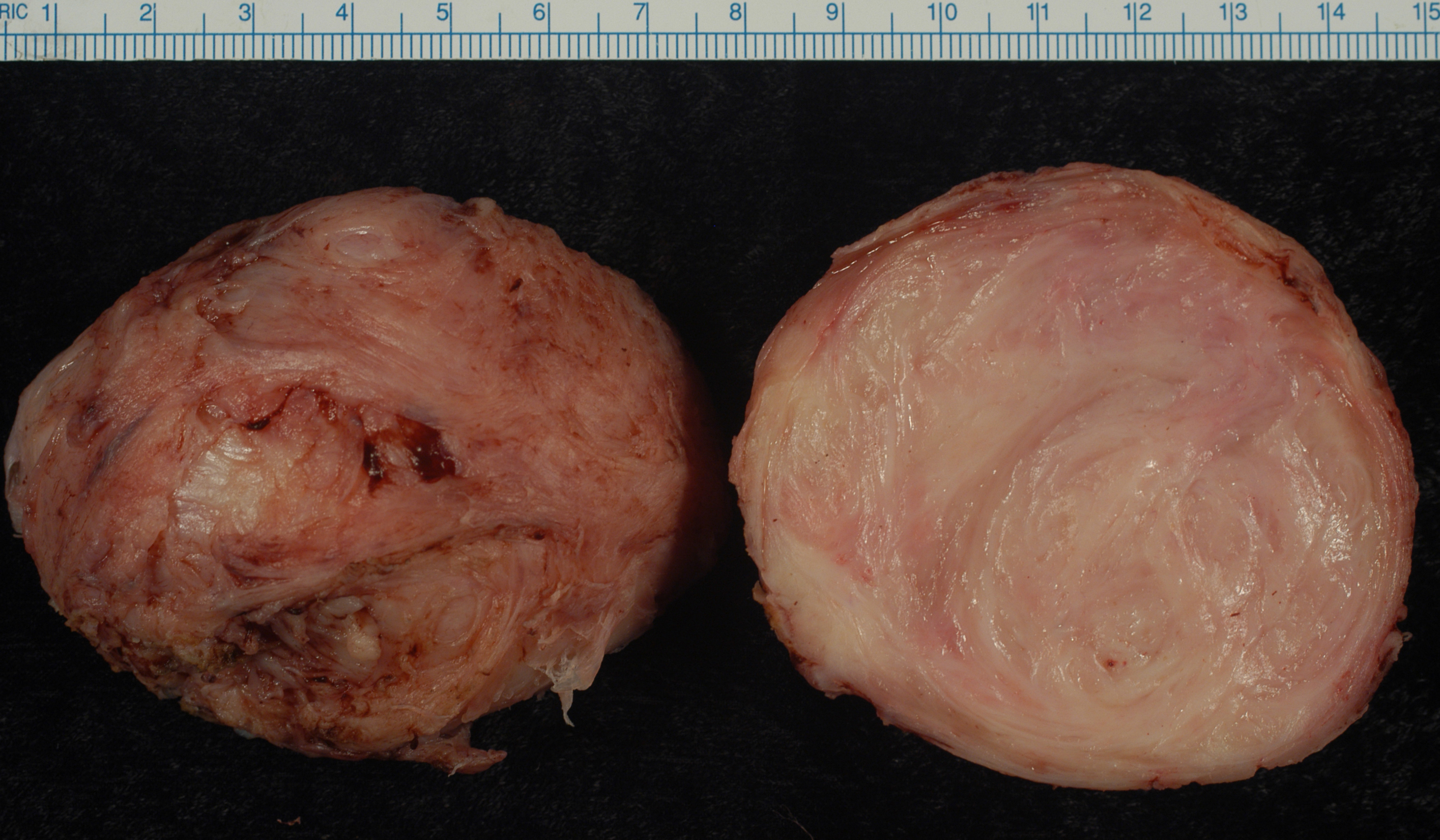
Uterine fibroids, also known as uterine leiomyomas or fibroids, are benign smooth muscle tumors of the uterus. Most women with fibroids have no symptoms while others may have painful or heavy periods. If large enough, they may push on the bladder, causing a frequent need to urinate. They may also cause pain during penetrative sex or lower back pain. A woman can have one uterine fibroid or many. Occasionally, fibroids may make it difficult to become pregnant, although this is uncommon. The exact cause of uterine fibroids is unclear. However, fibroids run in families and appear to be partly determined by hormone levels. Risk factors include obesity and eating red meat. Diagnosis can be performed by pelvic examination or medical imaging. Treatment is typically not needed if there are no symptoms. NSAIDs, such as ibuprofen, may help with pain and bleeding while paracetamol (acetaminophen) may help with pain. Iron supplements may be needed in those with heavy periods. Medic ... [...More Info...] [...Related Items...] OR: [Wikipedia] [Google] [Baidu] |
Uterine Artery Embolization
Uterine artery embolization is a procedure in which an interventional radiologist uses a catheter to deliver small particles that block the blood supply to the uterine body. The procedure is done for the treatment of uterine fibroids and adenomyosis. This minimally invasive procedure is commonly used in the treatment of uterine fibroids and is also called uterine fibroid embolization. Medical uses Uterine artery embolization is used to treat bothersome bulk-related symptoms or abnormal or heavy uterine bleeding due to uterine fibroids or for the treatment of adenomyosis. Fibroid size, number, and location are three potential predictors of a successful outcome. Long-term patient satisfaction outcomes are similar to that of surgery. There is tentative evidence that traditional surgery may result in better fertility. Uterine artery embolization also appears to require more repeat procedures than if surgery was done initially. It has shorter recovery times. Uterine artery emboliza ... [...More Info...] [...Related Items...] OR: [Wikipedia] [Google] [Baidu] |
Hysterectomy
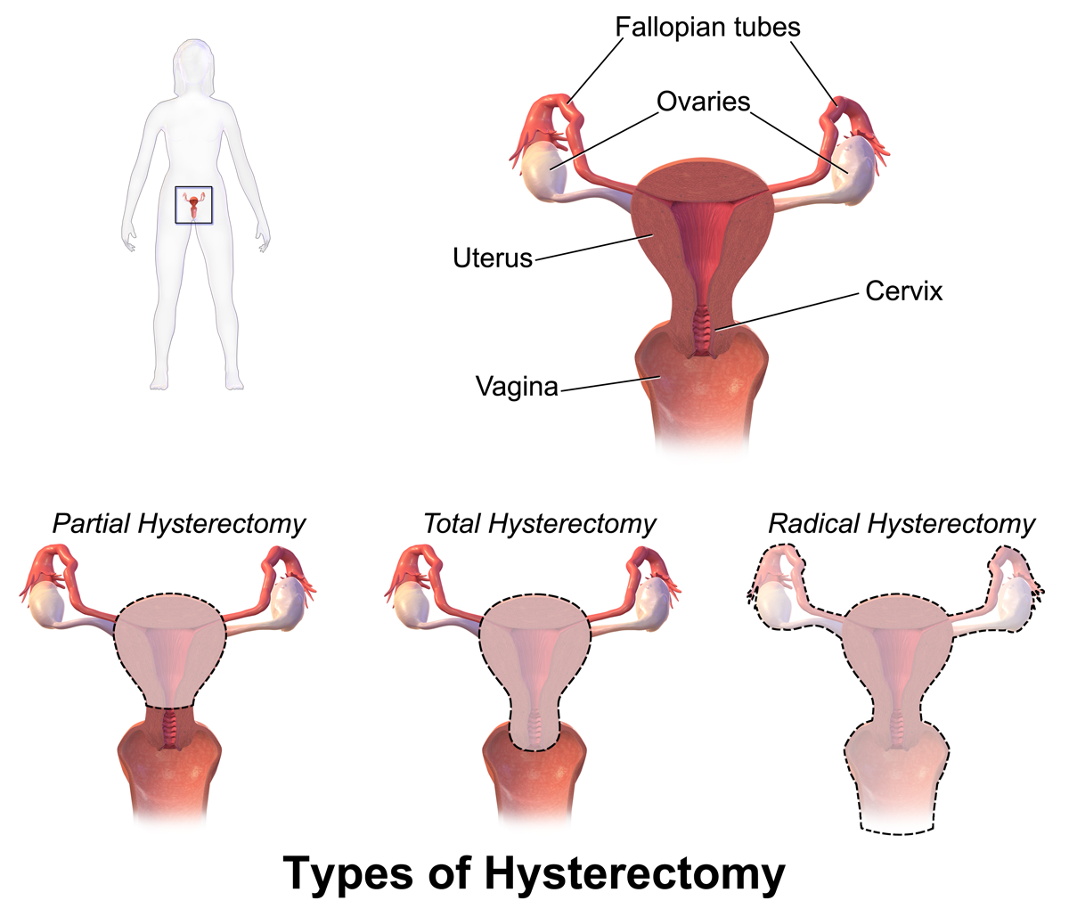
Hysterectomy is the surgical removal of the uterus. It may also involve removal of the cervix, ovaries (oophorectomy), Fallopian tubes (salpingectomy), and other surrounding structures. Usually performed by a gynecologist, a hysterectomy may be total (removing the body, fundus, and cervix of the uterus; often called "complete") or partial (removal of the uterine body while leaving the cervix intact; also called "supracervical"). Removal of the uterus renders the patient unable to bear children (as does removal of ovaries and fallopian tubes) and has surgical risks as well as long-term effects, so the surgery is normally recommended only when other treatment options are not available or have failed. It is the second most commonly performed gynecological surgical procedure, after cesarean section, in the United States. Nearly 68 percent were performed for conditions such as endometriosis, irregular bleeding, and uterine fibroids. It is expected that the frequency of hysterectom ... [...More Info...] [...Related Items...] OR: [Wikipedia] [Google] [Baidu] |
Ligature (medicine)
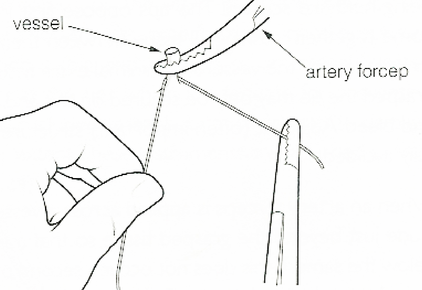
In surgery or medical procedure, a ligature consists of a piece of thread ( suture) tied around an anatomical structure, usually a blood vessel or another hollow structure (e.g. urethra) to shut it off. History The principle of ligation is attributed to Hippocrates and Galen. In ancient Rome, ligatures were used to treat hemorrhoids. The concept of a ligature was reintroduced some 1,500 years later by Ambroise Paré, and finally it found its modern use in 1870–80, made popular by Jules-Émile Péan. Procedure With a blood vessel the surgeon will clamp the vessel perpendicular to the axis of the artery or vein with a hemostat, then secure it by ligating it; i.e. using a piece of suture around it before dividing the structure and releasing the hemostat. It is different from a tourniquet in that the tourniquet will not be secured by knots and it can therefore be released/tightened at will. Ligature is one of the remedies to treat skin tag, or acrochorda. It is done by tying str ... [...More Info...] [...Related Items...] OR: [Wikipedia] [Google] [Baidu] |
Vaginal Branches Of Uterine Artery
The uterine artery supplies branches to the cervix uteri and others which descend on the vagina; the latter anastomose with branches of the vaginal arteries and form with them two median longitudinal vessels—the vaginal branches of uterine artery (or azygos arteries of the vagina)—one of which runs down in front of and the other behind the vagina In mammals, the vagina is the elastic, muscular part of the female genital tract. In humans, it extends from the vestibule to the cervix. The outer vaginal opening is normally partly covered by a thin layer of mucosal tissue called the hymen .... References Arteries of the abdomen {{circulatory-stub ... [...More Info...] [...Related Items...] OR: [Wikipedia] [Google] [Baidu] |
Arcuate Vessels Of Uterus
The arcuate vessels of the uterus are a component of the blood supply of the uterus. They are arteries and veins that branch from the uterine arteries and veins, respectively, with additional anastomoses from the ovarian arteries and veins, and penetrate and assume a circumferential course in the myometrium. They have also been called helicine branches of the uterus (or helicine arterioles), as they are spiral-shaped, but they should not be confused with the spiral arteries Spiral arteries are small arteries which temporarily supply blood to the endometrium of the uterus during the luteal phase of the menstrual cycle. In histology, identifying the presence of these arteries is one of the most useful techniques in i ... that penetrate the endometrium in the inner uterus. The radial arteries branch off from the arcuate artery through the myometrium. References Arteries of the abdomen {{circulatory-stub ... [...More Info...] [...Related Items...] OR: [Wikipedia] [Google] [Baidu] |
Anastomosis
An anastomosis (, plural anastomoses) is a connection or opening between two things (especially cavities or passages) that are normally diverging or branching, such as between blood vessels, leaf#Veins, leaf veins, or streams. Such a connection may be normal (such as the foramen ovale (heart), foramen ovale in a fetus's heart) or abnormal (such as the atrial septal defect#Patent foramen ovale, patent foramen ovale in an adult's heart); it may be acquired (such as an arteriovenous fistula) or innate (such as the arteriovenous shunt of a metarteriole); and it may be natural (such as the aforementioned examples) or artificial (such as a surgical anastomosis). The reestablishment of an anastomosis that had become blocked is called a reanastomosis. Anastomoses that are abnormal, whether congenital disorder, congenital or acquired, are often called fistulas. The term is used in medicine, biology, mycology, geology, and geography. Etymology Anastomosis: medical or Modern Latin, from Gre ... [...More Info...] [...Related Items...] OR: [Wikipedia] [Google] [Baidu] |
Broad Ligament Of The Uterus
The broad ligament of the uterus is the wide fold of peritoneum that connects the sides of the uterus to the walls and floor of the pelvis. Structure Subdivisions Contents The contents of the broad ligament include the following: * Reproductive ** uterine tubes (or Fallopian tube) ** ovary (some sources consider the ovary to be on the broad ligament, but not in it.) * vessels ** ovarian artery (in the suspensory ligament) ** uterine artery (in reality, travels in the cardinal ligament) * ligaments ** ovarian ligament ** round ligament of uterus ** suspensory ligament of the ovary (Some sources consider it a part of the broad ligament, while other sources just consider it a "termination" of the ligament.) Relations The peritoneum surrounds the uterus like a flat sheet that folds over its fundus, covering it anteriorly and posteriorly; on the sides of the uterus, this sheet of peritoneum comes in direct contact with itself, forming the double layer of peritoneum known as ... [...More Info...] [...Related Items...] OR: [Wikipedia] [Google] [Baidu] |
Parametrium
The parametrium is the fibrous and fatty connective tissue that surrounds the uterus. This tissue separates the supravaginal portion of the cervix from the bladder. The parametrium lies in front of the cervix and extends laterally between the layers of the broad ligaments. It connects the uterus to other tissues in the pelvis. It is different from the perimetrium, which is the outermost layer of the uterus. The uterine artery and ovarian ligament are located in the parametrium. An associated form of pelvic inflammatory disease is inflammation of the parametrium known as parametritis Parametritis (also known as pelvic cellulitis) is an infection of the parametrium (connective tissue adjacent to the uterus The uterus (from Latin ''uterus'', plural ''uteri'') or womb () is the organ in the reproductive system of most female .... References * External links Mammal female reproductive system {{genitourinary-stub ... [...More Info...] [...Related Items...] OR: [Wikipedia] [Google] [Baidu] |
Cardinal Ligament
The cardinal ligament (or Mackenrodt's ligament, lateral cervical ligament, or transverse cervical ligament) is a major ligament of the uterus. It is located at the base of the broad ligament of the uterus. There are a pair of cardinal ligaments in the female human body. Structure The cardinal ligament is a paired structure on the lateral side of the uterus. It originates from the lateral part of the cervix. It attaches to the uterosacral ligament. It attaches the cervix to the lateral pelvic wall by its attachment to the Obturator fascia of the Obturator internus muscle, and is continuous externally with the fibrous tissue that surrounds the pelvic blood vessels. It thus provides support to the uterus. It carries the uterine arteries to provide the primary blood supply to the uterus. Clinical significance The cardinal ligament may be affected in hysterectomy. Due to its close proximity to the ureters, it can get damaged during ligation of the ligament. It is routinely ... [...More Info...] [...Related Items...] OR: [Wikipedia] [Google] [Baidu] |


