|
Ulnar Canal
The ulnar canal or ulnar tunnel (also known as Guyon's canal or tunnel) is a semi-rigid longitudinal canal in the wrist that allows passage of the ulnar artery and ulnar nerve into the hand. The roof of the canal is made up of the superficial palmar carpal ligament, while the deeper flexor retinaculum and hypothenar muscles comprise the floor. The space is medially bounded by the pisiform and pisohamate ligament more proximally, and laterally bounded by the hook of the hamate more distally. It is approximately 4 cm long, beginning proximally at the transverse carpal ligament and ending at the aponeurotic arch of the hypothenar muscles. Eponym The ulnar tunnel is eponymously named after the French surgeon Jean Casimir Félix Guyon, who originally described the canal in 1861. Clinical significance Entrapment of the ulnar nerve at the ulnar canal can result in symptoms of ulnar neuropathy, including numbness or weakness of certain parts of the hand. (''See full article on ulna ... [...More Info...] [...Related Items...] OR: [Wikipedia] [Google] [Baidu] |
Ulnar Artery
The ulnar artery is the main blood vessel, with oxygenated blood, of the medial aspects of the forearm. It arises from the brachial artery and terminates in the superficial palmar arch, which joins with the superficial branch of the radial artery. It is palpable on the anterior and medial aspect of the wrist. Along its course, it is accompanied by a similarly named vein or veins, the ulnar vein or ulnar veins. The ulnar artery, the larger of the two terminal branches of the brachial, begins a little below the bend of the elbow in the cubital fossa, and, passing obliquely downward, reaches the ulnar side of the forearm at a point about midway between the elbow and the wrist. It then runs along the ulnar border to the wrist, crosses the transverse carpal ligament on the radial side of the pisiform bone, and immediately beyond this bone divides into two branches, which enter into the formation of the superficial and deep volar arches. Branches Forearm: Anterior ulnar recurrent ... [...More Info...] [...Related Items...] OR: [Wikipedia] [Google] [Baidu] |
Ulnar Nerve
In human anatomy, the ulnar nerve is a nerve that runs near the ulna bone. The ulnar collateral ligament of elbow joint is in relation with the ulnar nerve. The nerve is the largest in the human body unprotected by muscle or bone, so injury is common. This nerve is directly connected to the little finger, and the adjacent half of the ring finger, innervating the palmar aspect of these fingers, including both front and back of the tips, perhaps as far back as the fingernail beds. This nerve can cause an electric shock-like sensation by striking the medial epicondyle of the humerus posteriorly, or inferiorly with the elbow flexed. The ulnar nerve is trapped between the bone and the overlying skin at this point. This is commonly referred to as bumping one's "funny bone". This name is thought to be a pun, based on the sound resemblance between the name of the bone of the upper arm, the humerus, and the word "humorous". Alternatively, according to the Oxford English Dictionary, i ... [...More Info...] [...Related Items...] OR: [Wikipedia] [Google] [Baidu] |
Palmar Carpal Ligament
The palmar carpal ligament (also volar carpal ligament or ''Guyon's Tunnel'') is the thickened portion of antebrachial fascia on the anterior of the wrist. It is officially unnamed.Moore, Keith L., Arthur F. Dalley II: ''Clinically Oriented Anatomy'', 4th ed. Lippincott, Williams & Wilkins,1999. The palmar carpal ligament is a different structure than the flexor retinaculum of the hand, but the two are frequently confused. The palmar carpal ligament lies superficial and proximal to the flexor retinaculum. The ulnar nerve and the ulnar artery run through the ulnar canal, which is deep to the palmar carpal ligament and superficial to the flexor retinaculum. The palmar carpal ligament is continuous with the extensor retinaculum of the hand, which is located on the posterior side of the wrist. References See also * Flexor retinaculum of the hand * Extensor retinaculum of the hand * Antebrachial fascia The antebrachial fascia (antibrachial fascia or deep fascia of forearm) conti ... [...More Info...] [...Related Items...] OR: [Wikipedia] [Google] [Baidu] |
Flexor Retinaculum Of The Hand
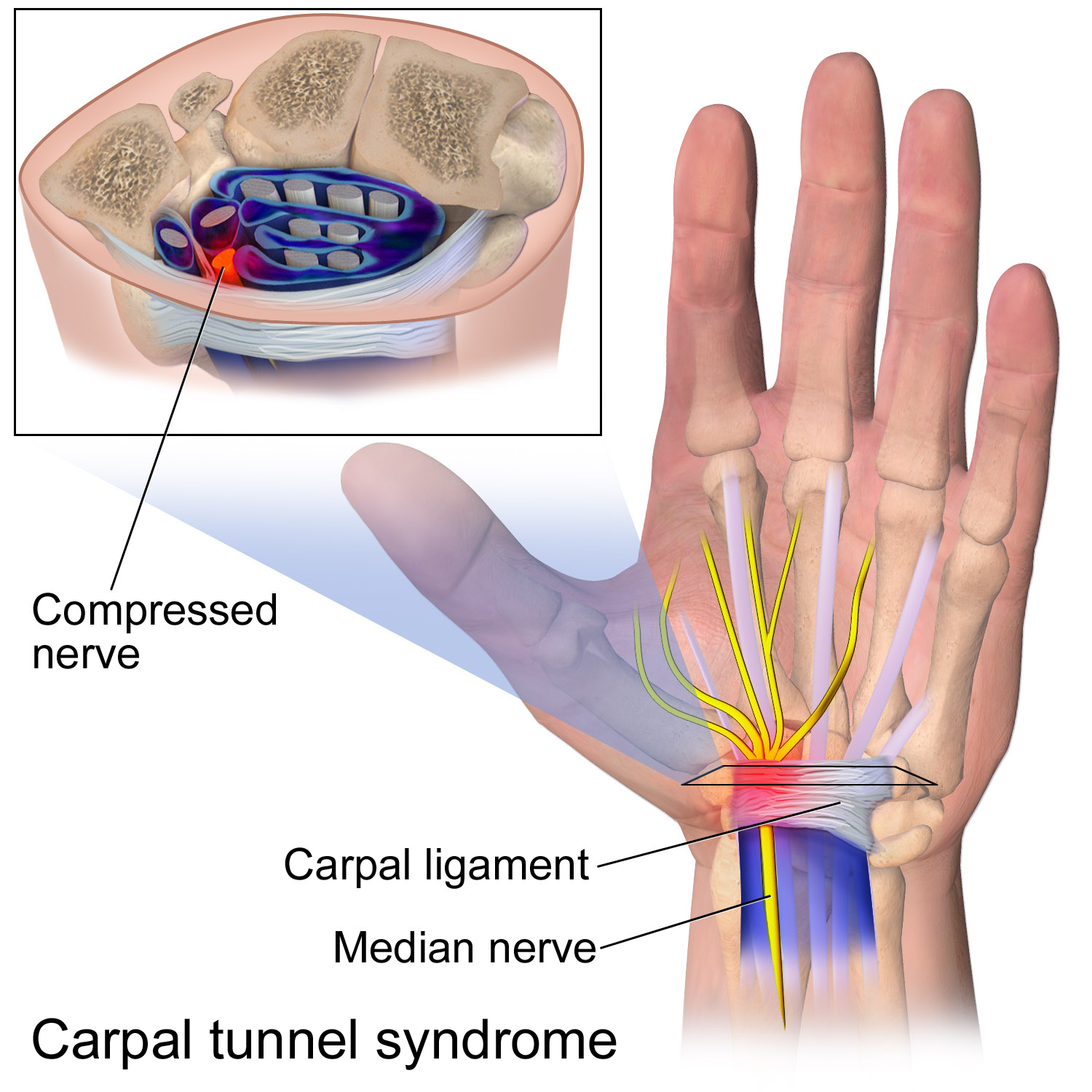
The flexor retinaculum (transverse carpal ligament, or anterior annular ligament) is a fibrous band on the palmar side of the hand near the wrist. It arches over the carpal bones of the hands, covering them and forming the carpal tunnel. Structure The flexor retinaculum is a strong, fibrous band that covers the carpal bones on the palmar side of the hand near the wrist. It attaches to the bones near the radius and ulna. On the ulnar side, the flexor retinaculum attaches to the pisiform bone and the hook of the hamate bone. On the radial side, it attaches to the tubercle of the scaphoid bone, and to the medial part of the palmar surface and the ridge of the trapezium bone. The flexor retinaculum is continuous with the palmar carpal ligament, and deeper with the palmar aponeurosis. The ulnar artery and ulnar nerve, and the cutaneous branches of the median and ulnar nerves, pass on top of the flexor retinaculum. On the radial side of the retinaculum is the tendon of the flexor c ... [...More Info...] [...Related Items...] OR: [Wikipedia] [Google] [Baidu] |
Hypothenar Muscles
The hypothenar muscles are a group of three muscles of the palm that control the motion of the little finger. The three muscles are: * Abductor digiti minimi * Flexor digiti minimi brevis * Opponens digiti minimi Structure The muscles of hypothenar eminence are from medial to lateral: * Opponens digiti minimi * Flexor digiti minimi brevis * Abductor digiti minimi The intrinsic muscles of hand can be remembered using the mnemonic, "A OF A OF A" for, Abductor pollicis brevis, Opponens pollicis, Flexor pollicis brevis (the three thenar muscles), Adductor pollicis, and the three hypothenar muscles, Opponens digiti minimi, Flexor digiti minimi brevis, Abductor digiti minimi. Clinical significance "Hypothenar atrophy" is associated with the lesion of the ulnar nerve, which supplies the three hypothenar muscles. Hypothenar hammer syndrome is a vascular occlusion of this region. See also * Thenar eminence * Palmaris brevis Palmaris brevis muscle is a thin, quadrilateral muscle, ... [...More Info...] [...Related Items...] OR: [Wikipedia] [Google] [Baidu] |
Pisiform
The pisiform bone ( or ), also spelled pisiforme (from the Latin ''pisifomis'', pea-shaped), is a small knobbly, sesamoid bone that is found in the wrist. It forms the ulnar border of the carpal tunnel. Structure The pisiform is a sesamoid bone, with no covering membrane of periosteum. It is the last carpal bone to ossify. The pisiform bone is a small bone found in the proximal row of the wrist (carpus). It is situated where the ulna joins the wrist, within the tendon of the flexor carpi ulnaris muscle. It only has one side that acts as a joint, articulating with the triquetral bone. It is on a plane anterior to the other carpal bones and is spheroidal in form. The pisiform bone has four surfaces: # The ''dorsal surface'' is smooth and oval, and articulates with the triquetral: this facet approaches the superior, but not the inferior border of the bone. # The ''palmar surface'' is rounded and rough, and gives attachment to the transverse carpal ligament, the flexor carpi ulnaris ... [...More Info...] [...Related Items...] OR: [Wikipedia] [Google] [Baidu] |
Pisohamate Ligament
The pisohamate ligament is a ligament in the hand. It connects the pisiform to the hook of the hamate. It is a prolongation of the tendon of the flexor carpi ulnaris. It serves as part of the origin for the abductor digiti minimi. It also forms the floor of the ulnar canal, a canal that allows the ulnar nerve and ulnar artery The ulnar artery is the main blood vessel, with oxygenated blood, of the medial aspects of the forearm. It arises from the brachial artery and terminates in the superficial palmar arch, which joins with the superficial branch of the radial ar ... into the hand. References Ligaments of the upper limb {{ligament-stub ... [...More Info...] [...Related Items...] OR: [Wikipedia] [Google] [Baidu] |
Hook Of The Hamate
The hamate bone (from Latin hamatus, "hooked"), or unciform bone (from Latin ''uncus'', "hook"), Latin os hamatum and occasionally abbreviated as just hamatum, is a bone in the human wrist readily distinguishable by its wedge shape and a hook-like process ("hamulus") projecting from its palmar surface. Structure The hamate is an irregularly shaped carpal bone found within the hand. The hamate is found within the distal row of carpal bones, and abuts the metacarpals of the little finger and ring finger. Adjacent to the hamate on the ulnar side, and slightly above it, is the pisiform bone. Adjacent on the radial side is the capitate, and proximal is the lunate bone. Surfaces The hamate bone has six surfaces: * The ''superior'', the apex of the wedge, is narrow, convex, smooth, and articulates with the lunate. * The ''inferior'' articulates with the fourth and fifth metacarpal bones, by concave facets which are separated by a ridge. * The ''dorsal'' is triangular and rough for l ... [...More Info...] [...Related Items...] OR: [Wikipedia] [Google] [Baidu] |
Jean Casimir Félix Guyon
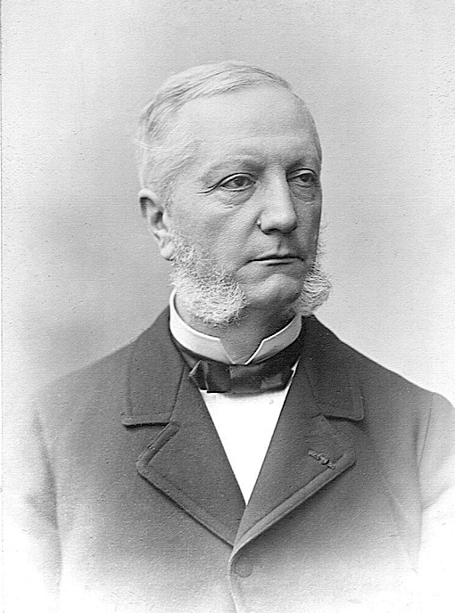
Jean Casimir Félix Guyon (21 July 1831 – 2 August 1920) was a French surgeon and urologist born in Saint-Denis, Ile-Bourbon ( Réunion). He studied medicine in Paris, receiving his doctorate in 1858. He was appointed ''médecin des hôpitaux'' in 1864, and was later a professor of surgical pathology (from 1877) and genitourinary surgery (from 1890) at the University of Paris. In 1878 he became a member of the ''Académie de Médecine''. At Hôpital Necker he held clinics that were attended by students worldwide In 1907, he along with urologists from Europe, the United States and South America established the ''Association Internationale d'Urologie''. In 1979 he was commemorated on a postage stamp, issued by France on the occasion of the 18th Congress of the ''Association Internationale d'Urologie'', held in Paris. The Hôpital Félix Guyon, located in Saint-Denis, Réunion, is named in his honour. Although he was primarily known for work with genitourinary anatomy, Guyon i ... [...More Info...] [...Related Items...] OR: [Wikipedia] [Google] [Baidu] |
Ulnar Nerve Entrapment
Ulnar nerve entrapment is a condition where the ulnar nerve becomes physically trapped or pinched, resulting in pain, numbness, or weakness, primarily affecting the little finger and ring finger of the hand. Entrapment may occur at any point from the spine at cervical vertebra C7 to the wrist; the most common point of entrapment is in the elbow (Cubital tunnel syndrome). Prevention is mostly through correct posture and avoiding repetitive or constant strain (e.g. "cell phone elbow"). Treatment is usually conservative, including medication, activity modification, and exercise, but may sometimes include surgery. Prognosis is generally good, with mild to moderate symptoms often resolving spontaneously. Signs and symptoms In general, ulnar neuropathy will result in symptoms in a specific anatomic distribution, affecting the little finger, the ulnar half of the ring finger, and the intrinsic muscles of the hand. The specific symptoms experienced in the characteristic distribution d ... [...More Info...] [...Related Items...] OR: [Wikipedia] [Google] [Baidu] |
Ulnar Neuropathy
Ulnar neuropathy is a disorder involving the ulnar nerve. Ulnar neuropathy may be caused by entrapment of the ulnar nerve with resultant numbness and tingling. It may also cause weakness or and paralysis of the muscles supplied by the nerve. Signs and symptoms In terms of the signs/symptoms of ulnar neuropathy trauma and pressure to the arm and wrist, especially the elbow, the medial side of the wrist, and other sites close to the course of the ulnar nerve are of interest in this condition. Many people complain of sensory changes in the fourth and fifth digits. Rarely, an individual actually notices that the unusual sensations are mainly in the medial side of the ring finger (fourth digit). Sometimes the third digit is also involved, especially on the ulnar ( medial) side. The sensory changes can be a feeling of numbness or a tingling, pain rarely occurs in the hand. Complaints of pain tend to be more common in the arm, up to and including the elbow area, which is probably the m ... [...More Info...] [...Related Items...] OR: [Wikipedia] [Google] [Baidu] |
Ganglion Cyst
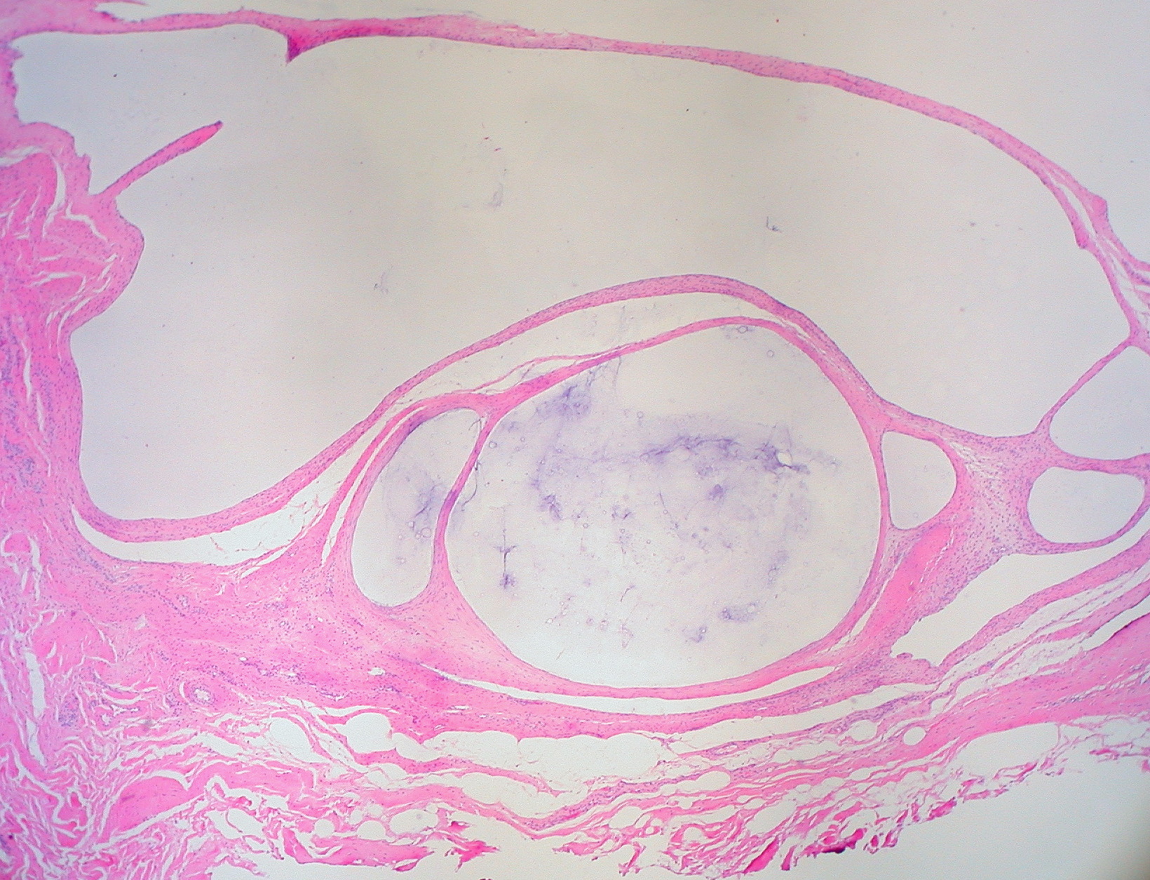
A ganglion cyst is a fluid-filled bump associated with a joint or tendon sheath. It most often occurs at the back of the wrist, followed by the front of the wrist. Onset is often over several months, typically with no further symptoms. Occasionally, pain or numbness may occur. Complications may include carpal tunnel syndrome. The cause is unknown. The underlying mechanism is believed to involve an outpouching of the synovial membrane. Risk factors include gymnastics activity. Diagnosis is typically based on examination with light shining through the lesion being supportive. Medical imaging may be done to rule out other potential causes. Treatment options include watchful waiting, splinting the affected joint, needle aspiration, or surgery. About half the time, they resolve on their own. About three per 10,000 people newly develop ganglion of the wrist or hand a year. They most commonly occur in young and middle-aged females. Presentation The average size of these cysts is 2.0 ... [...More Info...] [...Related Items...] OR: [Wikipedia] [Google] [Baidu] |


_-_animation02.gif)


