|
Ulnar Nerve Entrapment
Ulnar nerve entrapment is a condition where the ulnar nerve becomes physically trapped or pinched, resulting in pain, numbness, or weakness, primarily affecting the little finger and ring finger of the hand. Entrapment may occur at any point from the spine at cervical vertebra C7 to the wrist; the most common point of entrapment is in the elbow (Cubital tunnel syndrome). Prevention is mostly through correct posture and avoiding repetitive or constant strain (e.g. "cell phone elbow"). Treatment is usually conservative, including medication, activity modification, and exercise, but may sometimes include surgery. Prognosis is generally good, with mild to moderate symptoms often resolving spontaneously. Signs and symptoms In general, ulnar neuropathy will result in symptoms in a specific anatomic distribution, affecting the little finger, the ulnar half of the ring finger, and the intrinsic muscles of the hand. The specific symptoms experienced in the characteristic distribution d ... [...More Info...] [...Related Items...] OR: [Wikipedia] [Google] [Baidu] |
Ulnar Nerve
In human anatomy, the ulnar nerve is a nerve that runs near the ulna bone. The ulnar collateral ligament of elbow joint is in relation with the ulnar nerve. The nerve is the largest in the human body unprotected by muscle or bone, so injury is common. This nerve is directly connected to the little finger, and the adjacent half of the ring finger, innervating the palmar aspect of these fingers, including both front and back of the tips, perhaps as far back as the fingernail beds. This nerve can cause an electric shock-like sensation by striking the medial epicondyle of the humerus posteriorly, or inferiorly with the elbow flexed. The ulnar nerve is trapped between the bone and the overlying skin at this point. This is commonly referred to as bumping one's "funny bone". This name is thought to be a pun, based on the sound resemblance between the name of the bone of the upper arm, the humerus, and the word "humorous". Alternatively, according to the Oxford English Dictionary, i ... [...More Info...] [...Related Items...] OR: [Wikipedia] [Google] [Baidu] |
Pectoralis Minor Muscle
Pectoralis minor muscle () is a thin, triangular muscle, situated at the upper part of the chest, beneath the pectoralis major in the human body. Structure Attachments Pectoralis minor muscle arises from the upper margins and outer surfaces of the third, fourth, and fifth ribs, near their costal cartilages and from the aponeuroses covering the intercostalis. The fibers pass superior and lateral and converge to form a flat tendon. This tendon inserts onto the medial border and upper surface of the coracoid process of the scapula. Relations Pectoralis minor muscle forms part of the anterior wall of the axilla. It is covered anteriorly (superficially) by the clavipectoral fascia. The medial pectoral nerve pierces the pectoralis minor and the clavipectoral fascia. In attaching to the coracoid process, the pectoralis minor forms a 'bridge' - structures passing into the upper limb from the thorax will pass directly underneath.http://www.teachmeanatomy.com/muscles-of-the-pector ... [...More Info...] [...Related Items...] OR: [Wikipedia] [Google] [Baidu] |
Infection
An infection is the invasion of tissues by pathogens, their multiplication, and the reaction of host tissues to the infectious agent and the toxins they produce. An infectious disease, also known as a transmissible disease or communicable disease, is an illness resulting from an infection. Infections can be caused by a wide range of pathogens, most prominently bacteria and viruses. Hosts can fight infections using their immune system. Mammalian hosts react to infections with an innate response, often involving inflammation, followed by an adaptive response. Specific medications used to treat infections include antibiotics, antivirals, antifungals, antiprotozoals, and antihelminthics. Infectious diseases resulted in 9.2 million deaths in 2013 (about 17% of all deaths). The branch of medicine that focuses on infections is referred to as infectious disease. Types Infections are caused by infectious agents (pathogens) including: * Bacteria (e.g. ''Mycobacterium tuberculosis'', ... [...More Info...] [...Related Items...] OR: [Wikipedia] [Google] [Baidu] |
Thrombosis
Thrombosis (from Ancient Greek "clotting") is the formation of a blood clot inside a blood vessel, obstructing the flow of blood through the circulatory system. When a blood vessel (a vein or an artery) is injured, the body uses platelets (thrombocytes) and fibrin to form a blood clot to prevent blood loss. Even when a blood vessel is not injured, blood clots may form in the body under certain conditions. A clot, or a piece of the clot, that breaks free and begins to travel around the body is known as an embolus. Thrombosis may occur in veins (venous thrombosis) or in arteries (arterial thrombosis). Venous thrombosis (sometimes called DVT, deep vein thrombosis) leads to a blood clot in the affected part of the body, while arterial thrombosis (and, rarely, severe venous thrombosis) affects the blood supply and leads to damage of the tissue supplied by that artery (ischemia and necrosis). A piece of either an arterial or a venous thrombus can break off as an embolus, which could ... [...More Info...] [...Related Items...] OR: [Wikipedia] [Google] [Baidu] |
Aneurysms
An aneurysm is an outward bulging, likened to a bubble or balloon, caused by a localized, abnormal, weak spot on a blood vessel wall. Aneurysms may be a result of a hereditary condition or an acquired disease. Aneurysms can also be a nidus (starting point) for clot formation (thrombosis) and embolization. As an aneurysm increases in size, the risk of rupture, which leads to uncontrolled bleeding, increases. Although they may occur in any blood vessel, particularly lethal examples include aneurysms of the Circle of Willis in the brain, aortic aneurysms affecting the thoracic aorta, and abdominal aortic aneurysms. Aneurysms can arise in the heart itself following a heart attack, including both ventricular and atrial septal aneurysms. There are congenital atrial septal aneurysms, a rare heart defect. Etymology The word is from Greek: ἀνεύρυσμα, aneurysma, "dilation", from ἀνευρύνειν, aneurynein, "to dilate". Classification Aneurysms are classified by type, ... [...More Info...] [...Related Items...] OR: [Wikipedia] [Google] [Baidu] |
Hypothenar Hammer Syndrome
Hypothenar hammer syndrome (HHS) is a vascular occlusion in humans in the region of the ulna. It is caused by repetitive trauma to the hand or wrist (such as that caused by the use of a hammer) by the vulnerable portion of the ulnar artery as it passes over the hamate bone, which may result in thrombosis, irregularity or aneurysm formation. HHS is a potentially curable cause of Raynaud's syndrome, distinct from hand–arm vibration syndrome. Cause Diagnosis A physical examination of the hand may show discoloration (blanching, mottling, and/ or cyanosis; gangrene may be present in advanced cases), unusual tenderness/ a callous over the hypothenar eminence, and fingertip ulcerations and splinter hemorrhages over ulnar digits; if an aneurysm is present, there may also be a pulsatile mass. Allen's test will be positive if an occlusion is present and negative if an aneurysm is present. An angiogram may show a "corkscrew" ulnar artery or an occlusion or aneurysm at the hook of the hama ... [...More Info...] [...Related Items...] OR: [Wikipedia] [Google] [Baidu] |
Ulnar Tunnel Syndrome
Ulnar tunnel syndrome, also known as Guyon's canal syndrome or Handlebar palsy, is caused by entrapment of the ulnar nerve in the Guyon canal as it passes through the wrist. Symptoms usually begin with a feeling of pins and needles in the ring and little fingers before progressing to a loss of sensation and/or impaired motor function of the intrinsic muscles of the hand which are innervated by the ulnar nerve. Ulnar tunnel syndrome is commonly seen in regular cyclists due to prolonged pressure of the Guyon's canal against bicycle handlebars. Another very common cause of sensory loss in the ring and pink finger is due to ulnar nerve entrapment at the cubital tunnel near the elbow, which is known as cubital tunnel syndrome. Causes While being idiopathic in some cases, causative factors of the ulnar tunnel syndrome include tumors, ganglion cysts, repetitive use, anatomical variations, and diseases of the neighboring blood vessels (thrombosis or aneurysm of the ulnar artery). Dia ... [...More Info...] [...Related Items...] OR: [Wikipedia] [Google] [Baidu] |
Wrist
In human anatomy, the wrist is variously defined as (1) the Carpal bones, carpus or carpal bones, the complex of eight bones forming the proximal skeletal segment of the hand; "The wrist contains eight bones, roughly aligned in two rows, known as the carpal bones." (2) the wrist joint or radiocarpal joint, the joint between the radius (bone), radius and the Carpal bones, carpus and; (3) the anatomical region surrounding the carpus including the distal parts of the bones of the forearm and the proximal parts of the metacarpus or five metacarpal bones and the series of joints between these bones, thus referred to as ''wrist joints''. "With the large number of bones composing the wrist (ulna, radius, eight carpas, and five metacarpals), it makes sense that there are many, many joints that make up the structure known as the wrist." This region also includes the carpal tunnel, the anatomical snuff box, bracelet lines, the Flexor retinaculum of the hand, flexor retinaculum, and the ex ... [...More Info...] [...Related Items...] OR: [Wikipedia] [Google] [Baidu] |
Flexor Carpi Ulnaris
The flexor carpi ulnaris (FCU) is a muscle of the forearm that flexes and adducts at the wrist joint. Structure Origin The flexor carpi ulnaris has two heads; a humeral head and ulnar head. The humeral head originates from the medial epicondyle of the humerus via the common flexor tendon. The ulnar head originates from the medial margin of the olecranon of the ulnar and the upper two-thirds of the dorsal border of the ulnar by an aponeurosis. Between the two heads passes the ulnar nerve and ulnar artery. Insertion The flexor carpi ulnaris inserts onto the pisiform, hook of the hamate (via the pisohamate ligament) and the anterior surface of the base of the fifth metacarpal (via the pisometacarpal ligament). Action The flexor carpi ulnaris flexes and adducts at the wrist joint. Innervation The flexor carpi ulnaris is innervated by the ulnar nerve. The corresponding spinal nerves are C8 and T1. Tendon The tendon of flexor carpi ulnaris can be seen on the anterior surface of th ... [...More Info...] [...Related Items...] OR: [Wikipedia] [Google] [Baidu] |
Forearm
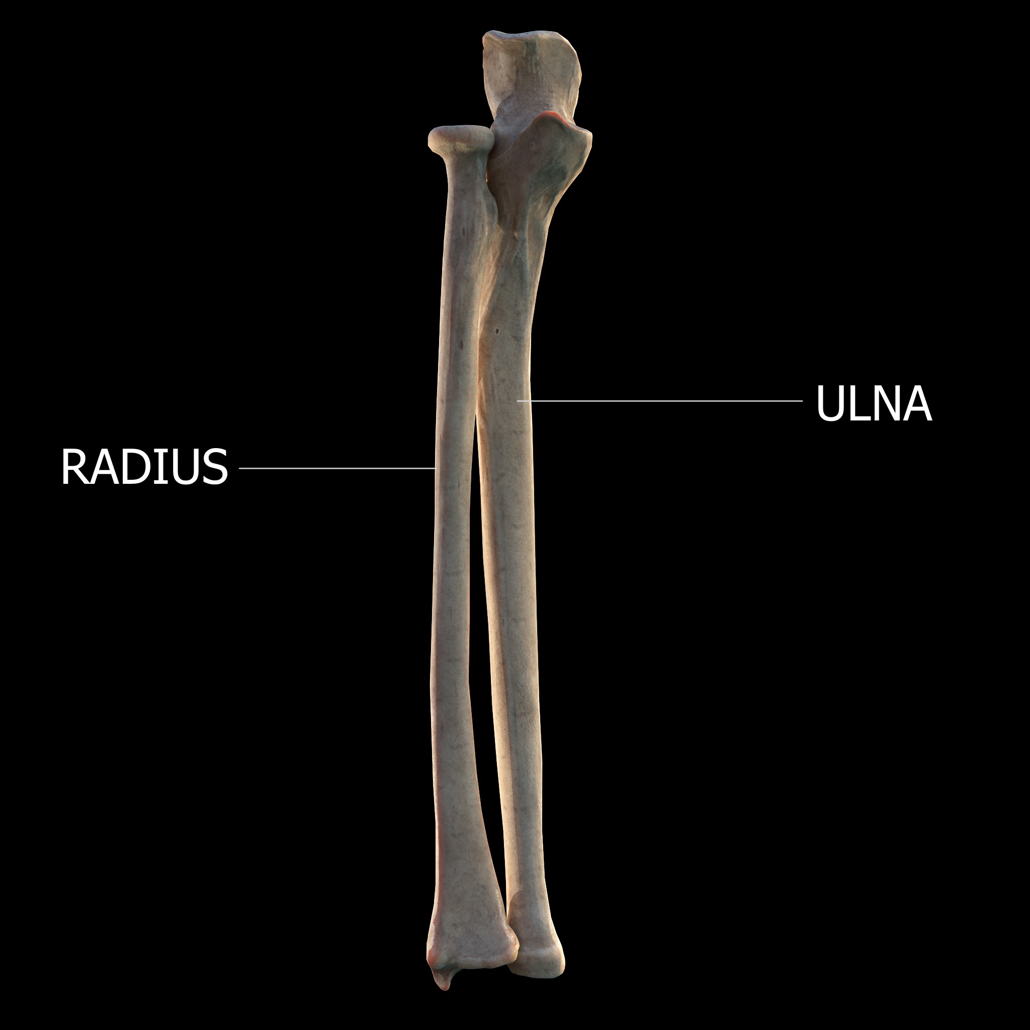
The forearm is the region of the upper limb between the elbow and the wrist. The term forearm is used in anatomy to distinguish it from the arm, a word which is most often used to describe the entire appendage of the upper limb, but which in anatomy, technically, means only the region of the upper arm, whereas the lower "arm" is called the forearm. It is homologous to the region of the leg that lies between the knee and the ankle joints, the crus. The forearm contains two long bones, the radius and the ulna, forming the two radioulnar joints. The interosseous membrane connects these bones. Ultimately, the forearm is covered by skin, the anterior surface usually being less hairy than the posterior surface. The forearm contains many muscles, including the flexors and extensors of the wrist, flexors and extensors of the digits, a flexor of the elbow (brachioradialis), and pronators and supinators that turn the hand to face down or upwards, respectively. In cross-section, the for ... [...More Info...] [...Related Items...] OR: [Wikipedia] [Google] [Baidu] |
Struthers' Ligament
Struthers' ligament is a feature of human anatomy consisting of a band of connective tissue at the medial aspect of the distal humerus. It courses from the supracondylar process of the humerus (also known as ''avian spur'') to the medial humeral epicondyle. It is not a constant ligament, and can be acquired or congenital. The structure was highlighted by John Struthers, who discussed the feature's evolutionary significance with Charles Darwin. Struthers originally reported that the ligament usually arose at a position 3.2 to 6.4 cm from the medial condyle, being 1.2 to 1.9 cm in length, and nearer to the anterior than the medial border of the humerus. The clinical significance of this structure is due to the median nerve and brachial artery which may pass underneath the "arch" formed by the process and ligament over the humeral body. Within this space the nerve may be compressed leading to supracondylar process syndrome. The ligament may also affect the ulnar nerve a ... [...More Info...] [...Related Items...] OR: [Wikipedia] [Google] [Baidu] |


