|
Upper Motor Neuron Lesion
An upper motor neuron lesion (also known as pyramidal insufficiency) Is an injury or abnormality that occurs in the neural pathway above the anterior horn cell of the spinal cord or motor nuclei of the cranial nerves. Conversely, a lower motor neuron lesion affects nerve fibers traveling from the anterior horn of the spinal cord or the cranial motor nuclei to the relevant muscle(s). Upper motor neuron lesions occur in the brain or the spinal cord as the result of stroke, multiple sclerosis, traumatic brain injury, cerebral palsy, atypical parkinsonisms, multiple system atrophy, and amyotrophic lateral sclerosis. Symptoms Changes in muscle performance can be broadly described as the upper motor neuron syndrome. These changes vary depending on the site and the extent of the lesion, and may include: * Muscle weakness. known as 'pyramidal weakness' * Decreased control of active movement, particularly slowness * Spasticity, a velocity-dependent change in muscle tone * Clasp-knif ... [...More Info...] [...Related Items...] OR: [Wikipedia] [Google] [Baidu] |
Anterior Horn Of Spinal Cord
The anterior grey column (also called the anterior cornu, anterior horn of spinal cord, motor horn or ventral horn) is the front column of grey matter in the spinal cord. It is one of the three grey columns. The anterior grey column contains motor neurons that affect the skeletal muscles while the posterior grey column receives information regarding touch and sensation. The anterior grey column is the column where the cell bodies of alpha motor neurons are located. Structure The anterior grey column, directed forward, is broad and of a rounded or quadrangular shape. Its posterior part is termed the base, and its anterior part the head, but these are not differentiated from each other by any well-defined constriction. It is separated from the surface of the medulla spinalis by a layer of white substance which is traversed by the bundles of the anterior nerve roots. In the thoracic region, the postero-lateral part of the anterior column projects laterally as a triangular field, whi ... [...More Info...] [...Related Items...] OR: [Wikipedia] [Google] [Baidu] |
Deep Tendon Reflex
The stretch reflex (myotatic reflex), or more accurately "muscle stretch reflex", is a muscle contraction in response to stretching within the muscle. The reflex functions to maintain the muscle at a constant length. The term deep tendon reflex is often used by many health workers and students to refer to this reflex. "Tendons have little to do with the response, other than being responsible for mechanically transmitting the sudden stretch from the reflex hammer to the muscle spindle. In addition, some muscles with stretch reflexes have no tendons (e.g., "jaw jerk" of the masseter muscle)". As an example of a spinal reflex, it results in a fast response that involves an afferent signal into the spinal cord and an efferent signal out to the muscle. The stretch reflex can be a Reflex arc#Monosynaptic vs. polysynaptic, monosynaptic reflex which provides automatic regulation of skeletal striated muscle, skeletal muscle length, whereby the signal entering the spinal cord arises from a ... [...More Info...] [...Related Items...] OR: [Wikipedia] [Google] [Baidu] |
Lower Motor Neuron
Lower motor neurons (LMNs) are motor neurons located in either the anterior grey column, anterior nerve roots (spinal lower motor neurons) or the cranial nerve nuclei of the brainstem and cranial nerves with motor function (cranial nerve lower motor neurons). Many voluntary movements rely on spinal lower motor neurons, which innervate skeletal muscle fibers and act as a link between upper motor neurons and muscles. Cranial nerve lower motor neurons also control some voluntary movements of the eyes, face and tongue, and contribute to chewing, swallowing and vocalization. Damage to the lower motor neurons can lead to flaccid paralysis, absent deep tendon reflexes and muscle atrophy. Classification Lower motor neurons are classified based on the type of muscle fiber they innervate: * Alpha motor neurons (α-MNs) innervate extrafusal muscle fibers, the most numerous type of muscle fiber and the one involved in muscle contraction. * Beta motor neurons (β-MNs) innervate intrafusal fi ... [...More Info...] [...Related Items...] OR: [Wikipedia] [Google] [Baidu] |
Upper Motor Neuron
Upper motor neurons (UMNs) is a term introduced by William Gowers in 1886. They are found in the cerebral cortex and brainstem and carry information down to activate interneurons and lower motor neurons, which in turn directly signal muscles to contract or relax. UMNs in the cerebral cortex are the main source of voluntary movement. They are the larger pyramidal cells in the cerebral cortex. There is a type of giant pyramidal cell called ''Betz cells'' and are found just below the surface of the cerebral cortex within layer V of the primary motor cortex. The cell bodies of Betz cell neurons are the largest in the brain, approaching nearly 0.1 mm in diameter. The primary motor cortex, or precentral gyrus, is the most posterior gyrus of the frontal lobe and lies anterior to the central sulcus. The pyramidal cells of the precentral gyrus are also called upper motor neurons. The fibers of the upper motor neurons project out of the precentral gyrus ending in the brainstem ... [...More Info...] [...Related Items...] OR: [Wikipedia] [Google] [Baidu] |
Nerve Biopsy
In medicine, a nerve biopsy is an invasive procedure in which a piece of nerve is removed from an organism and examined under a microscope. A nerve biopsy can lead to the discovery of various necrotizing vasculitis, amyloidosis, sarcoidosis, leprosy, metabolic neuropathies, inflammation of the nerve, loss of axon tissue, and demyelination. Biopsy literally means an examination of tissue removed from a living body to discover the presence, cause, or extent of a disease. A nerve biopsy may be necessary when a patient experiences numbness, pain, or weakness in places such as the fingers or toes. A nerve biopsy can help to determine the cause of such symptoms. The procedure is usually only performed when all other options have failed in determining the cause of a disease. It is an outpatient procedure that is performed under local anesthetic. Uses A nerve biopsy can potentially find the cause of the numbness and/or pain experienced in the limbs. It can reveal if these symptoms are c ... [...More Info...] [...Related Items...] OR: [Wikipedia] [Google] [Baidu] |
Lumbar Puncture
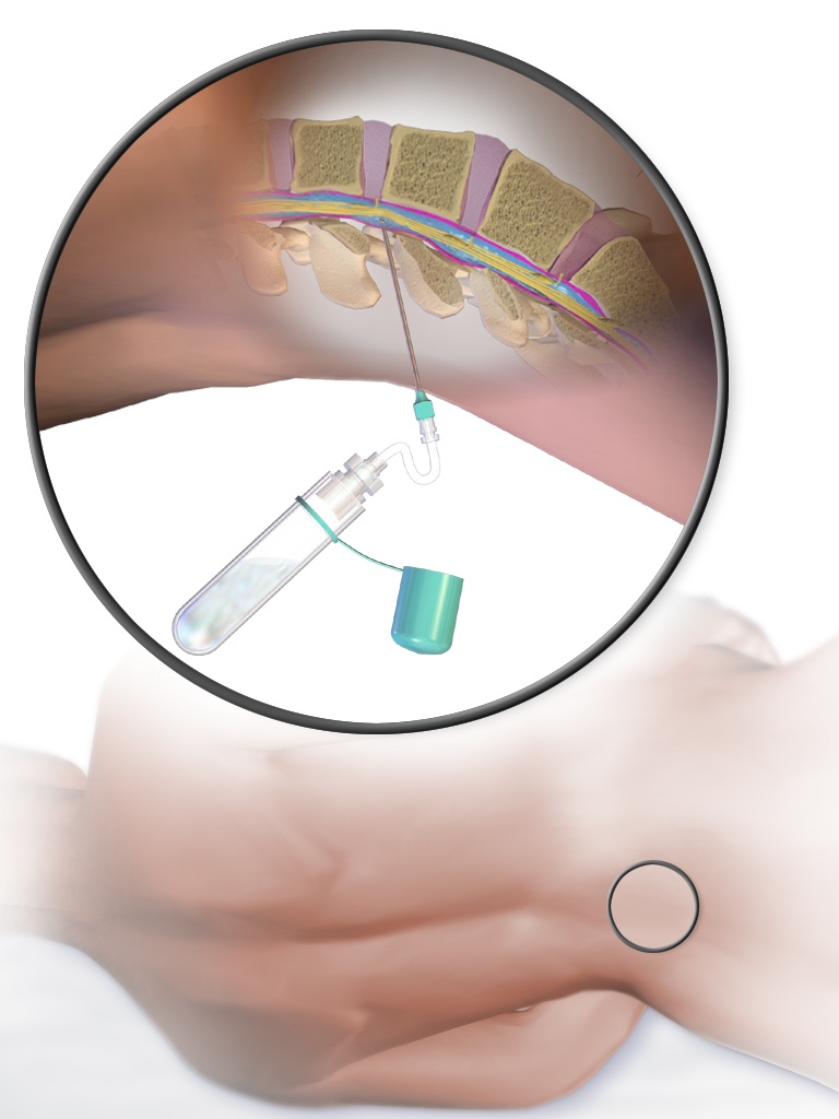
Lumbar puncture (LP), also known as a spinal tap, is a medical procedure in which a needle is inserted into the spinal canal, most commonly to collect cerebrospinal fluid (CSF) for diagnostic testing. The main reason for a lumbar puncture is to help diagnose diseases of the central nervous system, including the brain and spine. Examples of these conditions include meningitis and subarachnoid hemorrhage. It may also be used therapeutically in some conditions. Increased intracranial pressure (pressure in the skull) is a contraindication, due to risk of brain matter being compressed and pushed toward the spine. Sometimes, lumbar puncture cannot be performed safely (for example due to a bleeding diathesis, severe bleeding tendency). It is regarded as a safe procedure, but post-dural-puncture headache is a common side effect if a small atraumatic needle is not used. The procedure is typically performed under local anesthesia using a aseptic technique, sterile technique. A hypodermic ... [...More Info...] [...Related Items...] OR: [Wikipedia] [Google] [Baidu] |
Nerve Conduction Study
A nerve conduction study (NCS) is a medical diagnostic test commonly used to evaluate the function, especially the ability of electrical conduction, of the motor and sensory nerves of the human body. These tests may be performed by medical specialists such as clinical neurophysiologists, physical therapists, chiropractors, physiatrists (physical medicine and rehabilitation physicians), and neurologists who subspecialize in electrodiagnostic medicine. In the United States, neurologists and physiatrists receive training in electrodiagnostic medicine (performing needle electromyography (EMG) and NCSs) as part of residency training and in some cases acquire additional expertise during a fellowship in clinical neurophysiology, electrodiagnostic medicine, or neuromuscular medicine. Outside the US, clinical neurophysiologists learn needle EMG and NCS testing. Nerve conduction velocity (NCV) is a common measurement made during this test. The term NCV often is used to mean the actual tes ... [...More Info...] [...Related Items...] OR: [Wikipedia] [Google] [Baidu] |
Lower Motor Neuron
Lower motor neurons (LMNs) are motor neurons located in either the anterior grey column, anterior nerve roots (spinal lower motor neurons) or the cranial nerve nuclei of the brainstem and cranial nerves with motor function (cranial nerve lower motor neurons). Many voluntary movements rely on spinal lower motor neurons, which innervate skeletal muscle fibers and act as a link between upper motor neurons and muscles. Cranial nerve lower motor neurons also control some voluntary movements of the eyes, face and tongue, and contribute to chewing, swallowing and vocalization. Damage to the lower motor neurons can lead to flaccid paralysis, absent deep tendon reflexes and muscle atrophy. Classification Lower motor neurons are classified based on the type of muscle fiber they innervate: * Alpha motor neurons (α-MNs) innervate extrafusal muscle fibers, the most numerous type of muscle fiber and the one involved in muscle contraction. * Beta motor neurons (β-MNs) innervate intrafusal fi ... [...More Info...] [...Related Items...] OR: [Wikipedia] [Google] [Baidu] |
Medullary Pyramids
In neuroanatomy, the medullary pyramids are paired white matter structures of the brainstem's medulla oblongata that contain motor fibers of the corticospinal and corticobulbar tracts – known together as the pyramidal tracts. The lower limit of the pyramids is marked when the fibers cross ( decussate). Structure The ventral portion of the medulla oblongata contains the medullary pyramids. These two ridge-like structures travel along the length of the medulla oblongata and are bordered medially by the anterior median fissure. They each have an anterolateral sulcus along their lateral borders, where the hypoglossal nerve emerges from. Also at the side of each pyramid there is a pronounced bulge known as an olive. Fibers of the posterior column, which transmit sensory and proprioceptive information, are located behind the pyramids on the medulla oblongata. The medullary pyramids contain motor fibers that are known as the corticobulbar and corticospinal tracts. The cortico ... [...More Info...] [...Related Items...] OR: [Wikipedia] [Google] [Baidu] |
Decussation
Decussation is used in biological contexts to describe a crossing (due to the shape of the Roman numeral for ten, an uppercase 'X' (), ). In Latin anatomical terms, the form is used, e.g. . Similarly, the anatomical term chiasma is named after the Greek uppercase 'Χ' (chi). Whereas a decussation refers to a crossing within the central nervous system, various kinds of crossings in the peripheral nervous system are called chiasma. Examples include: * In the brain, where nerve fibers obliquely cross from one lateral side of the brain to the other, that is to say they cross at a level other than their origin. See for examples Decussation of pyramids and sensory decussation. In neuroanatomy, the term ''chiasma'' is reserved for crossing of- or within nerves such as in the optic chiasm. * In botanical leaf taxology, the word ''decussate'' describes an opposite pattern of leaves which has successive pairs at right angles to each other (i.e. rotated 90 degrees along the stem when ... [...More Info...] [...Related Items...] OR: [Wikipedia] [Google] [Baidu] |
Internal Capsule
The internal capsule is a white matter structure situated in the inferomedial part of each cerebral hemisphere of the brain. It carries information past the basal ganglia, separating the caudate nucleus and the thalamus from the putamen and the globus pallidus. The internal capsule contains both ascending and descending axons, going to and coming from the cerebral cortex. It also separates the caudate nucleus and the putamen in the dorsal striatum, a brain region involved in motor and reward pathways. The corticospinal tract constitutes a large part of the internal capsule, carrying motor information from the primary motor cortex to the lower motor neurons in the spinal cord. Above the basal ganglia the corticospinal tract is a part of the corona radiata. Below the basal ganglia the tract is called cerebral crus (a part of the cerebral peduncle) and below the pons it is referred to as the corticospinal tract. Structure The internal capsule consists of three parts and is V-shap ... [...More Info...] [...Related Items...] OR: [Wikipedia] [Google] [Baidu] |
Corona Radiata
In neuroanatomy, the corona radiata is a white matter sheet that continues inferiorly as the internal capsule and superiorly as the centrum semiovale. This sheet of both ascending and descending axons carries most of the neural traffic from and to the cerebral cortex. The corona radiata is associated with the corticopontine tract, the corticobulbar tract, and the corticospinal tract. Structure Projection fibers are afferents carrying information to the cerebral cortex, and efferents carrying information away from it. The most prominent projection fibers are the corona radiata, which radiate out from the cortex and then come together in the brain stem. The projection fibers that make up the corona radiata also radiate out of the brain stem via the internal capsule. Cerebral white matter is commonly regarded today as an intricately organized system of fasciculi that facilitate the highest expression of cerebral activity. Function Motor pathway Evidence from subcortical small i ... [...More Info...] [...Related Items...] OR: [Wikipedia] [Google] [Baidu] |


