|
Unilateral Nevoid Telangiectasia
Unilateral nevoid telangiectasia presents with fine thread veins, typically over a segment of skin supplied by a particular nerve on one side of the body. It most frequently involves the trigeminal, C3 and C4, or nearby areas. The condition was named in 1970 by Victor Selmanowitz. Signs and symptoms Unilateral nevoid telangiectasia is characterized by multiple chronic asymptomatic superficial blanching telangiectasias along dermatomes or Blaschko's lines, with asymmetric skin involvement, while symmetric instances have been infrequently recorded. The trunk and upper extremities' third and fourth cervical dermatomes are the most severely impacted. Causes Unilateral nevoid telangiectasia can be congenital or acquired. Rare congenital type manifests at or soon after the neonatal period; it is more common in males and occurs in an autosomal dominant pattern. Conversely, the acquired form is nearly exclusively found in young female patients who have physiologic problems. D ... [...More Info...] [...Related Items...] OR: [Wikipedia] [Google] [Baidu] |
Dermatology
Dermatology is the branch of medicine dealing with the skin.''Random House Webster's Unabridged Dictionary.'' Random House, Inc. 2001. Page 537. . It is a speciality with both medical and surgical aspects. A dermatologist is a specialist medical doctor who manages diseases related to skin, hair, nails, and some cosmetic problems. Etymology Attested in English in 1819, the word "dermatology" derives from the Greek δέρματος (''dermatos''), genitive of δέρμα (''derma''), "skin" (itself from δέρω ''dero'', "to flay") and -λογία '' -logia''. Neo-Latin ''dermatologia'' was coined in 1630, an anatomical term with various French and German uses attested from the 1730s. History In 1708, the first great school of dermatology became a reality at the famous Hôpital Saint-Louis in Paris, and the first textbooks (Willan's, 1798–1808) and atlases ( Alibert's, 1806–1816) appeared in print around the same time.Freedberg, et al. (2003). ''Fitzpatrick's Dermatology in ... [...More Info...] [...Related Items...] OR: [Wikipedia] [Google] [Baidu] |
Telangiectasia
Telangiectasias, also known as spider veins, are small dilated blood vessels that can occur near the surface of the skin or mucous membranes, measuring between 0.5 and 1 millimeter in diameter. These dilated blood vessels can develop anywhere on the body, but are commonly seen on the face around the nose, cheeks and chin. Dilated blood vessels can also develop on the legs, although when they occur on the legs, they often have underlying venous reflux or "hidden varicose veins" (see Venous hypertension section below). When found on the legs, they are found specifically on the upper thigh, below the knee joint and around the ankles. Many patients with spider veins seek the assistance of physicians who specialize in vein care or peripheral vascular disease. These physicians are called vascular surgeons or phlebologists. More recently, interventional radiologists have started treating venous problems. Some telangiectasias are due to developmental abnormalities that can closely mimic ... [...More Info...] [...Related Items...] OR: [Wikipedia] [Google] [Baidu] |
Dermatome (anatomy)
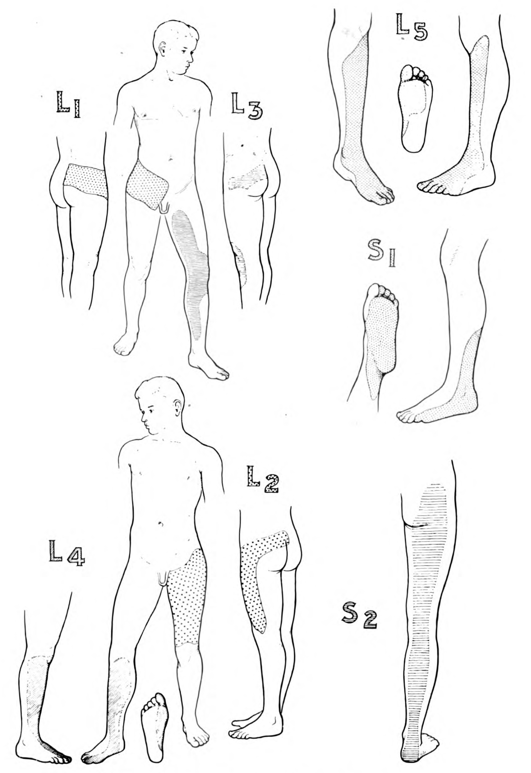
A dermatome is an area of skin that is mainly supplied by afferent nerve fibres from the dorsal root of any given spinal nerve. There are 8 cervical nerves (C1 being an exception with no dermatome), 12 thoracic nerves, 5 lumbar nerves and 5 sacral nerves. Each of these nerves relays sensation (including pain) from a particular region of skin to the brain. The term is also used to refer to a part of an embryonic somite. Along the thorax and abdomen the dermatomes are like a stack of discs forming a human, each supplied by a different spinal nerve. Along the arms and the legs, the pattern is different: the dermatomes run longitudinally along the limbs. Although the general pattern is similar in all people, the precise areas of innervation are as unique to an individual as fingerprints. An area of skin innervated by a single nerve is called a peripheral nerve field. The word ''dermatome'' is formed from Ancient Greek δέρμα 'skin, hide' and τέμνω 'cut'. Clinical signif ... [...More Info...] [...Related Items...] OR: [Wikipedia] [Google] [Baidu] |
Trigeminal Nerve
In neuroanatomy, the trigeminal nerve ( lit. ''triplet'' nerve), also known as the fifth cranial nerve, cranial nerve V, or simply CN V, is a cranial nerve responsible for sensation in the face and motor functions such as biting and chewing; it is the most complex of the cranial nerves. Its name ("trigeminal", ) derives from each of the two nerves (one on each side of the pons) having three major branches: the ophthalmic nerve (V), the maxillary nerve (V), and the mandibular nerve (V). The ophthalmic and maxillary nerves are purely sensory, whereas the mandibular nerve supplies motor as well as sensory (or "cutaneous") functions. Adding to the complexity of this nerve is that autonomic nerve fibers as well as special sensory fibers (taste) are contained within it. The motor division of the trigeminal nerve derives from the basal plate of the embryonic pons, and the sensory division originates in the cranial neural crest. Sensory information from the face and body is proc ... [...More Info...] [...Related Items...] OR: [Wikipedia] [Google] [Baidu] |
Cervical Spinal Nerve 3
The cervical spinal nerve 3 (C3) is a spinal nerve of the cervical segment. Nervous System -- Groups of Nerves It originates from the spinal column from above the cervical vertebra 3
In tetrapods, cervical vertebrae (singular: vertebra) are the vertebrae of the neck, immediately below the skull. Truncal vertebrae (divided into thoracic and lumbar vertebrae in mammals) lie caudal (toward the tail) of cervical vertebrae. In sau ... (C3).
References Spinal nerves {{neuroanatomy-stub ...[...More Info...] [...Related Items...] OR: [Wikipedia] [Google] [Baidu] |
Cervical Spinal Nerve 4
Cervical spinal nerve 4, also called C4, is a spinal nerve of the cervical segment. It originates from the spinal cord above the 4th cervical vertebra (C4). It contributes nerve fibers to the phrenic nerve, the motor nerve to the thoracoabdominal diaphragm. It also provides motor nerves for the longus capitis, longus colli, anterior scalene, middle scalene, and levator scapulae muscles. C4 contributes some sensory fibers to the supraclavicular nerves, responsible for sensation from the skin above the clavicle. Additional Images File:Slide1y.JPG, Cervical spinal nerve 4 File:Projectional radiograph of cervical foraminal stenosis, annotated.jpg, Projectional radiograph of a man presenting with pain by the nape and left shoulder, showing a stenosis in the intervertebral foramen of cervical spinal nerve 4, corresponding with the affected dermatome Dermatome may refer to: * Dermatome (anatomy), an area of skin that is supplied by a single pair of dorsal roots * Dermatome (embryo ... [...More Info...] [...Related Items...] OR: [Wikipedia] [Google] [Baidu] |
Victor Selmanowitz
Victor Joel Selmanowitz was an American dermatologist. In 1970, he coined the term unilateral nevoid telangiectasia Unilateral nevoid telangiectasia presents with fine thread veins, typically over a segment of skin supplied by a particular nerve on one side of the body. It most frequently involves the trigeminal, C3 and C4, or nearby areas. The condition was .... The Victor J. Selmanowitz Memorial Lecture and chair of modern Jewish history at the Touro College Graduate School of Jewish Studies is named for him. Selected publications * * * References {{DEFAULTSORT:Selmanowitz, Victor J. American physicians ... [...More Info...] [...Related Items...] OR: [Wikipedia] [Google] [Baidu] |
Dermatome (anatomy)
A dermatome is an area of skin that is mainly supplied by afferent nerve fibres from the dorsal root of any given spinal nerve. There are 8 cervical nerves (C1 being an exception with no dermatome), 12 thoracic nerves, 5 lumbar nerves and 5 sacral nerves. Each of these nerves relays sensation (including pain) from a particular region of skin to the brain. The term is also used to refer to a part of an embryonic somite. Along the thorax and abdomen the dermatomes are like a stack of discs forming a human, each supplied by a different spinal nerve. Along the arms and the legs, the pattern is different: the dermatomes run longitudinally along the limbs. Although the general pattern is similar in all people, the precise areas of innervation are as unique to an individual as fingerprints. An area of skin innervated by a single nerve is called a peripheral nerve field. The word ''dermatome'' is formed from Ancient Greek δέρμα 'skin, hide' and τέμνω 'cut'. Clinical signif ... [...More Info...] [...Related Items...] OR: [Wikipedia] [Google] [Baidu] |
Blaschko's Lines
Blaschko's lines, also called the lines of Blaschko, are lines of normal cell development in the skin. These lines are only visible in those with a mosaic skin condition or in chimeras where different cell lines contain different genes. These lines may express different amounts of melanin, or become visible due to a differing susceptibility to disease. In such individuals, they can become apparent as whorls, patches, streaks or lines in a linear or segmental distribution over the skin. They follow a ''V'' shape over the back, ''S''-shaped whirls over the chest and sides, and wavy shapes on the head. Not all mosaic skin conditions follow Blascko's lines. The lines are believed to trace the migration of embryonic cells. They do not correspond to nervous, muscular, or lymphatic systems. The lines are not unique to humans and can be observed in other non-human animals with mosaicism. Alfred Blaschko is credited with the first demonstration of these lines in 1901. Signs and sym ... [...More Info...] [...Related Items...] OR: [Wikipedia] [Google] [Baidu] |
Autosomal Dominant
In genetics, dominance is the phenomenon of one variant (allele) of a gene on a chromosome masking or overriding the effect of a different variant of the same gene on the other copy of the chromosome. The first variant is termed dominant and the second recessive. This state of having two different variants of the same gene on each chromosome is originally caused by a mutation in one of the genes, either new (''de novo'') or inherited. The terms autosomal dominant or autosomal recessive are used to describe gene variants on non-sex chromosomes ( autosomes) and their associated traits, while those on sex chromosomes (allosomes) are termed X-linked dominant, X-linked recessive or Y-linked; these have an inheritance and presentation pattern that depends on the sex of both the parent and the child (see Sex linkage). Since there is only one copy of the Y chromosome, Y-linked traits cannot be dominant or recessive. Additionally, there are other forms of dominance such as incomplete d ... [...More Info...] [...Related Items...] OR: [Wikipedia] [Google] [Baidu] |
Epidermis
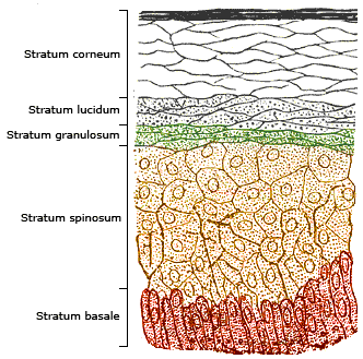
The epidermis is the outermost of the three layers that comprise the skin, the inner layers being the dermis and hypodermis. The epidermis layer provides a barrier to infection from environmental pathogens and regulates the amount of water released from the body into the atmosphere through transepidermal water loss. The epidermis is composed of multiple layers of flattened cells that overlie a base layer (stratum basale) composed of columnar cells arranged perpendicularly. The layers of cells develop from stem cells in the basal layer. The human epidermis is a familiar example of epithelium, particularly a stratified squamous epithelium. The word epidermis is derived through Latin , itself and . Something related to or part of the epidermis is termed epidermal. Structure Cellular components The epidermis primarily consists of keratinocytes ( proliferating basal and differentiated suprabasal), which comprise 90% of its cells, but also contains melanocytes, Langerhans ... [...More Info...] [...Related Items...] OR: [Wikipedia] [Google] [Baidu] |
Blood Vessel
The blood vessels are the components of the circulatory system that transport blood throughout the human body. These vessels transport blood cells, nutrients, and oxygen to the tissues of the body. They also take waste and carbon dioxide away from the tissues. Blood vessels are needed to sustain life, because all of the body's tissues rely on their functionality. There are five types of blood vessels: the arteries, which carry the blood away from the heart; the arterioles; the capillaries, where the exchange of water and chemicals between the blood and the tissues occurs; the venules; and the veins, which carry blood from the capillaries back towards the heart. The word ''vascular'', meaning relating to the blood vessels, is derived from the Latin ''vas'', meaning vessel. Some structures – such as cartilage, the epithelium, and the lens and cornea of the eye – do not contain blood vessels and are labeled ''avascular''. Etymology * artery: late Middle English; from Latin ... [...More Info...] [...Related Items...] OR: [Wikipedia] [Google] [Baidu] |


