|
TGFBR2
Transforming growth factor, beta receptor II (70/80kDa) is a TGF beta receptor. ''TGFBR2'' is its human gene. It is a tumor suppressor gene. Function This gene encodes a member of the serine/threonine protein kinase family and the TGFB receptor subfamily. The encoded protein is a transmembrane protein that has a protein kinase domain, forms a heterodimeric complex with another receptor protein, and binds TGF-beta. This receptor/ligand complex phosphorylates proteins, which then enter the nucleus and regulate the transcription of a subset of genes related to cell proliferation. Mutations in this gene have been associated with Marfan syndrome, Loeys-Deitz aortic aneurysm syndrome, Osler–Weber–Rendu syndrome, and the development of various types of tumors. At least 73 disease-causing mutations in this gene have been discovered. Alternatively spliced transcript variants encoding different isoforms have been characterized. Interactions TGF beta receptor 2 has been shown to Pr ... [...More Info...] [...Related Items...] OR: [Wikipedia] [Google] [Baidu] |
TGF Beta Receptor
Transforming growth factor beta (TGFβ) receptors are bitopic protein, single pass Receptor protein serine/threonine kinase, serine/threonine kinase receptors that belong to TGFβ superfamily receptor, TGFβ receptor family. They exist in several different isoforms that can be homodimeric, homo- or heterodimeric. (free full text) The number of characterized ligands in the TGFβ superfamily far exceeds the number of known receptors, suggesting the promiscuity that exists between the ligand and receptor interactions. TGFβ is a growth factor and cytokine involved in paracrine signalling and can be found in many different tissue (biology), tissue types, including brain, heart, kidney, liver, bone, and testes. Over-gene expression, expression of TGFβ can induce renal fibrosis, causing kidney disease, as well as diabetes, and ultimately end-stage renal disease. Recent developments have found that, using certain types of protein receptor antagonist, antagonists against TGFβ recepto ... [...More Info...] [...Related Items...] OR: [Wikipedia] [Google] [Baidu] |
TGF Beta Receptors
Transforming growth factor beta (TGFβ) receptors are single pass serine/threonine kinase receptors that belong to TGFβ receptor family. They exist in several different isoforms that can be homo- or heterodimeric. (free full text) The number of characterized ligands in the TGFβ superfamily far exceeds the number of known receptors, suggesting the promiscuity that exists between the ligand and receptor interactions. TGFβ is a growth factor and cytokine involved in paracrine signalling and can be found in many different tissue types, including brain, heart, kidney, liver, bone, and testes. Over-expression of TGFβ can induce renal fibrosis, causing kidney disease, as well as diabetes, and ultimately end-stage renal disease. Recent developments have found that, using certain types of protein antagonists against TGFβ receptors, can halt and in some cases reverse the effects of renal fibrosis. Three TGFβ superfamily receptors specific for TGFβ, the TGFβ receptors, can be ... [...More Info...] [...Related Items...] OR: [Wikipedia] [Google] [Baidu] |
Marfan Syndrome
Marfan syndrome (MFS) is a multi-systemic genetic disorder that affects the connective tissue. Those with the condition tend to be tall and thin, with long arms, legs, fingers, and toes. They also typically have exceptionally flexible joints and abnormally curved spines. The most serious complications involve the heart and aorta, with an increased risk of mitral valve prolapse and aortic aneurysm. The lungs, eyes, bones, and the covering of the spinal cord are also commonly affected. The severity of the symptoms is variable. MFS is caused by a mutation in ''FBN1'', one of the genes that makes fibrillin, which results in abnormal connective tissue. It is an autosomal dominant disorder. In about 75% of cases, it is inherited from a parent with the condition, while in about 25% it is a new mutation. Diagnosis is often based on the Ghent criteria. There is no known cure for MFS. Many of those with the disorder have a normal life expectancy with proper treatment. Management of ... [...More Info...] [...Related Items...] OR: [Wikipedia] [Google] [Baidu] |
Heat Shock Protein 90kDa Alpha (cytosolic), Member A1
Heat shock protein HSP 90-alpha is a protein that in humans is encoded by the ''HSP90AA1'' gene. Function The gene, HSP90AA1, encodes the human stress-inducible 90-kDa heat shock protein alpha (Hsp90A). Complemented by the constitutively expressed paralog Hsp90B which shares over 85% amino acid sequence identity, Hsp90A expression is initiated when a cell experiences proteotoxic stress. Once expressed Hsp90A dimers operate as molecular chaperones that bind and fold other proteins into their functional 3-dimensional structures. This molecular chaperoning ability of Hsp90A is driven by a cycle of structural rearrangements fueled by ATP hydrolysis. Current research on Hsp90A focuses in its role as a drug target due to its interaction with a large number of tumor promoting proteins and its role in cellular stress adaptation. Gene structure Human HSP90AA1 is encoded on the complement strand of Chromosome 14q32.33 and spans over 59 kbp. Several pseudogenes of HSP90AA1 exist th ... [...More Info...] [...Related Items...] OR: [Wikipedia] [Google] [Baidu] |
Protein Kinase Domain
The protein kinase domain is a structurally conserved protein domain containing the catalytic function of protein kinases. Protein kinases are a group of enzymes that move a phosphate group onto proteins, in a process called phosphorylation. This functions as an on/off switch for many cellular processes, including metabolism, transcription, cell cycle progression, cytoskeletal rearrangement and cell movement, apoptosis, and differentiation. They also function in embryonic development, physiological responses, and in the nervous and immune system. Abnormal phosphorylation causes many human diseases, including cancer, and drugs that affect phosphorylation can treat those diseases. Protein kinases possess a catalytic subunit which transfers the gamma phosphate from nucleoside triphosphates (almost always ATP) to the side chain of an amino acid in a protein, resulting in a conformational and/or dynamic changes affecting protein function. These enzymes fall into two broad classes, cha ... [...More Info...] [...Related Items...] OR: [Wikipedia] [Google] [Baidu] |
Tumor Suppressor Gene
A tumor suppressor gene (TSG), or anti-oncogene, is a gene that regulates a cell during cell division and replication. If the cell grows uncontrollably, it will result in cancer. When a tumor suppressor gene is mutated, it results in a loss or reduction in its function. In combination with other genetic mutations, this could allow the cell to grow abnormally. The loss of function for these genes may be even more significant in the development of human cancers, compared to the activation of oncogenes. TSGs can be grouped into the following categories: caretaker genes, gatekeeper genes, and more recently landscaper genes. Caretaker genes ensure stability of the genome via DNA repair and subsequently when mutated allow mutations to accumulate. Meanwhile, gatekeeper genes directly regulate cell growth by either inhibiting cell cycle progression or inducing apoptosis. Lastly landscaper genes regulate growth by contributing to the surrounding environment, when mutated can cause an envir ... [...More Info...] [...Related Items...] OR: [Wikipedia] [Google] [Baidu] |
C-terminal
The C-terminus (also known as the carboxyl-terminus, carboxy-terminus, C-terminal tail, C-terminal end, or COOH-terminus) is the end of an amino acid chain (protein or polypeptide), terminated by a free carboxyl group (-COOH). When the protein is translated from messenger RNA, it is created from N-terminus to C-terminus. The convention for writing peptide sequences is to put the C-terminal end on the right and write the sequence from N- to C-terminus. Chemistry Each amino acid has a carboxyl group and an amine group. Amino acids link to one another to form a chain by a dehydration reaction which joins the amine group of one amino acid to the carboxyl group of the next. Thus polypeptide chains have an end with an unbound carboxyl group, the C-terminus, and an end with an unbound amine group, the N-terminus. Proteins are naturally synthesized starting from the N-terminus and ending at the C-terminus. Function C-terminal retention signals While the N-terminus of a protein often cont ... [...More Info...] [...Related Items...] OR: [Wikipedia] [Google] [Baidu] |
Beta-sheet
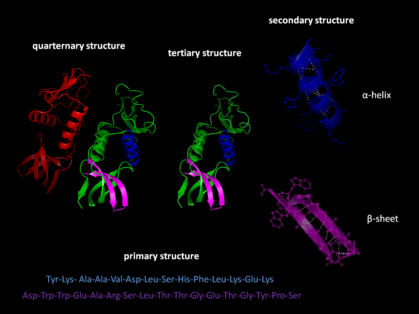
The beta sheet, (β-sheet) (also β-pleated sheet) is a common motif of the regular protein secondary structure. Beta sheets consist of beta strands (β-strands) connected laterally by at least two or three backbone hydrogen bonds, forming a generally twisted, pleated sheet. A β-strand is a stretch of polypeptide chain typically 3 to 10 amino acids long with backbone in an extended conformation. The supramolecular association of β-sheets has been implicated in the formation of the fibrils and protein aggregates observed in amyloidosis, notably Alzheimer's disease. History The first β-sheet structure was proposed by William Astbury in the 1930s. He proposed the idea of hydrogen bonding between the peptide bonds of parallel or antiparallel extended β-strands. However, Astbury did not have the necessary data on the bond geometry of the amino acids in order to build accurate models, especially since he did not then know that the peptide bond was planar. A refined versi ... [...More Info...] [...Related Items...] OR: [Wikipedia] [Google] [Baidu] |
Antiparallel (biochemistry)
In biochemistry, two biopolymers are antiparallel if they run parallel to each other but with opposite directionality (alignments). An example is the two complementary strands of a DNA double helix, which run in opposite directions alongside each other. Nucleic acids Nucleic acid molecules have a phosphoryl (5') end and a hydroxyl (3') end. This notation follows from organic chemistry nomenclature, and can be used to define the movement of enzymes such as DNA polymerases relative to the DNA strand in a non-arbitrary manner. G-quadruplexes G-quadruplexes, also known as G4 DNA are secondary structures found in nucleic acids that are rich in guanine. These structures are normally located at the telomeres (the ends of the chromosomes). The G-quadruplex can either be parallel or antiparallel depending on the loop configuration, which is a component of the structure. If all the DNA strands run in the same direction, it is termed to be a parallel quadruplex, and is known as a str ... [...More Info...] [...Related Items...] OR: [Wikipedia] [Google] [Baidu] |
Disulphide Bonds
In biochemistry, a disulfide (or disulphide in British English) refers to a functional group with the structure . The linkage is also called an SS-bond or sometimes a disulfide bridge and is usually derived by the coupling of two thiol groups. In biology, disulfide bridges formed between thiol groups in two cysteine residues are an important component of the secondary and tertiary structure of proteins. ''Persulfide'' usually refers to compounds. In inorganic chemistry disulfide usually refers to the corresponding anion (−S−S−). Organic disulfides Symmetrical disulfides are compounds of the formula . Most disulfides encountered in organo sulfur chemistry are symmetrical disulfides. Unsymmetrical disulfides (also called heterodisulfides) are compounds of the formula . They are less common in organic chemistry, but most disulfides in nature are unsymmetrical. Properties The disulfide bonds are strong, with a typical bond dissociation energy of 60 kcal/mol (251&nbs ... [...More Info...] [...Related Items...] OR: [Wikipedia] [Google] [Baidu] |
Alpha Helix
The alpha helix (α-helix) is a common motif in the secondary structure of proteins and is a right hand-helix conformation in which every backbone N−H group hydrogen bonds to the backbone C=O group of the amino acid located four residues earlier along the protein sequence. The alpha helix is also called a classic Pauling–Corey–Branson α-helix. The name 3.613-helix is also used for this type of helix, denoting the average number of residues per helical turn, with 13 atoms being involved in the ring formed by the hydrogen bond. Among types of local structure in proteins, the α-helix is the most extreme and the most predictable from sequence, as well as the most prevalent. Discovery In the early 1930s, William Astbury showed that there were drastic changes in the X-ray fiber diffraction of moist wool or hair fibers upon significant stretching. The data suggested that the unstretched fibers had a coiled molecular structure with a characteristic repeat of ≈. Astb ... [...More Info...] [...Related Items...] OR: [Wikipedia] [Google] [Baidu] |
Protein Folding
Protein folding is the physical process by which a protein chain is translated to its native three-dimensional structure, typically a "folded" conformation by which the protein becomes biologically functional. Via an expeditious and reproducible process, a polypeptide folds into its characteristic three-dimensional structure from a random coil. Each protein exists first as an unfolded polypeptide or random coil after being translated from a sequence of mRNA to a linear chain of amino acids. At this stage the polypeptide lacks any stable (long-lasting) three-dimensional structure (the left hand side of the first figure). As the polypeptide chain is being synthesized by a ribosome, the linear chain begins to fold into its three-dimensional structure. Folding of many proteins begins even during translation of the polypeptide chain. Amino acids interact with each other to produce a well-defined three-dimensional structure, the folded protein (the right hand side of the figure), ... [...More Info...] [...Related Items...] OR: [Wikipedia] [Google] [Baidu] |


_structure.png)


