|
Tsutomu Sato (ophthalmologist)
Tsutomu Sato (1902 – June 9, 1960) was a Japanese ophthalmologist who performed an early version of the radial keratotomy and was the first professor at the Research Institute of Ophthalmology at Juntendo University School of Medicine. Biography Sato was the first professor at the Research Institute of Ophthalmology at Juntendo University School of Medicine, where he researched the treatment of eye conditions such as trachoma and myopia. In the mid-1930s, Sato devised a glass scleral lens, but it never came into wide usage. Within a few years, higher-quality glass scleral lenses from Carl Zeiss were available in Japan. In 1939, Sato became the first physician to report the use of radial keratotomy for myopia. Sato knew that the cornea flattened out in patients with keratoconus who suffered corneal hydrops from the leakage of fluid through Descemet's membrane. His idea was to create numerous breaks in that membrane through anterior and posterior approaches. His initial pati ... [...More Info...] [...Related Items...] OR: [Wikipedia] [Google] [Baidu] |
Ophthalmologist
Ophthalmology ( ) is a surgery, surgical subspecialty within medicine that deals with the diagnosis and treatment of eye disorders. An ophthalmologist is a physician who undergoes subspecialty training in medical and surgical eye care. Following a medical degree, a doctor specialising in ophthalmology must pursue additional postgraduate residency (medicine), residency training specific to that field. This may include a one-year integrated internship that involves more general medical training in other fields such as internal medicine or general surgery. Following residency, additional specialty training (or fellowship) may be sought in a particular aspect of eye pathology. Ophthalmologists prescribe medications to treat eye diseases, implement laser therapy, and perform surgery when needed. Ophthalmologists provide both primary and specialty eye care - medical and surgical. Most ophthalmologists participate in academic research on eye diseases at some point in their training an ... [...More Info...] [...Related Items...] OR: [Wikipedia] [Google] [Baidu] |
Juntendo University
is a private university in Japan. Its headquarters are on its campus in Bunkyo, Tokyo, for the School of Medicine and in Inzai, Chiba, for the School of Health and Sports Science. The university was established in 1838 for medical and in 1946 for other departments. It is nicknamed ''Jundai''. Campuses *Hongō-Ochanomizu Campus: Bunkyo, Tokyo, *Sakura Campus: Inzai, Chiba, *Urayasu Campus:Urayasu, Chiba, *Mishima Campus: Mishima, Shizuoka, Faculties *Faculty of Medicine *Faculty of Health and Sports Science *Faculty of Health Care and Nursing *Faculty of Health Sciences and Nursing *Faculty of International Liberal Arts The Juntendo University Graduate School of Medicine has granted doctorates since 1963, and the total numbers of the two types doctorate holders (甲 Kou and 乙 Otsu) has reached reach 1,897 and 2,394, respectively, as of 2017. Notable alumni Athletes *Masatada Ishii, Manager of Buriram United *Takehiro Kashima, gymnast *Ryōhei Katō, gymnast *Hiroshi Na ... [...More Info...] [...Related Items...] OR: [Wikipedia] [Google] [Baidu] |
Trachoma
Trachoma is an infectious disease caused by bacterium ''Chlamydia trachomatis''. The infection causes a roughening of the inner surface of the eyelids. This roughening can lead to pain in the eyes, breakdown of the outer surface or cornea of the eyes, and eventual blindness. Untreated, repeated trachoma infections can result in a form of permanent blindness when the eyelids turn inward. The bacteria that cause the disease can be spread by both direct and indirect contact with an affected person's eyes or nose. Indirect contact includes through clothing or flies that have come into contact with an affected person's eyes or nose. Children spread the disease more often than adults. Poor sanitation, crowded living conditions, and not enough clean water and toilets also increase spread. Efforts to prevent the disease include improving access to clean water and treatment with antibiotics to decrease the number of people infected with the bacterium. This may include treating, all ... [...More Info...] [...Related Items...] OR: [Wikipedia] [Google] [Baidu] |
Myopia
Near-sightedness, also known as myopia and short-sightedness, is an eye disease where light focuses in front of, instead of on, the retina. As a result, distant objects appear blurry while close objects appear normal. Other symptoms may include headaches and eye strain. Severe near-sightedness is associated with an increased risk of retinal detachment, cataracts, and glaucoma. The underlying mechanism involves the length of the eyeball growing too long or less commonly the lens being too strong. It is a type of refractive error. Diagnosis is by eye examination. Tentative evidence indicates that the risk of near-sightedness can be decreased by having young children spend more time outside. This decrease in risk may be related to natural light exposure. Near-sightedness can be corrected with eyeglasses, contact lenses, or a refractive surgery. Eyeglasses are the easiest and safest method of correction. Contact lenses can provide a wider field of vision, but are associated with ... [...More Info...] [...Related Items...] OR: [Wikipedia] [Google] [Baidu] |
Scleral Lens
A scleral lens, also known as a scleral contact lens, is a large contact lens that rests on the sclera and creates a tears, tear-filled vault over the cornea. Scleral lenses are designed to treat a variety of eye conditions, many of which do not respond to other forms of treatment. Uses Medical uses Scleral lenses may be used to improve vision and reduce pain and light sensitivity for people with a growing number of disorders or injuries to the eye, such as severe dry eye syndrome, microphthalmia, keratoconus, corneal ectasia, Stevens–Johnson syndrome, Sjögren's syndrome, aniridia, neurotrophic keratitis (anesthetic corneas), complications post-LASIK, higher-order aberrations of the eye, complications post-corneal transplant and Keratoconus#Related disorders, pellucid degeneration. Injuries to the eye such as surgical complications, distorted corneal implants, as well as chemical and burn injuries also may be treated by the use of scleral lenses. Sclerals may also be use ... [...More Info...] [...Related Items...] OR: [Wikipedia] [Google] [Baidu] |
Carl Zeiss AG
Carl Zeiss AG (), branded as ZEISS, is a German manufacturer of optical systems and optoelectronics, founded in Jena, Germany in 1846 by optician Carl Zeiss. Together with Ernst Abbe (joined 1866) and Otto Schott (joined 1884) he laid the foundation for today's multi-national company. The current company emerged from a reunification of Carl Zeiss companies in East and West Germany with a consolidation phase in the 1990s. ZEISS is active in four business segments with approximately equal revenue (Industrial Quality and Research, Medical Technology, Consumer Markets and Semiconductor Manufacturing Technology) in almost 50 countries, has 30 production sites and around 25 development sites worldwide. Carl Zeiss AG is the holding of all subsidiaries within Zeiss Group, of which Carl Zeiss Meditec AG is the only one that is traded at the stock market. Carl Zeiss AG is owned by the foundation Carl-Zeiss-Stiftung. The Zeiss Group has its headquarters in southern Germany, in the smal ... [...More Info...] [...Related Items...] OR: [Wikipedia] [Google] [Baidu] |
Keratoconus
Keratoconus (KC) is a disorder of the eye that results in progressive thinning of the cornea. This may result in blurry vision, double vision, nearsightedness, irregular astigmatism, and light sensitivity leading to poor quality-of-life. Usually both eyes are affected. In more severe cases a scarring or a circle may be seen within the cornea. While the cause is unknown, it is believed to occur due to a combination of genetic, environmental, and hormonal factors. Patients with a parent, sibling, or child who has keratoconus have 15 to 67 times higher risk in developing corneal ectasia compared to patients with no affected relatives. Proposed environmental factors include rubbing the eyes and allergies. The underlying mechanism involves changes of the cornea to a cone shape. Diagnosis is most often by topography. Topography measures the curvature of the cornea and creates a colored "map" of the cornea. Keratoconus causes very distinctive changes in the appearance of these ma ... [...More Info...] [...Related Items...] OR: [Wikipedia] [Google] [Baidu] |
Corneal Hydrops
Corneal hydrops is an uncommon complication seen in people with advanced keratoconus or other corneal ectatic disorders, and is characterized by stromal edema due to leakage of aqueous humor through a tear in Descemet's membrane. Although a hydrops usually causes increased scarring of the cornea, occasionally it will benefit a patient by creating a flatter cone, aiding the fitting of contact lenses. Corneal transplantation is not usually indicated during corneal hydrops. Signs and symptoms The person experiences pain and a sudden severe clouding of vision, with the cornea taking on a translucent milky-white appearance known as a corneal hydrops. Diagnosis Patients are recommended to take a Sodium Chloride eye drop solution as well as a Dexamethasone solution for a period of 4-6 weeks, timeframes may vary depending on the severity of a patients condition. Once the medication cycle is complete and the cloud clears, scarring will be left on the cornea. Management The effect is ... [...More Info...] [...Related Items...] OR: [Wikipedia] [Google] [Baidu] |
Descemet's Membrane
Descemet's membrane ( or the Descemet membrane) is the basement membrane that lies between the corneal proper substance, also called stroma, and the endothelial layer of the cornea. It is composed of different kinds of collagen (Type IV and VIII) than the stroma. The endothelial layer is located at the posterior of the cornea. Descemet's membrane, as the basement membrane for the endothelial layer, is secreted by the single layer of squamous epithelial cells that compose the endothelial layer of the cornea. Structure Its thickness ranges from 3 μm at birth to 8–10 μm in adults.Johnson DH, Bourne WM, Campbell RJ: The ultrastructure of Descemet's membrane. I. Changes with age in normal cornea. Arch Ophthalmol 100:1942, 1982 The corneal endothelium is a single layer of squamous cells covering the surface of the cornea that faces the anterior chamber. Clinical significance Significant damage to the membrane may require a corneal transplant. Damage caused by the hereditary co ... [...More Info...] [...Related Items...] OR: [Wikipedia] [Google] [Baidu] |
Bullous Keratopathy
Bullous keratopathy, also known as pseudophakic bullous keratopathy (PBK), is a pathological condition in which small vesicles, or '' bullae'', are formed in the cornea due to endothelial dysfunction. In a healthy cornea, endothelial cells keeps the tissue from excess fluid absorption, pumping it back into the aqueous humor. When affected by some reason, such as Fuchs' dystrophy Fuchs dystrophy, also referred to as Fuchs endothelial corneal dystrophy (FECD) and Fuchs endothelial dystrophy (FED), is a slowly progressing corneal dystrophy that usually affects both eyes and is slightly more common in women than in men. Althou ... or a trauma during cataract removal, endothelial cells suffer mortality or damage. The corneal endothelial cells normally do not undergo mitotic cell division, and cell loss results in permanent loss of function. When endothelial cell counts drop too low, the pump starts failing to function and fluid moves anterior into the stroma and epithelium. The excess ... [...More Info...] [...Related Items...] OR: [Wikipedia] [Google] [Baidu] |
Corneal Endothelium
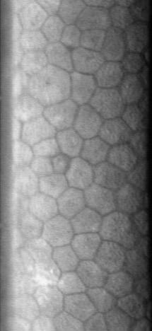
The corneal endothelium is a single layer of endothelial cells on the inner surface of the cornea. It faces the chamber formed between the cornea and the iris. The corneal endothelium are specialized, flattened, mitochondria-rich cells that line the posterior surface of the cornea and face the anterior chamber of the eye. The corneal endothelium governs fluid and solute transport across the posterior surface of the cornea and maintains the cornea in the slightly dehydrated state that is required for optical transparency. Embryology and anatomy The corneal endothelium is embryologically derived from the neural crest. The postnatal total endothelial cellularity of the cornea (approximately 300,000 cells per cornea) is achieved as early as the second trimester of gestation. Thereafter the endothelial cell density (but not the absolute number of cells) rapidly declines, as the fetal cornea grows in surface area, achieving a final adult density of approximately 2400 - 3200 cells ... [...More Info...] [...Related Items...] OR: [Wikipedia] [Google] [Baidu] |


.jpg)

.jpg)

