Corneal Endothelium on:
[Wikipedia]
[Google]
[Amazon]
The corneal endothelium is a single layer of endothelial cells on the inner surface of the
 The principal physiological function of the corneal endothelium is to allow leakage of solutes and nutrients from the aqueous humor to the more superficial layers of the cornea while at the same time pumping water in the opposite direction, from the stroma to the aqueous. This dual function of the corneal endothelium is described by the "pump-leak hypothesis." Since the cornea is avascular, which renders it optimally transparent, the nutrition of the corneal epithelium, stromal keratocytes, and corneal endothelium must occur via diffusion of glucose and other solutes from the aqueous humor, across the corneal endothelium. The corneal endothelium then transports water from the stromal-facing surface to the aqueous-facing surface by an interrelated series of active and passive ion exchangers. Critical to this energy-driven process is the role of Na+/K+ATPase and carbonic anhydrase. Bicarbonate ions formed by the action of carbonic anhydrase are translocated across the cell membrane, allowing water to passively follow.
cornea
The cornea is the transparent front part of the eye that covers the iris, pupil, and anterior chamber. Along with the anterior chamber and lens, the cornea refracts light, accounting for approximately two-thirds of the eye's total optical power ...
. It faces the chamber formed between the cornea and the iris.
The corneal endothelium are specialized, flattened, mitochondria-rich cell
Cell most often refers to:
* Cell (biology), the functional basic unit of life
Cell may also refer to:
Locations
* Monastic cell, a small room, hut, or cave in which a religious recluse lives, alternatively the small precursor of a monastery ...
s that line the posterior surface of the cornea and face the anterior chamber
The anterior chamber ( AC) is the aqueous humor-filled space inside the eye between the iris and the cornea's innermost surface, the endothelium. Hyphema, anterior uveitis and glaucoma are three main pathologies in this area. In hyphema, blood f ...
of the eye. The corneal endothelium governs fluid and solute transport across the posterior surface of the cornea and maintains the cornea in the slightly dehydrated state that is required for optical transparency.
Embryology and anatomy
The corneal endothelium is embryologically derived from theneural crest
Neural crest cells are a temporary group of cells unique to vertebrates that arise from the embryonic ectoderm germ layer, and in turn give rise to a diverse cell lineage—including melanocytes, craniofacial cartilage and bone, smooth muscle, per ...
. The postnatal total endothelial cellularity of the cornea (approximately 300,000 cells per cornea) is achieved as early as the second trimester of gestation. Thereafter the endothelial cell density (but not the absolute number of cells) rapidly declines, as the fetal cornea grows in surface area, achieving a final adult density of approximately 2400 - 3200 cells/mm². The number of endothelial cells in the fully developed cornea decreases with age up until early adulthood, stabilizing around 50 years of age.
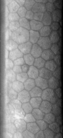
The normal corneal endothelium is a single layer of uniformly sized cells with a predominantly hexagonal shape. This honeycomb
A honeycomb is a mass of Triangular prismatic honeycomb#Hexagonal prismatic honeycomb, hexagonal prismatic Beeswax, wax cells built by honey bees in their beehive, nests to contain their larvae and stores of honey and pollen.
beekeeping, Beekee ...
tiling scheme yields the greatest efficiency, in terms of total perimeter, of packing the posterior corneal surface with cells of a given area. The corneal endothelium is attached to the rest of the cornea through Descemet's membrane
Descemet's membrane ( or the Descemet membrane) is the basement membrane that lies between the corneal proper substance, also called stroma, and the endothelial layer of the cornea. It is composed of different kinds of collagen (Type IV and VIII) ...
, which is an acellular layer composed mostly of collagen IV.
Physiology
 The principal physiological function of the corneal endothelium is to allow leakage of solutes and nutrients from the aqueous humor to the more superficial layers of the cornea while at the same time pumping water in the opposite direction, from the stroma to the aqueous. This dual function of the corneal endothelium is described by the "pump-leak hypothesis." Since the cornea is avascular, which renders it optimally transparent, the nutrition of the corneal epithelium, stromal keratocytes, and corneal endothelium must occur via diffusion of glucose and other solutes from the aqueous humor, across the corneal endothelium. The corneal endothelium then transports water from the stromal-facing surface to the aqueous-facing surface by an interrelated series of active and passive ion exchangers. Critical to this energy-driven process is the role of Na+/K+ATPase and carbonic anhydrase. Bicarbonate ions formed by the action of carbonic anhydrase are translocated across the cell membrane, allowing water to passively follow.
Mechanisms of corneal edema
Corneal endothelial cells are post-mitotic and divide rarely, if at all, in the post-natal human cornea. Wounding of the corneal endothelium, as from trauma or other insults, prompts healing of the endothelial monolayer by sliding and enlargement of adjacent endothelial cells, rather than mitosis. Endothelial cell loss, if sufficiently severe, can cause endothelial cell density to fall below the threshold level needed to maintain corneal deturgescence. This threshold of endothelial cell density varies considerably amongst individuals, but is typically in the range of 500 - 1000 cells/mm². Typically, loss of endothelial cell density is accompanied by increases in cell size variability (polymegathism) and cell shape variation (polymorphism). Corneal edema can also occur as the result of compromised endothelial function due to intraocular inflammation or other causes. Excess hydration of the corneal stroma disrupts the normally uniform periodic spacing ofType I collagen
Type I collagen is the most abundant collagen of the human body. It forms large, eosinophilic fibers known as collagen fibers.
It is present in scar tissue, the end product when tissue heals by repair, as well as tendons, ligaments, the endomy ...
fibrils, creating light scatter. In addition, excessive corneal hydration can result in edema of the corneal epithelial layer, which creates irregularity at the optically critical tear film-air interface. Both stromal light scatter and surface epithelial irregularity contribute to degraded optical performance of the cornea and can compromise visual acuity.
Causes of endothelial disease
Leading causes of endothelial failure include inadvertent endothelial trauma from intraocular surgery (such ascataract surgery
Cataract surgery, also called lens replacement surgery, is the removal of the natural lens of the eye (also called "crystalline lens") that has developed an opacification, which is referred to as a cataract, and its replacement with an intra ...
) and Fuchs' dystrophy
Fuchs dystrophy, also referred to as Fuchs endothelial corneal dystrophy (FECD) and Fuchs endothelial dystrophy (FED), is a slowly progressing corneal dystrophy that usually affects both eyes and is slightly more common in women than in men. Althou ...
. Surgical causes of endothelial failure include both acute intraoperative trauma as well as chronic postoperative trauma, such as from a malpositioned intraocular lens
Intraocular lens (IOL) is a lens (optics), lens implanted in the human eye, eye as part of a treatment for cataracts or myopia. If the natural lens is left in the eye, the IOL is known as Phakic intraocular lens, phakic, otherwise it is a pseudop ...
or retained nuclear fragment in the anterior chamber. Other risk factors include narrow-angle glaucoma
Glaucoma is a group of eye diseases that result in damage to the optic nerve (or retina) and cause vision loss. The most common type is open-angle (wide angle, chronic simple) glaucoma, in which the drainage angle for fluid within the eye rem ...
, aging
Ageing ( BE) or aging ( AE) is the process of becoming older. The term refers mainly to humans, many other animals, and fungi, whereas for example, bacteria, perennial plants and some simple animals are potentially biologically immortal. In ...
, and iritis
Uveitis () is inflammation of the uvea, the pigmented layer of the eye between the inner retina and the outer fibrous layer composed of the sclera and cornea. The uvea consists of the middle layer of pigmented vascular structures of the eye and ...
.
A rare disease called X-linked endothelial corneal dystrophy
X-linked endothelial corneal dystrophy (XECD) is a rare form of corneal dystrophy described first in 2006, based on a 4-generation family of 60 members with 9 affected males and 35 trait carriers, which led to mapping the XECD locus to Xq25. It man ...
was described in 2006.
Treatment for endothelial disease
There is no medical treatment that can promote wound healing or regeneration of the corneal endothelium. In early stages of corneal edema, symptoms of blurred vision and episodic ocular pain predominate, due to edema and blistering (bullae) of the corneal epithelium. Partial palliation of these symptoms can sometimes be obtained through the instillation of topical hypertonic saline drops, use of bandage soft contact lenses, and/or application of anterior stromal micropuncture. In cases in which irreversible corneal endothelial failure develops, severe corneal edema ensues, and the only effective remedy is replacement of the diseased corneal endothelium through the surgical approach of corneal transplantation. Historically, penetrating keratoplasty, or full thickness corneal transplantation, was the treatment of choice for irreversible endothelial failure. More recently, new corneal transplant techniques have been developed to enable more selective replacement of the diseased corneal endothelium. This approach, termed endokeratoplasty, is most appropriate for disease processes that exclusively or predominantly involve the corneal endothelium. Penetrating keratoplasty is preferred when the disease process involves irreversible damage not just to the corneal endothelium, but to other layers of the cornea as well. Compared to full-thickness keratoplasty, endokeratoplasty techniques are associated with shorter recovery times, improved visual results, and greater resistance to wound rupture. Although instrumentation and surgical techniques for endokeratoplasty are still in evolution, one commonly performed form of endokeratoplasty at present is Descemet's Stripping (Automated) Endothelial Keratoplasty (DSEK r DSAEK. In this form of endokeratoplasty, the diseased host endothelium and associatedDescemet's membrane
Descemet's membrane ( or the Descemet membrane) is the basement membrane that lies between the corneal proper substance, also called stroma, and the endothelial layer of the cornea. It is composed of different kinds of collagen (Type IV and VIII) ...
are removed from the central cornea, and in their place a specially harvested layer of healthy donor tissue is grafted. This layer consists of posterior stroma, Descemet's membrane, and endothelium that has been dissected from cadaveric donor corneal tissue, typically using a mechanized (or "automated") instrument.
Investigational methods of corneal endothelial surgical replacement include Descemet's Membrane Endothelial Keratoplasty (DMEK), in which the donor tissue consists only of Descemet's membrane and endothelium, and corneal endothelial cell replacement therapy, in which in vitro cultivated endothelial cells are transplanted. These techniques, although still in an early developmental stage, aim to improve the selectivity of the transplantation approach by eliminating the presence of posterior stromal tissue from the grafted tissue.
References
Further reading
* {{DEFAULTSORT:Corneal Endothelium Human eye anatomy Visual system Human head and neck Ophthalmology