|
Neural Crest
Neural crest cells are a temporary group of cells unique to vertebrates that arise from the embryonic ectoderm germ layer, and in turn give rise to a diverse cell lineage—including melanocytes, craniofacial cartilage and bone, smooth muscle, peripheral and enteric neurons and glia. After gastrulation, neural crest cells are specified at the border of the neural plate and the non-neural ectoderm. During neurulation, the borders of the neural plate, also known as the neural folds, converge at the dorsal midline to form the neural tube. Subsequently, neural crest cells from the roof plate of the neural tube undergo an epithelial to mesenchymal transition, delaminating from the neuroepithelium and migrating through the periphery where they differentiate into varied cell types. The emergence of neural crest was important in vertebrate evolution because many of its structural derivatives are defining features of the vertebrate clade. Underlying the development of neura ... [...More Info...] [...Related Items...] OR: [Wikipedia] [Google] [Baidu] |
Neural Plate
The neural plate is a key developmental structure that serves as the basis for the nervous system. Cranial to the primitive node of the embryonic primitive streak, ectodermal tissue thickens and flattens to become the neural plate. The region anterior to the primitive node can be generally referred to as the neural plate. Cells take on a columnar appearance in the process as they continue to lengthen and narrow. The ends of the neural plate, known as the neural folds, push the ends of the plate up and together, folding into the neural tube, a structure critical to brain and spinal cord development. This process as a whole is termed primary neurulation. Signaling proteins are also important in neural plate development, and aid in differentiating the tissue destined to become the neural plate. Examples of such proteins include bone morphogenetic proteins and cadherins. Expression of these proteins is essential to neural plate folding and subsequent neural tube formation. Invol ... [...More Info...] [...Related Items...] OR: [Wikipedia] [Google] [Baidu] |
Gene Regulatory Network
A gene (or genetic) regulatory network (GRN) is a collection of molecular regulators that interact with each other and with other substances in the cell to govern the gene expression levels of mRNA and proteins which, in turn, determine the function of the cell. GRN also play a central role in morphogenesis, the creation of body structures, which in turn is central to evolutionary developmental biology (evo-devo). The regulator can be DNA, RNA, protein or any combination of two or more of these three that form a complex, such as a specific sequence of DNA and a transcription factor to activate that sequence. The interaction can be direct or indirect (through transcribed RNA or translated protein). In general, each mRNA molecule goes on to make a specific protein (or set of proteins). In some cases this protein will be structural, and will accumulate at the cell membrane or within the cell to give it particular structural properties. In other cases the protein will be an enzyme, ... [...More Info...] [...Related Items...] OR: [Wikipedia] [Google] [Baidu] |
Mesoderm
The mesoderm is the middle layer of the three germ layers that develops during gastrulation in the very early development of the embryo of most animals. The outer layer is the ectoderm, and the inner layer is the endoderm.Langman's Medical Embryology, 11th edition. 2010. The mesoderm forms mesenchyme, mesothelium, non-epithelial blood cells and coelomocytes. Mesothelium lines coeloms. Mesoderm forms the muscles in a process known as myogenesis, septa (cross-wise partitions) and mesenteries (length-wise partitions); and forms part of the gonads (the rest being the gametes). Myogenesis is specifically a function of mesenchyme. The mesoderm differentiates from the rest of the embryo through intercellular signaling, after which the mesoderm is polarized by an organizing center. The position of the organizing center is in turn determined by the regions in which beta-catenin is protected from degradation by GSK-3. Beta-catenin acts as a co-factor that alters the activity of ... [...More Info...] [...Related Items...] OR: [Wikipedia] [Google] [Baidu] |
Epidermis
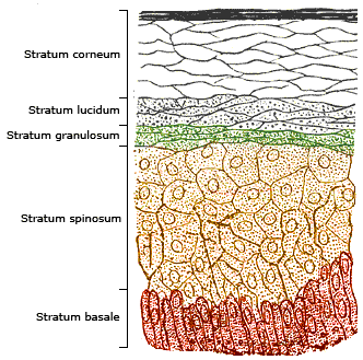
The epidermis is the outermost of the three layers that comprise the skin, the inner layers being the dermis and Subcutaneous tissue, hypodermis. The epidermis layer provides a barrier to infection from environmental pathogens and regulates the amount of water released from the body into the atmosphere through transepidermal water loss. The epidermis is composed of stratified squamous epithelium, multiple layers of flattened cells that overlie a base layer (stratum basale) composed of Epithelium#Cell types, columnar cells arranged perpendicularly. The layers of cells develop from stem cells in the basal layer. The human epidermis is a familiar example of epithelium, particularly a stratified squamous epithelium. The word epidermis is derived through Latin , itself and . Something related to or part of the epidermis is termed epidermal. Structure Cellular components The epidermis primarily consists of keratinocytes (cell proliferation, proliferating basal and cell differen ... [...More Info...] [...Related Items...] OR: [Wikipedia] [Google] [Baidu] |
Ontogeny
Ontogeny (also ontogenesis) is the origination and development of an organism (both physical and psychological, e.g., moral development), usually from the time of fertilization of the egg to adult. The term can also be used to refer to the study of the entirety of an organism's lifespan. Ontogeny is the developmental history of an organism within its own lifetime, as distinct from phylogeny, which refers to the evolutionary history of a species. Another way to think of ontogeny is that it is the process of an organism going through all of the developmental stages over its lifetime. The developmental history includes all the developmental events that occur during the existence of an organism, beginning with the changes in the egg at the time of fertilization and events from the time of birth or hatching and afterward (i.e., growth, remolding of body shape, development of secondary sexual characteristics, etc.). While developmental (i.e., ontogenetic) processes can influence sub ... [...More Info...] [...Related Items...] OR: [Wikipedia] [Google] [Baidu] |
Chimera (genetics)
A genetic chimerism or chimera ( ) is a single organism composed of cells with more than one distinct genotype. In animals, this means an individual derived from two or more zygotes, which can include possessing blood cells of different blood types, subtle variations in form (phenotype) and, if the zygotes were of differing sexes, then even the possession of both female and male sex organs. Animal chimeras are produced by the merger of two (or more) embryos. In plant chimeras, however, the distinct types of tissue may originate from the same zygote, and the difference is often due to mutation during ordinary cell division. Normally, genetic chimerism is not visible on casual inspection; however, it has been detected in the course of proving parentage. Another way that chimerism can occur in animals is by organ transplantation, giving one individual tissues that developed from a different genome. For example, transplantation of bone marrow often determines the recipient's ensuin ... [...More Info...] [...Related Items...] OR: [Wikipedia] [Google] [Baidu] |
Sven Hörstadius
Sven Hörstadius (1898–1996) was a Swedish embryologist known for his work on sea urchin embryos. He was responsible for an increased understanding of the neural crest. Hörstadius studied under John Runnström at Stockholm University College and was awarded his Ph.D. in 1930. He was appointed professor of zoology at Uppsala University 1942, where he remained until his retirement in 1964, but continued to lecture as an emeritus. He was elected a member of the Royal Swedish Academy of Sciences in 1946 and of the Royal Society The Royal Society, formally The Royal Society of London for Improving Natural Knowledge, is a learned society and the United Kingdom's national academy of sciences. The society fulfils a number of roles: promoting science and its benefits, r ... in 1952. References 1898 births 1996 deaths 20th-century Swedish zoologists Uppsala University faculty Members of the Royal Swedish Academy of Sciences Foreign Members of the Royal Society Buria ... [...More Info...] [...Related Items...] OR: [Wikipedia] [Google] [Baidu] |
Wilhelm His Sr , the Dutch national anthem
{{Disambiguation ...
Wilhelm may refer to: People and fictional characters * William Charles John Pitcher, costume designer known professionally as "Wilhelm" * Wilhelm (name), a list of people and fictional characters with the given name or surname Other uses * Mount Wilhelm, the highest mountain in Papua New Guinea * Wilhelm Archipelago, Antarctica * Wilhelm (crater), a lunar crater See also * Wilhelm scream, a stock sound effect * SS ''Kaiser Wilhelm II'', or USS ''Agamemnon'', a German steam ship * Wilhelmus "Wilhelmus van Nassouwe", usually known just as "Wilhelmus" ( nl, Het Wilhelmus, italic=no; ; English translation: "The William"), is the national anthem of both the Netherlands and the Kingdom of the Netherlands. It dates back to at least 157 ... [...More Info...] [...Related Items...] OR: [Wikipedia] [Google] [Baidu] |
DiGeorge Syndrome
DiGeorge syndrome, also known as 22q11.2 deletion syndrome, is a syndrome caused by a microdeletion on the long arm of chromosome 22. While the symptoms can vary, they often include congenital heart problems, specific facial features, frequent infections, developmental delay, learning problems and cleft palate. Associated conditions include kidney problems, schizophrenia, hearing loss and autoimmune disorders such as rheumatoid arthritis or Graves' disease. DiGeorge syndrome is typically due to the deletion of 30 to 40 genes in the middle of chromosome 22 at a location known as ''22q11.2''. About 90% of cases occur due to a new mutation during early development, while 10% are inherited from a person's parents. It is autosomal dominant, meaning that only one affected chromosome is needed for the condition to occur. Diagnosis is suspected based on the symptoms and confirmed by genetic testing. Although there is no cure, treatment can improve symptoms. This often includes ... [...More Info...] [...Related Items...] OR: [Wikipedia] [Google] [Baidu] |
Waardenburg–Shah Syndrome
Waardenburg syndrome is a group of rare genetic conditions characterised by at least some degree of congenital hearing loss and pigmentation deficiencies, which can include bright blue eyes (or one blue eye and one brown eye), a white forelock or patches of light skin. These basic features constitute type 2 of the condition; in type 1, there is also a wider gap between the inner corners of the eyes called telecanthus, or dystopia canthorum. In type 3, which is rare, the arms and hands are also malformed, with permanent finger contractures or fused fingers, while in type 4, the person also has Hirschsprung's disease. There also exist at least two types (2E and PCWH) that can result in central nervous system (CNS) symptoms such as developmental delay and muscle tone abnormalities. The syndrome is caused by mutations in any of several genes that affect the division and migration of neural crest cells during embryonic development (though some of the genes involved also affect ... [...More Info...] [...Related Items...] OR: [Wikipedia] [Google] [Baidu] |
Frontonasal Dysplasia
Frontonasal dysplasia (FND) is a congenital malformation of the midface. For the diagnosis of FND, a patient should present at least two of the following characteristics: hypertelorism (an increased distance between the eyes), a wide nasal root, vertical midline cleft of the nose and/or upper lip, cleft of the wings of the nose, malformed nasal tip, encephalocele (an opening of the skull with protrusion of the brain) or V-shaped hair pattern on the forehead. The cause of FND remains unknown. FND seems to be sporadic (random) and multiple environmental factors are suggested as possible causes for the syndrome. However, in some families multiple cases of FND were reported, which suggests a genetic cause of FND.Wu E, Vargevik K, Slavotinek AM (2007) Subtypes of frontonasal dysplasia are useful in determining clinical prognosis. Am J Med Genet A. 143A(24):3069-78. Classification There are multiple classification systems for FND. None of these classification systems have unraveled any ... [...More Info...] [...Related Items...] OR: [Wikipedia] [Google] [Baidu] |
Neurocristopathy
Neurocristopathy is a diverse class of pathologies that may arise from defects in the development of tissues containing cells commonly derived from the embryonic neural crest cell lineage. The term was coined by Robert P. Bolande in 1974. After the induction of the neural crest, the newly formed neural crest cells (NCC) delaminate from their tissue of origin and migrate from the entire neural axis of the vertebrate embryo to specific locations where they will give rise to different cell derivatives. The formation of this cell population therefore requires a timely and spatially controlled interplay of inter- and intra-cellular signals. An alteration in the occurrence and timing of these signals leads to a set of syndromes called Neurocristopathies (NCP), which comprises a broad spectrum of congenital malformations affecting an appreciable percentage of newborns. Moreover, since NCC migrate along the embryo, they are susceptible to subtle changes in the environment both during their ... [...More Info...] [...Related Items...] OR: [Wikipedia] [Google] [Baidu] |
.jpg)


