|
Tropoelastin
Elastin is a protein that in humans is encoded by the ''ELN'' gene. Elastin is a key component of the extracellular matrix in gnathostomes (jawed vertebrates). It is highly elastic and present in connective tissue allowing many tissues in the body to resume their shape after stretching or contracting. Elastin helps skin to return to its original position when it is poked or pinched. Elastin is also an important load-bearing tissue in the bodies of vertebrates and used in places where mechanical energy is required to be stored. Function The ''ELN'' gene encodes a protein that is one of the two components of elastic fibers. The encoded protein is rich in hydrophobic amino acids such as glycine and proline, which form mobile hydrophobic regions bounded by crosslinks between lysine residues. Multiple transcript variants encoding different isoforms have been found for this gene. Elastin's soluble precursor is tropoelastin. The characterization of disorder is consistent with an en ... [...More Info...] [...Related Items...] OR: [Wikipedia] [Google] [Baidu] |
Elastic Fibers
Elastic fibers (or yellow fibers) are an essential component of the extracellular matrix composed of bundles of proteins (elastin) which are produced by a number of different cell types including fibroblasts, endothelial, smooth muscle, and airway epithelial cells. These fibers are able to stretch many times their length, and snap back to their original length when relaxed without loss of energy. Elastic fibers include elastin, elaunin and oxytalan. Elastic tissue is classified as "connective tissue proper". Elastic fibers are formed via elastogenesis, a highly complex process involving several key proteins including fibulin-4, fibulin-5, latent transforming growth factor β binding protein 4, and microfibril associated protein 4. In this process tropoelastin, the soluble monomeric precursor to elastic fibers is produced by elastogenic cells and chaperoned to the cell surface. Following excretion from the cell, tropoelastin self associates into ~200 nm particles by coace ... [...More Info...] [...Related Items...] OR: [Wikipedia] [Google] [Baidu] |
Extracellular Matrix
In biology, the extracellular matrix (ECM), also called intercellular matrix, is a three-dimensional network consisting of extracellular macromolecules and minerals, such as collagen, enzymes, glycoproteins and hydroxyapatite that provide structural and biochemical support to surrounding cells. Because multicellularity evolved independently in different multicellular lineages, the composition of ECM varies between multicellular structures; however, cell adhesion, cell-to-cell communication and differentiation are common functions of the ECM. The animal extracellular matrix includes the interstitial matrix and the basement membrane. Interstitial matrix is present between various animal cells (i.e., in the intercellular spaces). Gels of polysaccharides and fibrous proteins fill the Interstitial fluid, interstitial space and act as a compression buffer against the stress placed on the ECM. Basement membranes are sheet-like depositions of ECM on which various epithelial cells rest ... [...More Info...] [...Related Items...] OR: [Wikipedia] [Google] [Baidu] |
Protein
Proteins are large biomolecules and macromolecules that comprise one or more long chains of amino acid residues. Proteins perform a vast array of functions within organisms, including catalysing metabolic reactions, DNA replication, responding to stimuli, providing structure to cells and organisms, and transporting molecules from one location to another. Proteins differ from one another primarily in their sequence of amino acids, which is dictated by the nucleotide sequence of their genes, and which usually results in protein folding into a specific 3D structure that determines its activity. A linear chain of amino acid residues is called a polypeptide. A protein contains at least one long polypeptide. Short polypeptides, containing less than 20–30 residues, are rarely considered to be proteins and are commonly called peptides. The individual amino acid residues are bonded together by peptide bonds and adjacent amino acid residues. The sequence of amino acid residue ... [...More Info...] [...Related Items...] OR: [Wikipedia] [Google] [Baidu] |
Menkes Syndrome
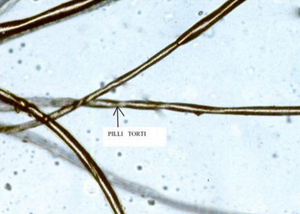
Menkes disease (MNK), also known as Menkes syndrome, is an X-linked recessive disorder caused by mutations in genes coding for the copper-transport protein ATP7A, leading to copper deficiency. Characteristic findings include kinky hair, growth failure, and nervous system deterioration. Like all X-linked recessive conditions, Menkes disease is more common in males than in females. The disorder was first described by John Hans Menkes in 1962. Onset occurs during infancy, with incidence of about 1 in 100,000 to 250,000 newborns; affected infants often do not live past the age of three years, though there are rare cases in which less severe symptoms emerge later in childhood. Signs and symptoms Affected infants may be born prematurely. Signs and symptoms appear during infancy, typically after a two- to three-month period of normal or slightly slowed development that is followed by a loss of early developmental skills and subsequent developmental delay. Patients exhibit hypoto ... [...More Info...] [...Related Items...] OR: [Wikipedia] [Google] [Baidu] |
Micrograph Of Solar Elastosis
A micrograph or photomicrograph is a photograph or digital image taken through a microscope or similar device to show a magnified image of an object. This is opposed to a macrograph or photomacrograph, an image which is also taken on a microscope but is only slightly magnified, usually less than 10 times. Micrography is the practice or art of using microscopes to make photographs. A micrograph contains extensive details of microstructure. A wealth of information can be obtained from a simple micrograph like behavior of the material under different conditions, the phases found in the system, failure analysis, grain size estimation, elemental analysis and so on. Micrographs are widely used in all fields of microscopy. Types Photomicrograph A light micrograph or photomicrograph is a micrograph prepared using an optical microscope, a process referred to as ''photomicroscopy''. At a basic level, photomicroscopy may be performed simply by connecting a camera to a microscope, th ... [...More Info...] [...Related Items...] OR: [Wikipedia] [Google] [Baidu] |
Reticular Dermis
The dermis or corium is a layer of skin between the epidermis (with which it makes up the cutis) and subcutaneous tissues, that primarily consists of dense irregular connective tissue and cushions the body from stress and strain. It is divided into two layers, the superficial area adjacent to the epidermis called the papillary region and a deep thicker area known as the reticular dermis.James, William; Berger, Timothy; Elston, Dirk (2005). ''Andrews' Diseases of the Skin: Clinical Dermatology'' (10th ed.). Saunders. Pages 1, 11–12. . The dermis is tightly connected to the epidermis through a basement membrane. Structural components of the dermis are collagen, elastic fibers, and extrafibrillar matrix.Marks, James G; Miller, Jeffery (2006). ''Lookingbill and Marks' Principles of Dermatology'' (4th ed.). Elsevier Inc. Page 8–9. . It also contains mechanoreceptors that provide the sense of touch and thermoreceptors that provide the sense of heat. In addition, hair follicles, swe ... [...More Info...] [...Related Items...] OR: [Wikipedia] [Google] [Baidu] |
Papillary Dermis
The dermis or corium is a layer of skin between the epidermis (with which it makes up the cutis) and subcutaneous tissues, that primarily consists of dense irregular connective tissue and cushions the body from stress and strain. It is divided into two layers, the superficial area adjacent to the epidermis called the papillary region and a deep thicker area known as the reticular dermis.James, William; Berger, Timothy; Elston, Dirk (2005). ''Andrews' Diseases of the Skin: Clinical Dermatology'' (10th ed.). Saunders. Pages 1, 11–12. . The dermis is tightly connected to the epidermis through a basement membrane. Structural components of the dermis are collagen, elastic fibers, and extrafibrillar matrix.Marks, James G; Miller, Jeffery (2006). ''Lookingbill and Marks' Principles of Dermatology'' (4th ed.). Elsevier Inc. Page 8–9. . It also contains mechanoreceptors that provide the sense of touch and thermoreceptors that provide the sense of heat. In addition, hair follicles, swe ... [...More Info...] [...Related Items...] OR: [Wikipedia] [Google] [Baidu] |
Actinic Elastosis
Actinic elastosis, also known as solar elastosis, is an accumulation of abnormal elastin (elastic tissue) in the dermis of the skin, or in the conjunctiva of the eye, which occurs as a result of the cumulative effects of prolonged and excessive sun exposure, a process known as photoaging. Signs and symptoms Actinic elastosis usually appears as thickened, dry, wrinkled skin. Several clinical variants have been recorded. One of the most readily identifiable is the thickened, deeply fissured skin seen on the back of the chronically sun-exposed neck, known as cutis rhomboidalis nuchae. These features are a part of the constellation of changes that are seen in photoaged skin. Causes The origin of the elastotic material in the dermis remains a subject of debate. Theories on the formation of the elastotic material include actinic stimulation of fibroblasts, promoting synthesis of this material, or that the material is a degradation product of collagen, elastin, or both. Diagnosis In ... [...More Info...] [...Related Items...] OR: [Wikipedia] [Google] [Baidu] |
Linear Focal Elastosis
Linear focal elastosis or elastotic striae is a skin condition that presents with asymptomatic, palpable or atrophic, yellow lines of the middle and lower back, thighs, arms and breasts.James, William; Berger, Timothy; Elston, Dirk (2005). ''Andrews' Diseases of the Skin: Clinical Dermatology''. (10th ed.). Saunders. Page 517. . File:Histopathology of linear focal elastosis.jpg, Histopathology: Accumulation of fragmented elastotic material within the papillary dermis and transcutaneous elimination of elastotic fibers. license See also *Skin lesion
A skin condition, also known as cutaneous condition, is any m ...
[...More Info...] [...Related Items...] OR: [Wikipedia] [Google] [Baidu] |
Perforating Calcific Elastosis
Perforating calcific elastosis is an acquired, localized cutaneous disorder, most frequently found in obese, multiparous, middle-aged women, characterized by lax, well-circumscribed, reticulated or cobble-stoned plaques occurring in the periumbilical region with keratotic surface papule A papule is a small, well-defined bump in the skin. It may have a rounded, pointed or flat top, and may have a dip. It can appear with a stalk, be thread-like or look warty. It can be soft or firm and its surface may be rough or smooth. Some h ...s.James, William; Berger, Timothy; Elston, Dirk (2005). ''Andrews' Diseases of the Skin: Clinical Dermatology''. (10th ed.). Saunders. Page 512. . See also * List of cutaneous conditions References Abnormalities of dermal fibrous and elastic tissue {{Cutaneous-condition-stub ... [...More Info...] [...Related Items...] OR: [Wikipedia] [Google] [Baidu] |
Elastosis Perforans Serpiginosa
Elastosis perforans serpiginosa is a unique perforating disorder characterized by transepidermal elimination of elastic fibers and distinctive clinical lesions, which are serpiginous in distribution and can be associated with specific diseases.Freedberg, et al. (2003). ''Fitzpatrick's Dermatology in General Medicine''. (6th ed.). Page 1041. McGraw-Hill. . File:Histopathology of elastosis perforans serpiginosa.jpg, Histopathology of elastosis perforans serpiginosa: Degenerated elastic fibers and transepidermal perforating canals (arrow points at one of them) File:Autosomal dominant - en.svg, This condition is inherited in an autosomal dominant manner. See also * List of cutaneous conditions * Poikiloderma vasculare atrophicans Poikiloderma vasculare atrophicans (PVA), is a cutaneous condition (skin disease) characterized by hypo- or hyperpigmentation (diminished or heightened skin pigmentation, respectively), telangiectasia and skin atrophy. Other names for the conditio ... ... [...More Info...] [...Related Items...] OR: [Wikipedia] [Google] [Baidu] |
Photoaging
Photoaging or photoageing (also known as "dermatoheliosis") is a term used for the characteristic changes to skin induced by chronic UVA and UVB exposure. Tretinoin is the best studied retinoid in the treatment of photoaging. The deterioration of biological functions and ability to manage metabolic stress is one of the major consequences of the aging process. Aging is a complex, progressive process that leads to functional and aesthetic changes in the skin. This process can result from both intrinsic (i.e., genetically determined) as well as extrinsic processes (i.e., environmental factors). Photoaging is attributed to continuous, long-term exposure to ultraviolet (UV) radiation of approximately 300–400 nm, either natural or synthetic, on an intrinsically aged skin. Effects of UV light Molecular and genetic changes UVB rays are a primary mutagen that can only penetrate through the epidermal (outermost) layer of the skin, resulting in DNA mutations. These mutations arise ... [...More Info...] [...Related Items...] OR: [Wikipedia] [Google] [Baidu] |


