|
Trisomy 9
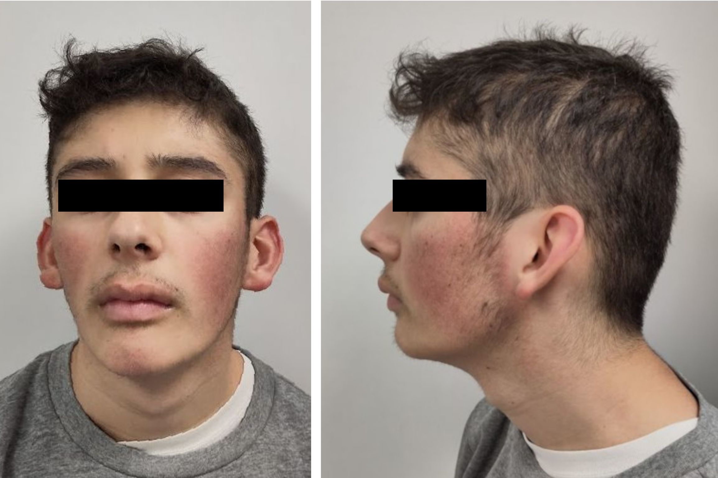
Full trisomy 9 is a lethal chromosomal disorder caused by having three copies (trisomy) of chromosome number 9. It can be a viable condition if trisomy affects only part of the cells of the body (mosaicism) or in cases of partial trisomy (trisomy 9p) in which cells have a normal set of two entire chromosomes 9 plus part of a third copy, usually of the short arm of the chromosome (arm p). Presentation Symptoms vary, but usually result in dysmorphisms in the skull, nervous system problems, and developmental delay. Dysmorphisms in the heart, kidneys, and musculoskeletal system may also occur. An infant with complete trisomy 9 surviving 20 days after birth showed clinical features including a small face, wide fontanelle, prominent occiput, micrognathia, low set ears, upslanting palpebral fissures, high-arched palate, short sternum, overlapping fingers, limited hip abduction, rocker bottom feet, heart murmurs and a webbed neck. Trisomy 9p is one of the most frequent autosomal anoma ... [...More Info...] [...Related Items...] OR: [Wikipedia] [Google] [Baidu] |
Chromosome 9 (human)
Chromosome 9 is one of the 23 pairs of chromosomes in humans. Humans normally have two copies of this chromosome, as they normally do with all chromosomes. Chromosome 9 spans about 138 million base pairs of nucleic acids (the building blocks of DNA) and represents between 4.0 and 4.5% of the total DNA in cells. Genes Number of genes These are some of the gene count estimates of human chromosome 9. Because researchers use different approaches to genome annotation, their predictions of the number of genes on each chromosome varies (for technical details, see gene prediction). Among various projects, the collaborative consensus coding sequence project ( CCDS) takes an extremely conservative strategy. So CCDS's gene number prediction represents a lower bound on the total number of human protein-coding genes. Gene list The following is a partial list of genes on human chromosome 9. For a complete list, see the link in the infobox on the right. Diseases and disorders The follo ... [...More Info...] [...Related Items...] OR: [Wikipedia] [Google] [Baidu] |
High-arched Palate
A high-arched palate (also termed high-vaulted palate) is where the palate is unusually high and narrow. It is usually a congenital developmental feature that results from the failure of the palatal shelves to fuse correctly in development, the same phenomenon that leads to cleft palate. It may occur in isolation or in association with a number of conditions. It may also be an acquired condition caused by chronic thumb-sucking. A high-arched palate may result in a narrowed airway and sleep disordered breathing. Examples of conditions which may be associated with a high-arched palate include: * Allergic rhinitis * Apert syndrome * Crouzon syndrome * Down syndrome * Ehlers-Danlos Syndrome * Fragile X syndrome * Incontinentia pigmenti * Marfan syndrome * Treacher Collins syndrome * Upper Airway Resistance Syndrome See also * Minor physical anomalies * Bardet–Biedl syndrome Bardet–Biedl syndrome (BBS) is a ciliopathic human genetic disorder that produces many effects a ... [...More Info...] [...Related Items...] OR: [Wikipedia] [Google] [Baidu] |
Karyotyping
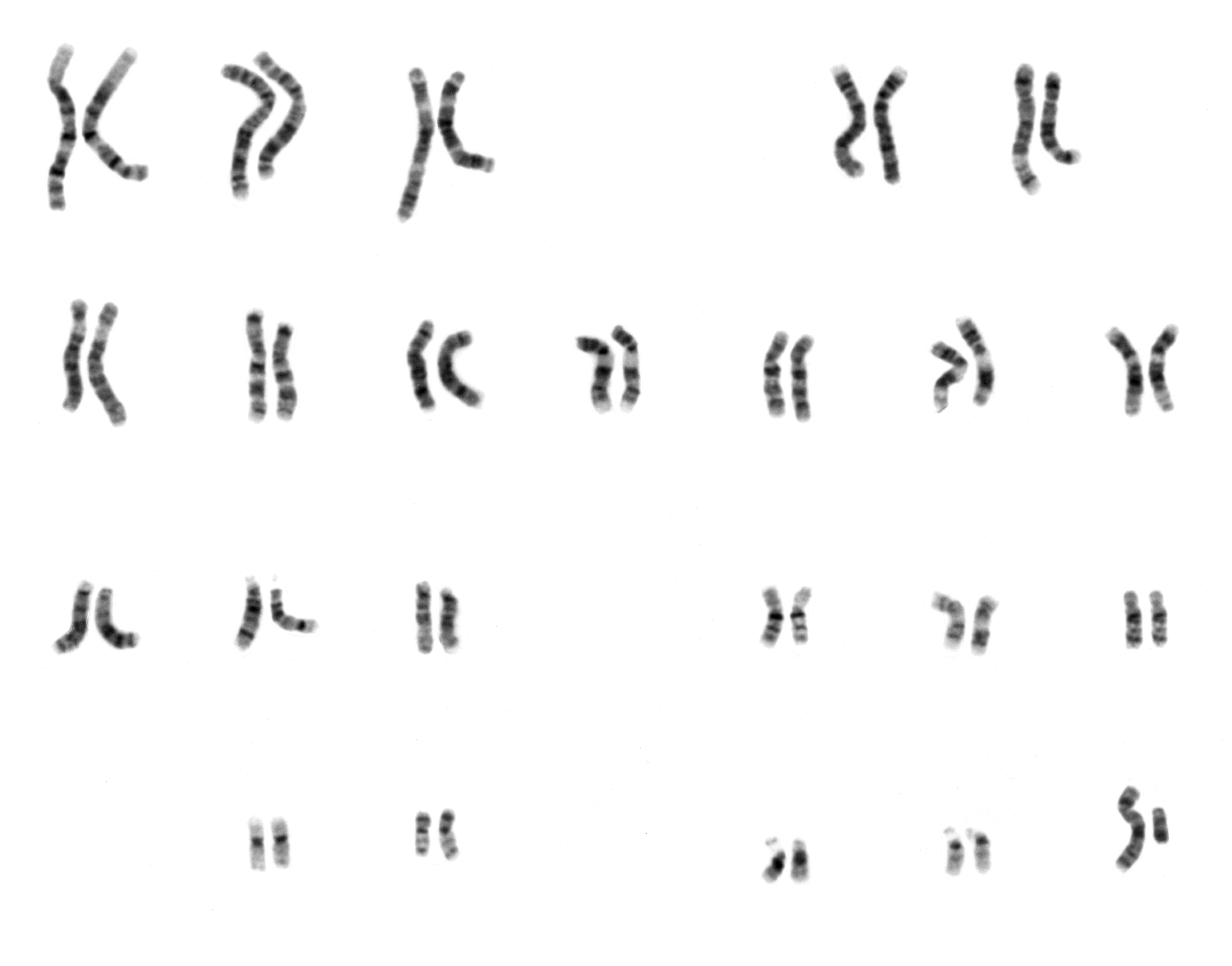
A karyotype is the general appearance of the complete set of metaphase chromosomes in the cells of a species or in an individual organism, mainly including their sizes, numbers, and shapes. Karyotyping is the process by which a karyotype is discerned by determining the chromosome complement of an individual, including the number of chromosomes and any abnormalities. A karyogram or idiogram is a graphical depiction of a karyotype, wherein chromosomes are organized in pairs, ordered by size and position of centromere for chromosomes of the same size. Karyotyping generally combines light microscopy and photography, and results in a photomicrographic (or simply micrographic) karyogram. In contrast, a schematic karyogram is a designed graphic representation of a karyotype. In schematic karyograms, just one of the sister chromatids of each chromosome is generally shown for brevity, and in reality they are generally so close together that they look as one on photomicrographs as well ... [...More Info...] [...Related Items...] OR: [Wikipedia] [Google] [Baidu] |
Obstetric Ultrasonography
Obstetric ultrasonography, or prenatal ultrasound, is the use of medical ultrasonography in pregnancy, in which sound waves are used to create real-time visual images of the developing embryo or fetus in the uterus (womb). The procedure is a standard part of prenatal care in many countries, as it can provide a variety of information about the health of the mother, the timing and progress of the pregnancy, and the health and development of the embryo or fetus. The International Society of Ultrasound in Obstetrics and Gynecology (ISUOG) recommends that pregnant women have routine obstetric ultrasounds between 18 weeks' and 22 weeks' gestational age (the anatomy scan) in order to confirm pregnancy dating, to measure the fetus so that growth abnormalities can be recognized quickly later in pregnancy, and to assess for congenital malformations and multiple pregnancies (twins, etc). Additionally, the ISUOG recommends that pregnant patients who desire genetic testing have obstetric ... [...More Info...] [...Related Items...] OR: [Wikipedia] [Google] [Baidu] |
Cordocentesis
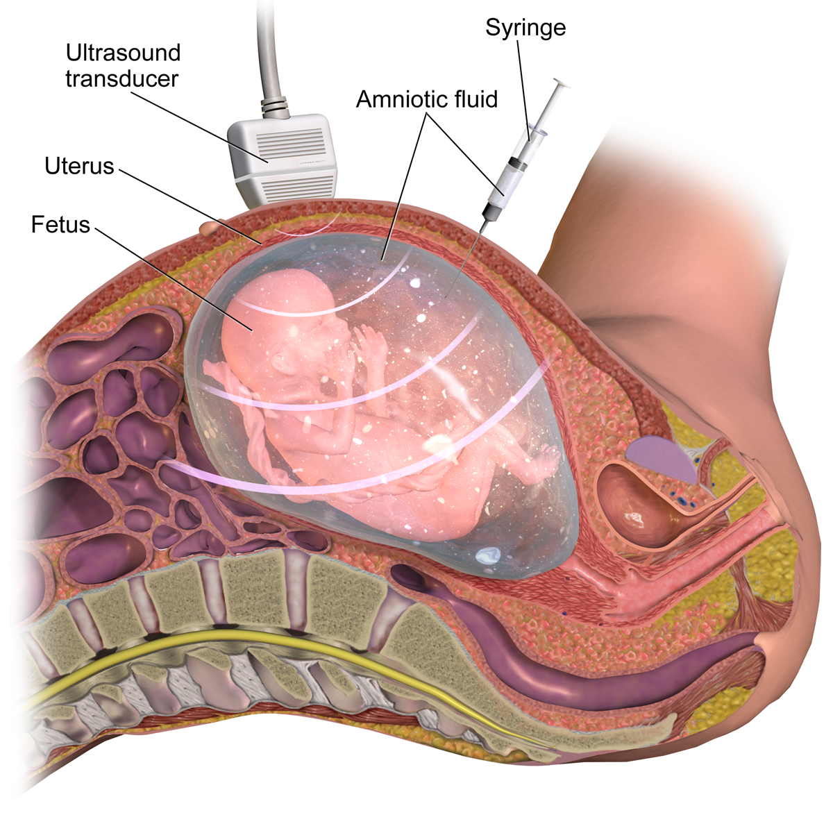
Percutaneous umbilical cord blood sampling (PUBS), also called cordocentesis, fetal blood sampling, or umbilical vein sampling is a diagnostic genetic test that examines blood from the fetal umbilical cord to detect fetal abnormalities. Fetal and maternal blood supply are typically connected in utero with one vein and two arteries to the fetus. The umbilical vein is responsible for delivering oxygen rich blood to the fetus from the mother; the umbilical arteries are responsible for removing oxygen poor blood from the fetus. This allows for the fetus’ tissues to properly perfuse. PUBS provides a means of rapid chromosome analysis and is useful when information cannot be obtained through amniocentesis, chorionic villus sampling, or ultrasound (or if the results of these tests were inconclusive); this test carries a significant risk of complication and is typically reserved for pregnancies determined to be at high risk for genetic defect. It has been used with mothers with immune ... [...More Info...] [...Related Items...] OR: [Wikipedia] [Google] [Baidu] |
Chorionic Villus Sampling
Chorionic villus sampling (CVS), sometimes called "chorionic ''villous'' sampling" (as "villous" is the adjectival form of the word "villus"), is a form of prenatal diagnosis done to determine chromosomal or genetic disorders in the fetus. It entails sampling of the chorionic villus (placental tissue) and testing it for chromosomal abnormalities, usually with FISH or PCR. CVS usually takes place at 10–12 weeks' gestation, earlier than amniocentesis or percutaneous umbilical cord blood sampling. It is the preferred technique before 15 weeks. CVS was performed for the first time in Milan by Italian biologist Giuseppe Simoni, scientific director of Biocell Center, in 1983. Use as early as eight weeks in special circumstances has been described. It can be performed in a transcervical or transabdominal manner. Although this procedure is mostly associated with testing for Down syndrome, overall, CVS can detect more than 200 disorders. Indications Possi ... [...More Info...] [...Related Items...] OR: [Wikipedia] [Google] [Baidu] |
Prenatal Diagnosis
Prenatal testing consists of prenatal screening and prenatal diagnosis, which are aspects of prenatal care that focus on detecting problems with the pregnancy as early as possible. These may be anatomic and physiologic problems with the health of the zygote, embryo, or fetus, either before gestation even starts (as in preimplantation genetic diagnosis) or as early in gestation as practicable. Screening can detect problems such as neural tube defects, chromosome abnormalities, and gene mutations that would lead to genetic disorders and birth defects, such as spina bifida, cleft palate, Down syndrome, Tay–Sachs disease, sickle cell anemia, thalassemia, cystic fibrosis, muscular dystrophy, and fragile X syndrome. Some tests are designed to discover problems which primarily affect the health of the mother, such as PAPP-A to detect pre-eclampsia or glucose tolerance tests to diagnose gestational diabetes. Screening can also detect anatomical defects such as hydrocephalus, anencepha ... [...More Info...] [...Related Items...] OR: [Wikipedia] [Google] [Baidu] |
Dandy–Walker Malformation
Dandy–Walker malformation (DWM), also known as Dandy–Walker syndrome (DWS), is a rare congenital brain malformation in which the part joining the two hemispheres of the cerebellum (the cerebellar vermis) does not fully form, and the fourth ventricle and space behind the cerebellum (the posterior fossa) are enlarged with cerebrospinal fluid. Most of those affected develop hydrocephalus within the first year of life, which can present as increasing head size, vomiting, excessive sleepiness, irritability, downward deviation of the eyes and seizures. Other, less common symptoms are generally associated with comorbid genetic conditions and can include congenital heart defects, eye abnormalities, intellectual disability, congenital tumours, other brain defects such as agenesis of the corpus callosum, skeletal abnormalities, an occipital encephalocele or underdeveloped genitalia or kidneys. It is sometimes discovered in adolescents or adults due to mental health problems. DWM ... [...More Info...] [...Related Items...] OR: [Wikipedia] [Google] [Baidu] |
Corpus Callosum
The corpus callosum (Latin for "tough body"), also callosal commissure, is a wide, thick nerve tract, consisting of a flat bundle of commissural fibers, beneath the cerebral cortex in the brain. The corpus callosum is only found in placental mammals. It spans part of the longitudinal fissure, connecting the left and right cerebral hemispheres, enabling communication between them. It is the largest white matter structure in the human brain, about in length and consisting of 200–300 million axonal projections. A number of separate nerve tracts, classed as subregions of the corpus callosum, connect different parts of the hemispheres. The main ones are known as the genu, the rostrum, the trunk or body, and the splenium. Structure The corpus callosum forms the floor of the longitudinal fissure that separates the two cerebral hemispheres. Part of the corpus callosum forms the roof of the lateral ventricles. The corpus callosum has four main parts – individual nerve ... [...More Info...] [...Related Items...] OR: [Wikipedia] [Google] [Baidu] |
Ventricular System
The ventricular system is a set of four interconnected cavities known as cerebral ventricles in the brain. Within each ventricle is a region of choroid plexus which produces the circulating cerebrospinal fluid (CSF). The ventricular system is continuous with the central canal of the spinal cord from the fourth ventricle, allowing for the flow of CSF to circulate. All of the ventricular system and the central canal of the spinal cord are lined with ependyma, a specialised form of epithelium connected by tight junctions that make up the blood–cerebrospinal fluid barrier. Structure The system comprises four ventricles: * lateral ventricles right and left (one for each hemisphere) * third ventricle * fourth ventricle There are several foramina, openings acting as channels, that connect the ventricles. The interventricular foramina (also called the foramina of Monro) connect the lateral ventricles to the third ventricle through which the cerebrospinal fluid can flow. V ... [...More Info...] [...Related Items...] OR: [Wikipedia] [Google] [Baidu] |
Coffin–Siris Syndrome
Coffin–Siris Syndrome (CSS), first described in 1970 by Dr Coffin and Dr Siris, is a rare genetic disorder that causes developmental delays and absent fifth finger and toe nails. There had been 31 reported cases by 1991. The number of occurrences since then has grown and is now reported to be around 200. The differential includes Nicolaides–Baraitser syndrome. Presentation * mild to moderate to severe intellectual disability, also called "developmental disability" * short fifth digits with hypoplastic or absent nails * low birth weight * feeding difficulties upon birth * frequent respiratory infections during infancy * hypotonia * joint laxity * delayed bone age * microcephaly * coarse facial features, including wide nose, wide mouth, and thick eyebrows and lashes Causes Disease can be inherited as an autosomal dominant trait, however most cases of CSS appear to be the result of a de novo mutation. This syndrome has been associated with mutations in the ARID1B gene, whic ... [...More Info...] [...Related Items...] OR: [Wikipedia] [Google] [Baidu] |
Heart Murmurs
Heart murmurs are unique heart sounds produced when blood flows across a heart valve or blood vessel. This occurs when turbulent blood flow creates a sound loud enough to hear with a stethoscope. Turbulent blood flow is not smooth. The sound differs from normal heart sounds by their characteristics. For example, heart murmurs may have a distinct pitch, duration and timing. The major way health care providers examine the heart on physical exam is heart auscultation; another clinical technique is palpation, which can detect by touch when such turbulence causes the vibrations called cardiac thrill. A murmur is a sign found during the cardiac exam. Murmurs are of various types and are important in the detection of cardiac and valvular pathologies (i.e. can be a sign of heart diseases or defects). There are two types of murmur. A functional murmur is a benign heart murmur that is primarily due to physiologic conditions outside the heart. The other type of heart murmur is due to ... [...More Info...] [...Related Items...] OR: [Wikipedia] [Google] [Baidu] |


