|
Thoracoacromial
The thoracoacromial artery (acromiothoracic artery; thoracic axis) is a short trunk that arises from the second part of the axillary artery, its origin being generally overlapped by the upper edge of the pectoralis minor. Structure Projecting forward to the upper border of the Pectoralis minor, it pierces the coracoclavicular fascia The clavipectoral fascia (costocoracoid membrane; coracoclavicular fascia) is a strong fascia situated under cover of the clavicular portion of the pectoralis major. It occupies the interval between the pectoralis minor and subclavius, and protec ... and divides into four branches—pectoral, acromial, clavicular, and deltoid. Additional images File:Gray523.png, The axillary artery and its branches. References External links * * * - "Pectoral Region: Thoracoacromial Artery and its Branches" * - "The axillary artery and its major branches shown in relation to major landmarks." {{Authority control Arteries of the upper limb ... [...More Info...] [...Related Items...] OR: [Wikipedia] [Google] [Baidu] |
Axillary Artery
In human anatomy, the axillary artery is a large blood vessel that conveys oxygenated blood to the lateral aspect of the thorax, the axilla (armpit) and the upper limb. Its origin is at the lateral margin of the first rib, before which it is called the subclavian artery. After passing the lower margin of teres major it becomes the brachial artery. Structure The axillary artery is often referred to as having three parts, with these divisions based on its location relative to the Pectoralis minor muscle, which is superficial to the artery. * First part – the part of the artery superior to the pectoralis minor * Second part – the part of the artery posterior to the pectoralis minor * Third part – the part of the artery inferior to the pectoralis minor. Relations The axillary artery is accompanied by the axillary vein, which lies medial to the artery, along its length. In the axilla, the axillary artery is surrounded by the brachial plexus. The second part of the axi ... [...More Info...] [...Related Items...] OR: [Wikipedia] [Google] [Baidu] |
Deltoideus
The deltoid muscle is the muscle forming the rounded contour of the human shoulder. It is also known as the 'common shoulder muscle', particularly in other animals such as the domestic cat. Anatomically, the deltoid muscle appears to be made up of three distinct sets of muscle fibers, namely the # anterior or clavicular part (pars clavicularis) # posterior or scapular part (pars scapularis) # intermediate or acromial part (pars acromialis) However, electromyography suggests that it consists of at least seven groups that can be independently coordinated by the nervous system. It was previously called the deltoideus (plural ''deltoidei'') and the name is still used by some anatomists. It is called so because it is in the shape of the Greek capital letter delta (Δ). Deltoid is also further shortened in slang as "delt". A study of 30 shoulders revealed an average mass of in humans, ranging from to . Structure Previous studies showed that the insertions of the tendons of the delto ... [...More Info...] [...Related Items...] OR: [Wikipedia] [Google] [Baidu] |
Cephalic Vein
In human anatomy, the cephalic vein is a superficial vein in the arm. It originates from the radial end of the dorsal venous network of hand, and ascends along the radial (lateral) side of the arm before emptying into the axillary vein. At the elbow, it communicates with the basilic vein via the median cubital vein. Anatomy The cephalic vein is situated within the superficial fascia along the anterolateral surface of the biceps. Origin The cephalic vein forms over the anatomical snuffbox at the radial end of the dorsal venous network of hand. Course and relations From its origin, it ascends ascends up the lateral aspect of the radius. Near the shoulder, the cephalic vein passes between the deltoid and pectoralis major muscles (deltopectoral groove) and through the clavipectoral triangle, where it empties into the axillary vein. Anastomoses It communicates with the basilic vein via the median cubital vein at the elbow. Clinical significance The cephalic vein ... [...More Info...] [...Related Items...] OR: [Wikipedia] [Google] [Baidu] |
Humeral
The humerus (; ) is a long bone in the arm that runs from the shoulder to the elbow. It connects the scapula and the two bones of the lower arm, the radius and ulna, and consists of three sections. The humeral upper extremity consists of a rounded head, a narrow neck, and two short processes (tubercles, sometimes called tuberosities). The body is cylindrical in its upper portion, and more prismatic below. The lower extremity consists of 2 epicondyles, 2 processes (trochlea & capitulum), and 3 fossae (radial fossa, coronoid fossa, and olecranon fossa). As well as its true anatomical neck, the constriction below the greater and lesser tubercles of the humerus is referred to as its surgical neck due to its tendency to fracture, thus often becoming the focus of surgeons. Etymology The word "humerus" is derived from la, humerus, umerus meaning upper arm, shoulder, and is linguistically related to Gothic ''ams'' shoulder and Greek ''ōmos''. Structure Upper extremity The upper or pro ... [...More Info...] [...Related Items...] OR: [Wikipedia] [Google] [Baidu] |
Deltoid Muscle
The deltoid muscle is the muscle forming the rounded contour of the human shoulder. It is also known as the 'common shoulder muscle', particularly in other animals such as the domestic cat. Anatomically, the deltoid muscle appears to be made up of three distinct sets of muscle fibers, namely the # anterior or clavicular part (pars clavicularis) # posterior or scapular part (pars scapularis) # intermediate or acromial part (pars acromialis) However, electromyography suggests that it consists of at least seven groups that can be independently coordinated by the nervous system. It was previously called the deltoideus (plural ''deltoidei'') and the name is still used by some anatomists. It is called so because it is in the shape of the Greek capital letter delta (Δ). Deltoid is also further shortened in slang as "delt". A study of 30 shoulders revealed an average mass of in humans, ranging from to . Structure Previous studies showed that the insertions of the tendons of the delto ... [...More Info...] [...Related Items...] OR: [Wikipedia] [Google] [Baidu] |
Subclavius
The subclavius is a small triangular muscle, placed between the clavicle and the first rib. Along with the pectoralis major and pectoralis minor muscles, the subclavius muscle makes up the anterior axioappendicular muscles, also known as anterior wall of the axilla. Structure It arises by a short, thick tendon from the first rib and its cartilage at their junction, in front of the costoclavicular ligament. The fleshy fibers proceed obliquely superolaterally, to be inserted into the groove on the under surface of the clavicle. Innervation The nerve to subclavius (or subclavian nerve) innervates the muscle. This arises from the junction of the fifth and sixth cervical nerves, from the superior/upper trunk of the brachial plexus. Variation Insertion into coracoid process instead of clavicle or into both clavicle and coracoid process. Sternoscapular fasciculus to the upper border of scapula. Sternoclavicularis from manubrium to clavicle between pectoralis major and coracoclavicular ... [...More Info...] [...Related Items...] OR: [Wikipedia] [Google] [Baidu] |
Sternoclavicular Joint
The sternoclavicular joint or sternoclavicular articulation is a synovial saddle joint between the manubrium of the sternum, and the clavicle, as well as the first rib. The joint possesses a joint capsule, and an articular disk, and is reinforced by multiple ligaments. Structure The joint is structurally classed as a synovial plane joint and functionally classed as a diarthrosis and multiaxial joint. It is composed of two portions separated by an articular disc of fibrocartilage. The joint is formed by the sternal end of the clavicle, the clavicular notch (the superior and lateral part of the sternum), and (the superior surface of) the cartilage of the first rib (visible from the outside as the suprasternal notch). The articular surface of the clavicle is larger than that of the sternum, and is invested with a layer of cartilage, which is considerably thicker than that of the sternum. The joint receives arterial supply via branches of the internal thoracic artery, and of th ... [...More Info...] [...Related Items...] OR: [Wikipedia] [Google] [Baidu] |
Clavicular
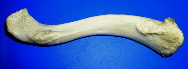
The clavicle, or collarbone, is a slender, S-shaped long bone approximately 6 inches (15 cm) long that serves as a strut between the shoulder blade and the sternum (breastbone). There are two clavicles, one on the left and one on the right. The clavicle is the only long bone in the body that lies horizontally. Together with the shoulder blade, it makes up the shoulder girdle. It is a palpable bone and, in people who have less fat in this region, the location of the bone is clearly visible. It receives its name from the Latin ''clavicula'' ("little key"), because the bone rotates along its axis like a key when the shoulder is abducted. The clavicle is the most commonly fractured bone. It can easily be fractured by impacts to the shoulder from the force of falling on outstretched arms or by a direct hit. Structure The collarbone is a thin doubly curved long bone that connects the arm to the trunk of the body. Located directly above the first rib, it acts as a strut to keep ... [...More Info...] [...Related Items...] OR: [Wikipedia] [Google] [Baidu] |
Posterior Humeral Circumflex
The posterior humeral circumflex artery (posterior circumflex artery, posterior circumflex humeral artery) arises from the third part of axillary artery at the lower border of the subscapularis, and runs posteriorly with the axillary nerve through the quadrangular space. It winds around the surgical neck of the humerus and is distributed to the deltoid muscle and shoulder-joint, anastomosing with the anterior humeral circumflex and deep artery of the arm. It supplies the teres major, teres minor, deltoid, and (long head only) triceps muscles. Additional images File:Gray810.png, Suprascapular and axillary nerves of right side, seen from behind. File:Slide4SSS.JPG, Posterior humeral circumflex artery See also * Anterior humeral circumflex artery The anterior humeral circumflex artery (anterior circumflex artery, anterior circumflex humeral artery) is an artery in the arm. It is one of two circumflexing arteries that branch from the axillary artery, the other being the posterio ... [...More Info...] [...Related Items...] OR: [Wikipedia] [Google] [Baidu] |
Suprascapular
The suprascapular artery is a branch of the thyrocervical trunk on the neck. Structure At first, it passes downward and laterally across the scalenus anterior and phrenic nerve, being covered by the sternocleidomastoid muscle; it then crosses the subclavian artery and the brachial plexus, running behind and parallel with the clavicle and subclavius muscle and beneath the inferior belly of the omohyoid to the superior border of the scapula. It passes over the superior transverse scapular ligament in most of the cases while below it through the suprascapular notch in some cases. The artery then enters the supraspinous fossa of the scapula. It travels close to the bone, running through the suprascapular canal underneath the supraspinatus muscle, to which it supplies branches. It then descends behind the neck of the scapula, through the great scapular notch and under cover of the inferior transverse ligament of scapula, inferior transverse ligament, to reach the infraspinatous fos ... [...More Info...] [...Related Items...] OR: [Wikipedia] [Google] [Baidu] |
Transverse Scapular
The suprascapular artery is a branch of the thyrocervical trunk on the neck. Structure At first, it passes downward and laterally across the scalenus anterior and phrenic nerve, being covered by the sternocleidomastoid muscle; it then crosses the subclavian artery and the brachial plexus, running behind and parallel with the clavicle and subclavius muscle and beneath the inferior belly of the omohyoid to the superior border of the scapula. It passes over the superior transverse scapular ligament in most of the cases while below it through the suprascapular notch in some cases. The artery then enters the supraspinous fossa of the scapula. It travels close to the bone, running through the suprascapular canal underneath the supraspinatus muscle, to which it supplies branches. It then descends behind the neck of the scapula, through the great scapular notch and under cover of the inferior transverse ligament, to reach the infraspinatous fossa, where it supplies infraspinatus and ... [...More Info...] [...Related Items...] OR: [Wikipedia] [Google] [Baidu] |

