|
Deltoideus
The deltoid muscle is the muscle forming the rounded contour of the human shoulder. It is also known as the 'common shoulder muscle', particularly in other animals such as the domestic cat. Anatomically, the deltoid muscle appears to be made up of three distinct sets of muscle fibers, namely the # anterior or clavicular part (pars clavicularis) # posterior or scapular part (pars scapularis) # intermediate or acromial part (pars acromialis) However, electromyography suggests that it consists of at least seven groups that can be independently coordinated by the nervous system. It was previously called the deltoideus (plural ''deltoidei'') and the name is still used by some anatomists. It is called so because it is in the shape of the Greek capital letter delta (Δ). Deltoid is also further shortened in slang as "delt". A study of 30 shoulders revealed an average mass of in humans, ranging from to . Structure Previous studies showed that the insertions of the tendons of the delto ... [...More Info...] [...Related Items...] OR: [Wikipedia] [Google] [Baidu] |
Spine Of Scapula
The spine of the scapula or scapular spine is a prominent plate of bone, which crosses obliquely the medial four-fifths of the scapula at its upper part, and separates the Supraspinatous fossa, supra- from the infraspinatous fossa. Structure It begins at the vertical [vertebral or medial border] border by a smooth, triangular area over which the tendon of insertion of the lower part of the Trapezius glides, and, gradually becoming more elevated, ends in the acromion, which overhangs the shoulder-joint. The spine is triangular, and flattened from above downward, its apex being directed toward the vertebral border. Root The ''root of the spine'' of the scapula is the most medial part of the scapular spine. It is termed "triangular area of the spine of scapula", based on its triangular shape giving it distinguishable visible shape on x-ray images. The root of the spine is on a level with the tip of the spinous process of the third thoracic vertebra.Gray's Anatomy (1918)p.1306/ref> ... [...More Info...] [...Related Items...] OR: [Wikipedia] [Google] [Baidu] |
Deltoid Tuberosity
In human anatomy, the deltoid tuberosity is a rough, triangular area on the anterolateral (front-side) surface of the middle of the humerus. It is a site of attachment of deltoid muscle. Structure Variation The deltoid tuberosity has been reported as very prominent in less than 10% of people. Development The deltoid tuberosity develops through endochondral ossification in a two-phase process. The initiating signal is tendon-dependent, whilst the growth phase is muscle-dependent. Clinical significance The deltoid tuberosity is at risk of avulsion fracture. These fractures may be managed conservatively with rest. Other animals In mammals, the humerus displays a wide morphological variation. The size and orientation of its functionally important features, including the deltoid tubercle, greater tubercle, and medial epicondyle, are pivotal to an animal's style of locomotion and habitat. In cursorial (running) animals such as the pronghorn, the deltoid tubercle is located ... [...More Info...] [...Related Items...] OR: [Wikipedia] [Google] [Baidu] |
Spine Of The Scapula
The spine of the scapula or scapular spine is a prominent plate of bone, which crosses obliquely the medial four-fifths of the scapula at its upper part, and separates the supra- from the infraspinatous fossa. Structure It begins at the vertical ertebral or medial borderborder by a smooth, triangular area over which the tendon of insertion of the lower part of the Trapezius glides, and, gradually becoming more elevated, ends in the acromion, which overhangs the shoulder-joint. The spine is triangular, and flattened from above downward, its apex being directed toward the vertebral border. Root The ''root of the spine'' of the scapula is the most medial part of the scapular spine. It is termed "triangular area of the spine of scapula", based on its triangular shape giving it distinguishable visible shape on x-ray images. The root of the spine is on a level with the tip of the spinous process of the third thoracic vertebra.Gray's Anatomy (1918)p.1306/ref> File:Root of spine - l ... [...More Info...] [...Related Items...] OR: [Wikipedia] [Google] [Baidu] |
Clavicle
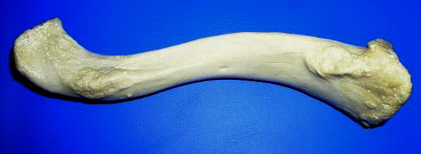
The clavicle, or collarbone, is a slender, S-shaped long bone approximately 6 inches (15 cm) long that serves as a strut between the shoulder blade and the sternum (breastbone). There are two clavicles, one on the left and one on the right. The clavicle is the only long bone in the body that lies horizontally. Together with the shoulder blade, it makes up the shoulder girdle. It is a palpable bone and, in people who have less fat in this region, the location of the bone is clearly visible. It receives its name from the Latin ''clavicula'' ("little key"), because the bone rotates along its axis like a key when the shoulder is abducted. The clavicle is the most commonly fractured bone. It can easily be fractured by impacts to the shoulder from the force of falling on outstretched arms or by a direct hit. Structure The collarbone is a thin doubly curved long bone that connects the arm to the trunk of the body. Located directly above the first rib, it acts as a strut to k ... [...More Info...] [...Related Items...] OR: [Wikipedia] [Google] [Baidu] |
Electromyography
Electromyography (EMG) is a technique for evaluating and recording the electrical activity produced by skeletal muscles. EMG is performed using an instrument called an electromyograph to produce a record called an electromyogram. An electromyograph detects the electric potential generated by muscle cells when these cells are electrically or neurologically activated. The signals can be analyzed to detect abnormalities, activation level, or recruitment order, or to analyze the biomechanics of human or animal movement. Needle EMG is an electrodiagnostic medicine technique commonly used by neurologists. Surface EMG is a non-medical procedure used to assess muscle activation by several professionals, including physiotherapists, kinesiologists and biomedical engineers. In Computer Science, EMG is also used as middleware in gesture recognition towards allowing the input of physical action to a computer as a form of human-computer interaction. Clinical uses EMG testing has a variety of ... [...More Info...] [...Related Items...] OR: [Wikipedia] [Google] [Baidu] |
Standard Anatomical Position
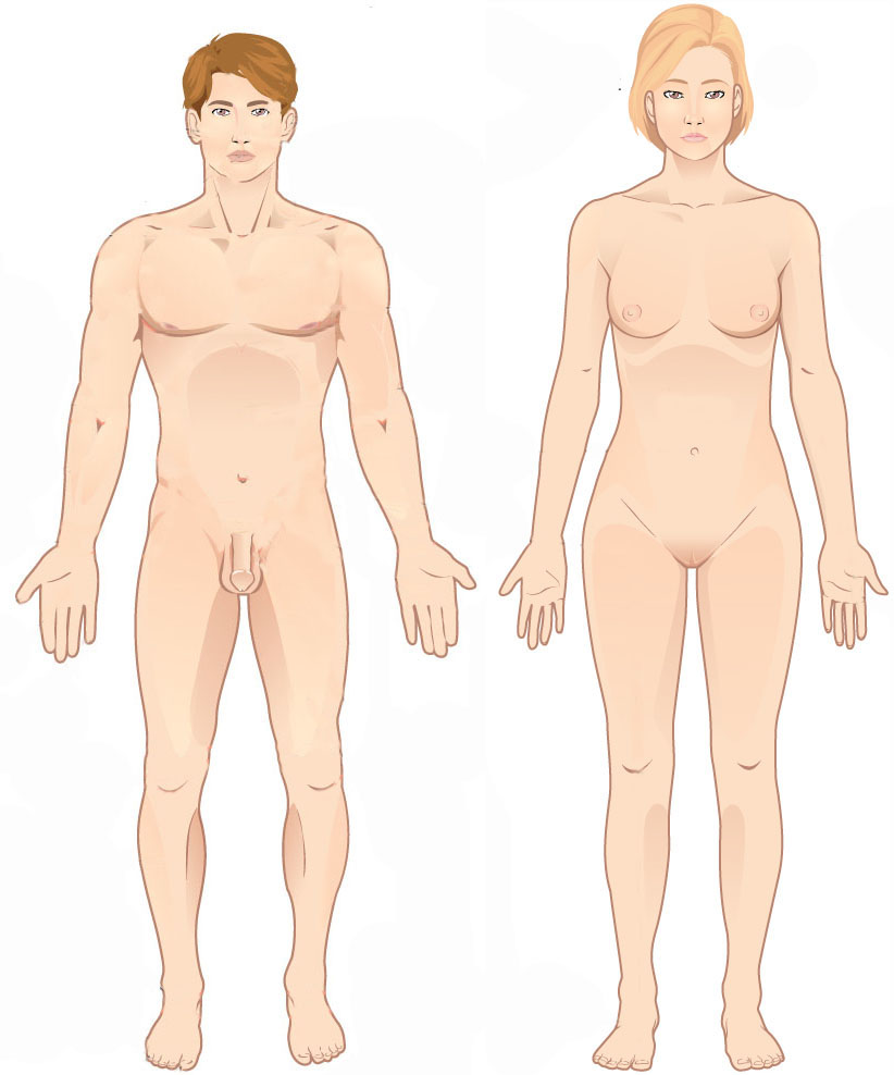
The standard anatomical position, or standard anatomical model, is the scientifically agreed upon reference position for Anatomical terms of location, anatomical location terms. Standard anatomical positions are used to standardise the position of Appendage, appendages of animals with respect to the main body of the organism. In medical disciplines, all references to a location on or in the body are made based upon the standard anatomical position. A straight position is assumed when describing a wikt:proximal, proximo-wikt:distal, distal axis (towards or away from a point of attachment). This helps avoid confusion in terminology when referring to the same organism in different postures. For example, if the elbow is flexed, the hand remains distal to the shoulder even if it approaches the shoulder. Human anatomy In standard anatomical position, the human body is standing erect and at rest. Unlike the situation in other vertebrates, the limbs are placed in positions reminiscent ... [...More Info...] [...Related Items...] OR: [Wikipedia] [Google] [Baidu] |
Posterior (anatomy)
Standard anatomical terms of location are used to unambiguously describe the anatomy of animals, including humans. The terms, typically derived from Latin or Greek language, Greek roots, describe something in its standard anatomical position. This position provides a definition of what is at the front ("anterior"), behind ("posterior") and so on. As part of defining and describing terms, the body is described through the use of anatomical planes and anatomical axis, anatomical axes. The meaning of terms that are used can change depending on whether an organism is bipedal or quadrupedal. Additionally, for some animals such as invertebrates, some terms may not have any meaning at all; for example, an animal that is radially symmetrical will have no anterior surface, but can still have a description that a part is close to the middle ("proximal") or further from the middle ("distal"). International organisations have determined vocabularies that are often used as standard vocabular ... [...More Info...] [...Related Items...] OR: [Wikipedia] [Google] [Baidu] |
Muscle And Fitness
''Muscle & Fitness'' is an American fitness and bodybuilding magazine founded in 1935 by Canadian entrepreneur Joe Weider. It was originally published under the title ''Your Physique'', before being renamed to ''Muscle Builder'' in 1954, and acquiring its current name in 1980. There is also a companion magazine called ''Muscle and Fitness Hers'', oriented toward women. History ''Muscle & Fitness'' has a more mainstream fitness and bodybuilding lifestyle focus than its companion publication, ''Flex'', which mainly covers more specialised "hardcore" and professional bodybuilding topics. It offers many exercise and nutrition tips, while at the same time advertising a variety of nutritional supplements from companies. Many professional bodybuilders are featured in each monthly issue of ''Muscle & Fitness'', such as Gustavo Badell, Darrem Charles, Ronnie Coleman, and Jay Cutler. Figure competitors such as Monica Brant, Jenny Lynn, and Davana Medina are also featured, as are enterta ... [...More Info...] [...Related Items...] OR: [Wikipedia] [Google] [Baidu] |
Cephalic Vein
In human anatomy, the cephalic vein is a superficial vein in the arm. It originates from the radial end of the dorsal venous network of hand, and ascends along the radial (lateral) side of the arm before emptying into the axillary vein. At the elbow, it communicates with the basilic vein via the median cubital vein. Anatomy The cephalic vein is situated within the superficial fascia along the anterolateral surface of the biceps. Origin The cephalic vein forms over the anatomical snuffbox at the radial end of the dorsal venous network of hand. Course and relations From its origin, it ascends ascends up the lateral aspect of the radius. Near the shoulder, the cephalic vein passes between the deltoid and pectoralis major muscles (deltopectoral groove) and through the clavipectoral triangle, where it empties into the axillary vein. Anastomoses It communicates with the basilic vein via the median cubital vein at the elbow. Clinical significance The cephalic vein ... [...More Info...] [...Related Items...] OR: [Wikipedia] [Google] [Baidu] |
Pectoralis Major
The pectoralis major () is a thick, fan-shaped or triangular convergent muscle, situated at the chest of the human body. It makes up the bulk of the chest muscles and lies under the breast. Beneath the pectoralis major is the pectoralis minor, a thin, triangular muscle. The pectoralis major's primary functions are flexion, adduction, and internal rotation of the humerus. The pectoral major may colloquially be referred to as "pecs", "pectoral muscle", or "chest muscle", because it is the largest and most superficial muscle in the chest area. Structure It arises from the anterior surface of the sternal half of the clavicle from breadth of the half of the anterior surface of the sternum, as low down as the attachment of the cartilage of the sixth or seventh rib; from the cartilages of all the true ribs, with the exception, frequently, of the first or seventh, and from the aponeurosis of the abdominal external oblique muscle. From this extensive origin the fibers converge toward the ... [...More Info...] [...Related Items...] OR: [Wikipedia] [Google] [Baidu] |
Anterior
Standard anatomical terms of location are used to unambiguously describe the anatomy of animals, including humans. The terms, typically derived from Latin or Greek roots, describe something in its standard anatomical position. This position provides a definition of what is at the front ("anterior"), behind ("posterior") and so on. As part of defining and describing terms, the body is described through the use of anatomical planes and anatomical axes. The meaning of terms that are used can change depending on whether an organism is bipedal or quadrupedal. Additionally, for some animals such as invertebrates, some terms may not have any meaning at all; for example, an animal that is radially symmetrical will have no anterior surface, but can still have a description that a part is close to the middle ("proximal") or further from the middle ("distal"). International organisations have determined vocabularies that are often used as standard vocabularies for subdisciplines of anatomy ... [...More Info...] [...Related Items...] OR: [Wikipedia] [Google] [Baidu] |
Muscle Fiber
A muscle cell is also known as a myocyte when referring to either a cardiac muscle cell (cardiomyocyte), or a smooth muscle cell as these are both small cells. A skeletal muscle cell is long and threadlike with many nuclei and is called a muscle fiber. Muscle cells (including myocytes and muscle fibers) develop from embryonic precursor cells called myoblasts. Myoblasts fuse to form multinucleated skeletal muscle cells known as syncytia in a process known as myogenesis. Skeletal muscle cells and cardiac muscle cells both contain myofibrils and sarcomeres and form a striated muscle tissue. Cardiac muscle cells form the cardiac muscle in the walls of the heart chambers, and have a single central nucleus. Cardiac muscle cells are joined to neighboring cells by intercalated discs, and when joined in a visible unit they are described as a ''cardiac muscle fiber''. Smooth muscle cells control involuntary movements such as the peristalsis contractions in the esophagus and stomach. Sm ... [...More Info...] [...Related Items...] OR: [Wikipedia] [Google] [Baidu] |


