|
Tanycytes
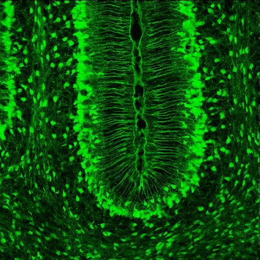
Tanycytes are special ependymal cells found in the third ventricle of the brain, and on the floor of the fourth ventricle and have processes extending deep into the hypothalamus. It is possible that their function is to transfer chemical signals from the cerebrospinal fluid to the central nervous system. The term ''tanycyte'' comes from the Greek word tanus which means elongated. Structure Tanycytes share some features with radial glial cells and astrocytes. Their form and location have led some authors to regard them as radial glial cells that remain in the hypothalamus throughout life. This has led some to believe that these cells share the same lineage. Even so, tanycytes also display certain characteristics that distinguish them from radial glia cells. Tanycytes in rats begin to develop in the last two days of gestation and continue on until they reach their full differentiation in the first month of life. Radial glia cells on the other hand, are a key component of the embryo ... [...More Info...] [...Related Items...] OR: [Wikipedia] [Google] [Baidu] |
Circumventricular Organs
Circumventricular organs (CVOs) ( circum-: around ; ventricular: of ventricle) are structures in the brain characterized by their extensive and highly permeable capillaries, unlike those in the rest of the brain where there exists a blood–brain barrier (BBB) at the capillary level. Although the term "circumventricular organs" was originally proposed in 1958 by Austrian anatomist Helmut O. Hofer concerning structures around the brain ventricular system, the penetration of blood-borne dyes into small specific CVO regions was discovered in the early 20th century. The ''permeable'' CVOs enabling rapid neurohumoral exchange include the subfornical organ (SFO), the area postrema (AP), the vascular organ of lamina terminalis (VOLT — also known as the ''organum vasculosum of the lamina terminalis'' (OVLT)), the median eminence, the pituitary neural lobe, and the pineal gland. The circumventricular organs are midline structures around the third and fourth ventricles that a ... [...More Info...] [...Related Items...] OR: [Wikipedia] [Google] [Baidu] |
Arcuate Nucleus
The arcuate nucleus of the hypothalamus (also known as ARH, ARC, or infundibular nucleus) is an aggregation of neurons in the mediobasal hypothalamus, adjacent to the third ventricle and the median eminence. The arcuate nucleus includes several important and diverse populations of neurons that help mediate different neuroendocrine and physiological functions, including neuroendocrine neurons, centrally projecting neurons, and astrocytes. The populations of neurons found in the arcuate nucleus are based on the hormones they secrete or interact with and are responsible for hypothalamic function, such as regulating hormones released from the pituitary gland or secreting their own hormones. Neurons in this region are also responsible for integrating information and providing inputs to other nuclei in the hypothalamus or inputs to areas outside this region of the brain. These neurons, generated from the ventral part of the periventricular epithelium during embryonic development, loca ... [...More Info...] [...Related Items...] OR: [Wikipedia] [Google] [Baidu] |
Third Ventricle
The third ventricle is one of the four connected ventricles of the ventricular system within the mammalian brain. It is a slit-like cavity formed in the diencephalon between the two thalami, in the midline between the right and left lateral ventricles, and is filled with cerebrospinal fluid (CSF). Running through the third ventricle is the interthalamic adhesion, which contains thalamic neurons and fibers that may connect the two thalami. Structure The third ventricle is a narrow, laterally flattened, vaguely rectangular region, filled with cerebrospinal fluid, and lined by ependyma. It is connected at the superior anterior corner to the lateral ventricles, by the interventricular foramina, and becomes the cerebral aqueduct (''aqueduct of Sylvius'') at the posterior caudal corner. Since the interventricular foramina are on the lateral edge, the corner of the third ventricle itself forms a bulb, known as the ''anterior recess'' (it is also known as the ''bulb of the ... [...More Info...] [...Related Items...] OR: [Wikipedia] [Google] [Baidu] |
Ventricular System
The ventricular system is a set of four interconnected cavities known as cerebral ventricles in the brain. Within each ventricle is a region of choroid plexus which produces the circulating cerebrospinal fluid (CSF). The ventricular system is continuous with the central canal of the spinal cord from the fourth ventricle, allowing for the flow of CSF to circulate. All of the ventricular system and the central canal of the spinal cord are lined with ependyma, a specialised form of epithelium connected by tight junctions that make up the blood–cerebrospinal fluid barrier. Structure The system comprises four ventricles: * lateral ventricles right and left (one for each hemisphere) * third ventricle * fourth ventricle There are several foramina, openings acting as channels, that connect the ventricles. The interventricular foramina (also called the foramina of Monro) connect the lateral ventricles to the third ventricle through which the cerebrospinal fluid can flow. ... [...More Info...] [...Related Items...] OR: [Wikipedia] [Google] [Baidu] |
Median Eminence
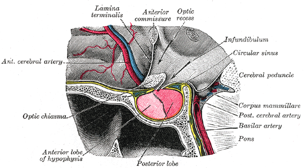
The median eminence, part of the inferior boundary of the hypothalamus in the brain, is attached to the infundibulum. The median eminence is a small swelling on the tuber cinereum, posterior to and atop the pituitary stalk; it lies in the area roughly bounded on its posterolateral region by the cerebral peduncles, and on its anterolateral region by the optic chiasm. As one of the seven areas of the brain devoid of a blood–brain barrier, the median eminence is a circumventricular organ having permeable capillaries. Its main function is as a gateway for release of hypothalamic hormones, although it does share contiguous perivascular spaces with the adjacent hypothalamic arcuate nucleus, indicating a potential sensory role. __TOC__ Physiology The median eminence is a part of the hypothalamus from which regulatory hormones are released. It is integral to the hypophyseal portal system, which connects the hypothalamus with the pituitary gland. The pars nervosa (part of the p ... [...More Info...] [...Related Items...] OR: [Wikipedia] [Google] [Baidu] |
Gonadotropin-releasing Hormone
Gonadotropin-releasing hormone (GnRH) is a releasing hormone responsible for the release of follicle-stimulating hormone (FSH) and luteinizing hormone (LH) from the anterior pituitary. GnRH is a tropic peptide hormone synthesized and released from GnRH neurons within the hypothalamus. The peptide belongs to gonadotropin-releasing hormone family. It constitutes the initial step in the hypothalamic–pituitary–gonadal axis. Structure The identity of GnRH was clarified by the 1977 Nobel Laureates Roger Guillemin and Andrew V. Schally: pyroGlu-His-Trp-Ser-Tyr-Gly-Leu-Arg-Pro-Gly-NH2 As is standard for peptide representation, the sequence is given from amino terminus to carboxyl terminus; also standard is omission of the designation of chirality, with assumption that all amino acids are in their L- form. The abbreviations are the standard abbreviations for the corresponding proteinogenic amino acids, except for ''pyroGlu'', which refers to pyroglutamic acid, a derivative ... [...More Info...] [...Related Items...] OR: [Wikipedia] [Google] [Baidu] |
Hypophyseal Portal System
The hypophyseal portal system is a system of blood vessels in the microcirculation at the base of the brain, connecting the hypothalamus with the anterior pituitary. Its main function is to quickly transport and exchange hormones between the hypothalamus arcuate nucleus and anterior pituitary gland. The capillaries in the portal system are fenestrated (have many small channels with high vascular permeability) which allows a rapid exchange between the hypothalamus and the pituitary. The main hormones transported by the system include gonadotropin-releasing hormone, corticotropin-releasing hormone, growth hormone–releasing hormone, and thyrotropin-releasing hormone. Structure The blood supply and direction of flow in the hypophyseal portal system has been studied over many years on laboratory animals and human cadaver specimens with injection and vascular corrosion casting methods. Short portal vessels between the neural and anterior pituitary lobes provide an avenue for rap ... [...More Info...] [...Related Items...] OR: [Wikipedia] [Google] [Baidu] |
Spinal Cord
The spinal cord is a long, thin, tubular structure made up of nervous tissue, which extends from the medulla oblongata in the brainstem to the lumbar region of the vertebral column (backbone). The backbone encloses the central canal of the spinal cord, which contains cerebrospinal fluid. The brain and spinal cord together make up the central nervous system (CNS). In humans, the spinal cord begins at the occipital bone, passing through the foramen magnum and then enters the spinal canal at the beginning of the cervical vertebrae. The spinal cord extends down to between the first and second lumbar vertebrae, where it ends. The enclosing bony vertebral column protects the relatively shorter spinal cord. It is around long in adult men and around long in adult women. The diameter of the spinal cord ranges from in the cervical and lumbar regions to in the thoracic area. The spinal cord functions primarily in the transmission of nerve signals from the motor cortex to the b ... [...More Info...] [...Related Items...] OR: [Wikipedia] [Google] [Baidu] |
Gestation
Gestation is the period of development during the carrying of an embryo, and later fetus, inside viviparous animals (the embryo develops within the parent). It is typical for mammals, but also occurs for some non-mammals. Mammals during pregnancy can have one or more gestations at the same time, for example in a multiple birth. The time interval of a gestation is called the ''gestation period''. In obstetrics, '' gestational age'' refers to the time since the onset of the last menses, which on average is fertilization age plus two weeks. Mammals In mammals, pregnancy begins when a zygote (fertilized ovum) implants in the female's uterus and ends once the fetus leaves the uterus during labor or an abortion (whether induced or spontaneous). Humans In humans, pregnancy can be defined clinically or biochemically. Clinically, pregnancy starts from first day of the mother's last period. Biochemically, pregnancy starts when a woman's human chorionic gonadotropin (hCG) le ... [...More Info...] [...Related Items...] OR: [Wikipedia] [Google] [Baidu] |
Brain
The brain is an organ that serves as the center of the nervous system in all vertebrate and most invertebrate animals. It consists of nervous tissue and is typically located in the head ( cephalization), usually near organs for special senses such as vision, hearing and olfaction. Being the most specialized organ, it is responsible for receiving information from the sensory nervous system, processing those information (thought, cognition, and intelligence) and the coordination of motor control (muscle activity and endocrine system). While invertebrate brains arise from paired segmental ganglia (each of which is only responsible for the respective body segment) of the ventral nerve cord, vertebrate brains develop axially from the midline dorsal nerve cord as a vesicular enlargement at the rostral end of the neural tube, with centralized control over all body segments. All vertebrate brains can be embryonically divided into three parts: the forebrain (prosencep ... [...More Info...] [...Related Items...] OR: [Wikipedia] [Google] [Baidu] |
Astrocyte
Astrocytes (from Ancient Greek , , "star" + , , "cavity", "cell"), also known collectively as astroglia, are characteristic star-shaped glial cells in the brain and spinal cord. They perform many functions, including biochemical control of endothelial cells that form the blood–brain barrier, provision of nutrients to the nervous tissue, maintenance of extracellular ion balance, regulation of cerebral blood flow, and a role in the repair and scarring process of the brain and spinal cord following infection and traumatic injuries. The proportion of astrocytes in the brain is not well defined; depending on the counting technique used, studies have found that the astrocyte proportion varies by region and ranges from 20% to 40% of all glia. Another study reports that astrocytes are the most numerous cell type in the brain. Astrocytes are the major source of cholesterol in the central nervous system. Apolipoprotein E transports cholesterol from astrocytes to neurons and other gli ... [...More Info...] [...Related Items...] OR: [Wikipedia] [Google] [Baidu] |
Radial Glial Cell
Radial glial cells, or radial glial progenitor cells (RGPs), are bipolar-shaped progenitor cells that are responsible for producing all of the neurons in the cerebral cortex. RGPs also produce certain lineages of glia, including astrocytes and oligodendrocytes. Their cell bodies ( somata) reside in the embryonic ventricular zone, which lies next to the developing ventricular system. During development, newborn neurons use radial glia as scaffolds, traveling along the radial glial fibers in order to reach their final destinations. Despite the various possible fates of the radial glial population, it has been demonstrated through clonal analysis that most radial glia have restricted, unipotent or multipotent, fates. Radial glia can be found during the neurogenic phase in all vertebrates (studied to date). The term "radial glia" refers to the morphological characteristics of these cells that were first observed: namely, their radial processes and their similarity to astrocytes, ... [...More Info...] [...Related Items...] OR: [Wikipedia] [Google] [Baidu] |


